[1] 李永耀,赵勇,程灏,等.夹板弹性固定桡骨远端FrykmanⅧ型骨折尺侧柱稳定性的有限元分析[J].中国组织工程研究,2020, 24(30):4769-4774.
[2] 毛昭冲, 李福伟,杨凤云,等.有限元分析在桡骨远端骨折研究中的应用进展[J].中医正骨,2023,35(2):30-32+45.
[3] PURUSHOTHAMAN Y, CHOI H, YOGANANDAN N, et al. A Comparison Study of Four Cervical Disk Arthroplasty Devices Using Finite Element Models. Asian Spine J. 2021;15(3):283-293.
[4] GHOSH R, CHANDA S, CHAKRABORTY D. Application of finite element analysis to tissue differentiation and bone remodelling approaches and their use in design optimization of orthopaedic implants: A review. Int J Numer Method Biomed Eng. 2022;38(10):e3637.
[5] SPANSWICK PJC, WHITTIER DE, KWONG C, et al. Improvements in radiographic and clinical assessment of distal radius fracture healing by FE-estimated bone stiffness. Bone Rep. 2021;14:100748.
[6] MALAKOUTIKHAH H, LATT LD. Disease-Specific Finite element Analysis of the Foot and Ankle. Foot Ankle Clin. 2023;28(1):155-172.
[7] LEWIS GS, MISCHLER D, WEE H, et al. Finite Element Analysis of Fracture Fixation. Curr Osteoporos Rep. 2021;19(4):403-416.
[8] 张宁,康颂科,李伟延,等.三种空心钉构型踝关节融合的有限元分析[J].中国矫形外科杂志,2024,32(8):744-749.
[9] 高升,辛世民,刘兴权,等.基于影像学腰椎退变的生物力学有限元分析[J].中国组织工程研究,2026,30(15):3810-3819.
[10] 张子峥,罗旺,刘长路,等.膝内侧间室骨关节炎单髁置换中有限元分析的应用价值[J].中国组织工程研究,2026,30(9): 2313-2322.
[11] 吴志鹏,高伟阳,吴立军.腕关节生物力学有限元分析研究进展[J].国际骨科学杂志,2008(5):307-309.
[12] 颜冰珊. 尺骨茎突骨折与下尺桡关节稳定性关系的有限元分析[D]. 上海:复旦大学,2009.
[13] 郭欣,樊瑜波,李宗明. 掌骨受轴向压力作用下的腕部生物力学分析[J]. 航天医学与医学工程,2008,21(1):45-49.
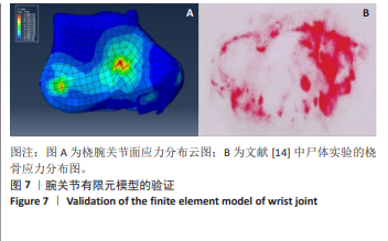
[14] 钦斌,黄永火,欧阳羽,等. 轴向应力作用下的舟骨有限元分析[J]. 第三军医大学学报,2010,32(11):1213-1215.
[15] 周晓宁.腕关节三维有限元模型的建立及桡骨远端骨折发生机制的生物力学分析[D]. 北京:北京中医药大学,2014.
[16] KAPANDJI AI. 顾冬云等译. 骨关节功能解剖学[M]. 北京:人民军医出版社,2011.
[17] 马志虎,于晓凤,孙玉亮,等.加速康复外科综合治疗在三角纤维软骨复合体损伤患者围术期中的应用[J].山东大学学报(医学版), 2025,63(3):28-35.
[18] 闫立强,胡坤然,王丰羽,等.带线骨锚钉重建腕背韧带治疗腕关节骨折脱位[J].临床骨科杂志,2024,27(1):70-74.
[19] 黄子阳,谢威,练克俭,等.桡骨远端骨折畸形愈合的外科治疗进展[J].中国中医骨伤科杂志,2022,30(11):80-84+88.
[20] 顾伟民,陆耀刚,王子平.桡骨远端骨折与腕关节不稳的临床研究[J].中华创伤骨科杂志,2006,8(3):212-215.
[21] KIJIMA Y, VIEGAS SF. Wrist anatomy and biomechanics. J Hand Surg Am. 2009;34(8):1555-63.
[22] LI ZM, JORDAN DB. Carpal tunnel mechanics and its relevance to carpal tunnel syndrome. Hum Mov Sci. 2023;87:103044.
[23] TAN J, ZHANG F, LIU Q, et al. Effect of different ulnar osteotomies on loading of the distal radioulnar joint: a finite element analysis. BMC Musculoskelet Disord. 2024;25(1):454.
[24] LIAO L, GU F, XIONG F, et al. Biomechanical Analysis of Transosseous Repair Versus Combined Transosseous With Capsular Repair for Triangular Fibrocartilage Complex Tears With Instability. J Hand Surg Am. 2025;50(6):752.e1-752.e8.
[25] CHAN YS, CHEN AC, CHEN CC, et al. The effect of lateral hinge fracture with hinge hole and protective K-wire for medial opening-wedge high tibial osteotomy by compression testing and finite element analysis. J Orthop Surg Res. 2025;20(1):598.
[26] GARAVELLI C, ALDIERI A, PALANCA M, et al. Comparing the predictions of CT-based subject-specific finite element models of human metastatic vertebrae with digital volume correlation measurements. Biomech Model Mechanobiol. 2025;24(3):1017-1030.
[27] MEJÍA RODRÍGUEZ M, GONZÁLEZ-ESTRADA OA, SÁNCHEZ-RESTREPO HD. Custom design of a temporomandibular joint prosthesis: A kinematic approach evaluated by finite element analysis. J Prosthet Dent. 2025;133(6):1580.e1-1580.e10.
[28] 岳肖华,李晏乐,程灏,等.尺骨茎突骨折有限元模型的建立与力学分析[J].中国中医骨伤科杂志,2018,26(10):6-9.
[29] 张必欢,蔡兴博,王斌,等.个性化3D打印钛合金舟骨部分置换术后有限元模型的建立及生物力学分析[J].中国临床解剖学杂志, 2024,42(3):293-303.
[30] 魏明杰,许育健,吴一芃,等.手腕部不同载荷状态下舟月骨间韧带应力分布分析[J].中国临床解剖学杂志,2021,39(5):
586-592.
[31] 谢澄铖,薛俊伟,黄晨阳,等.谢式夹板和解剖夹板外固定治疗伸直型桡骨远端骨折的生物力学特性比较[J].中国中医骨伤科杂志, 2024,32(1):42-47.
[32] MENA A, WOLLSTEIN R, BAUS J, et al. Finite Element Modeling of the Human Wrist: A Review. J Wrist Surg. 2023;12(6):478-487.
[33] MENA A, WOLLSTEIN R, YANG J. Development of a Finite Element Model of the Human Wrist Joint With Radial and Ulnar Axial Force Distribution and Radiocarpal Contact Validation. J Biomech Eng. 2025; 147(3):031006.
[34] 侯泽欣,许本柯,戴媛,等.老年人跌倒腕关节背伸损伤机制的有限元分析[J].中国组织工程研究,2024,28(6):886-890.
[35] 姜昆,陶宝琛,魏成建.小夹板治疗桡骨远端关节内骨折的有限元分析[J].医用生物力学,2018,33(3):206-211.
[36] AZAR FM, BEATY JH. 唐佩福等译. 坎贝尔骨科手术学[M]. 14版. 北京:北京大学医学出版社,2023.
[37] 王亦璁,姜保国.骨与关节损伤[M].北京:人民卫生出版社, 2012.
[38] 邢飞,马剑雄,马信龙. 软组织生物力学特性研究进展[J]. 中华骨科杂志,2017,37(22):1432-1440.
[39] 呙金海,黄富国.近排腕骨间不稳的生物力学研究进展[J].中国修复重建外科杂志,2015,29(1):108-112.
[40] ESCHWEILER J, LI J, QUACK V, et al. Anatomy, Biomechanics, and Loads of the Wrist Joint. Life (Basel). 2022;12(2):188.
[41] FORSTMANN M, YUDKIN DA, PROSSER AMB, et al. Transformative experience and social connectedness mediate the mood-enhancing effects of psychedelic use in naturalistic settings. Proc Natl Acad Sci U S A. 2020;117(5):2338-2346.
[42] 夏长江,袁志峰,方宁.基于尺桡骨三维有限元模型分析桡骨远端骨折的生物力学特征[J].中国组织工程研究,2020,24(6):893-897.
[43] 王爱国,陆贲琪,李沿鑫,等.桡骨远端骨折夹板外固定有限元模型的建立及其愈合过程的应力分析[J].中国中医骨伤科杂志, 2024,32(3):7-11.
[44] 陶宝琛,石耀武,夏均青,等.动力气囊压垫纠正桡骨远端AO C3.1型骨折手法复位后残余成角移位的有限元分析研究[J].山东中医杂志,2020,39(8):791-796.
[45] HALLGREN HB, NICOLESCU D, TÖRNQVIST L, et al. Ultrasonographic examination of acute soft tissue lesions in the elbow has good inter-rater reliability and acceptable agreement with magnetic resonance imaging. J Shoulder Elbow Surg. 2024;33(7):1615-1623.
[46] GARCIA-ELIAS M, ANANOS D, ESPLUGAS M, et al. Ligaments and muscles stabilizing the radio-ulno-carpal joint. J Hand Surg Eur Vol. 2022;47(1):65-72.
[47] WEBER A, REISSNER L, FRIEDL S, et al. Stability of the distal radioulnar joint with and without activation of forearm muscles. J Hand Surg Eur Vol. 2023;48(8):762-767.
[48] NAKAMURA T. Classifications of Triangular Fibrocartilage Complex Lesions. J Wrist Surg. 2024;13(1):1.
[49] BOECKSTYNS MEH, HERZBERG G. Complications after total wrist arthroplasty. J Hand Surg Eur Vol. 2024;49(2):177-187.
[50] REIGSTAD O. Total wrist arthroplasty: recent advances and current recommendations. J Hand Surg Eur Vol. 2025;50(1):34-41.
[51] 张浩,朱建民,马南,等. 腕关节有限元骨性建模及力学分析[J]. 江苏大学学报(医学版),2013,23(1):53-56.
[52] 李永耀,程灏,赵勇,等.夹板固定治疗尺骨茎突骨折的三维有限元分析[J].中国组织工程研究,2018,22(11):1737-1742. |