Chinese Journal of Tissue Engineering Research ›› 2026, Vol. 30 ›› Issue (18): 4582-4593.doi: 10.12307/2026.699
Previous Articles Next Articles
Finite element analysis of digitally designed free fibula flaps for repairing unilateral maxillary defects
Zhai Kun1, Liu Dongyang1, Ma Jian1, Lin Zhiyu2, Zheng Maosheng2, Ma Xiaoqin2, Jing Jie1
- 1Department of Oral and Maxillofacial Surgery, General Hospital of Ningxia Medical University, Yinchuan 750004, Ningxia Hui Autonomous Region, China; 2School of Stomatology, Ningxia Medical University, Yinchuan 750004, Ningxia Hui Autonomous Region, China
-
Received:2025-05-19Accepted:2025-09-17Online:2026-06-28Published:2025-12-02 -
Contact:Jing Jie, PhD., Chief physician, Professor, Master's supervisor, Department of Oral and Maxillofacial Surgery, General Hospital of Ningxia Medical University, Yinchuan 750004, Ningxia Hui Autonomous Region, China -
About author:Zhai Kun, MS, Attending physician, Department of Oral and Maxillofacial Surgery, General Hospital of Ningxia Medical University, Yinchuan 750004, Ningxia Hui Autonomous Region, China -
Supported by:Natural Science Foundation of Ningxia Hui Autonomous Region, Nos. 2021AAC03377 (to ZK), 2020AAC03357 (to JJ)
CLC Number:
Cite this article
Zhai Kun, Liu Dongyang, Ma Jian, Lin Zhiyu, Zheng Maosheng, Ma Xiaoqin, Jing Jie. Finite element analysis of digitally designed free fibula flaps for repairing unilateral maxillary defects[J]. Chinese Journal of Tissue Engineering Research, 2026, 30(18): 4582-4593.
share this article
Add to citation manager EndNote|Reference Manager|ProCite|BibTeX|RefWorks
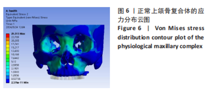
2.1 正常上颌骨复合体Von Mises应力分布情况 在双侧后牙区各施加300 N的垂直均布载荷下,正常颅上颌复合体(模型 A)Von Mises应力分布如图6示,其应力主要集中在双侧上颌骨近颧骨区域、双侧梨状孔外侧缘、双侧颧弓、眶内外侧缘及鼻根部区域且应力传导方向为向上传导,这与上颌骨在受到外力时,力沿着鼻旁支柱、颧突支柱、翼突支柱传导和分散的临床实际情况相符。"
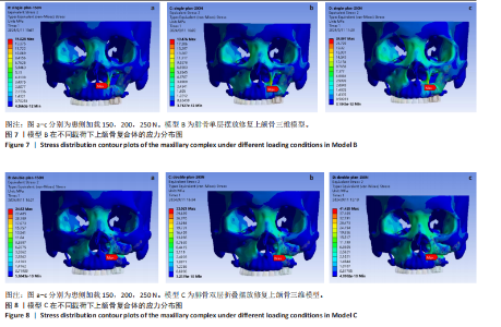
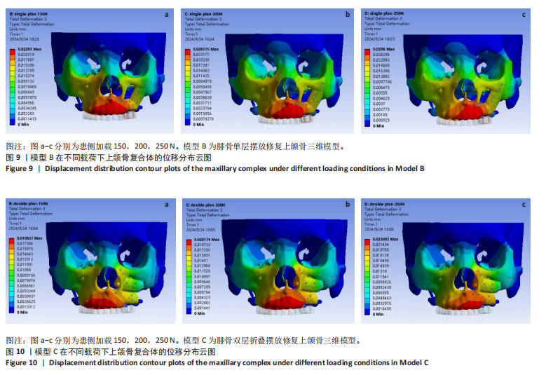
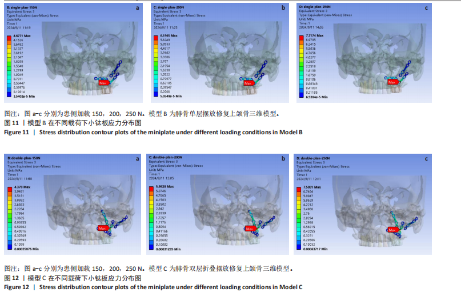
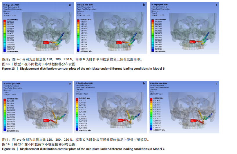
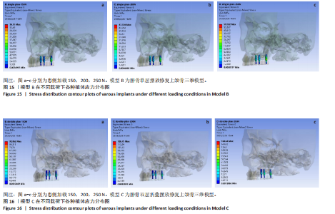
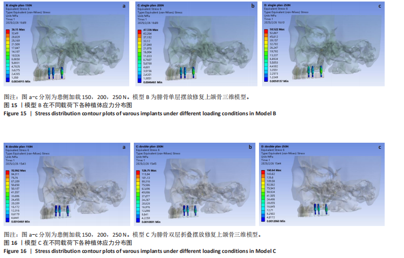
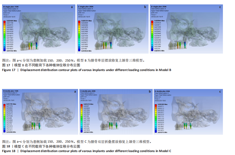
2.2 重建后模型上颌骨复合体Von Mises应力分布情况 重建后模型上颌骨在受到不同载荷下的应力分布如图7,8所示。从两模型应力分布云图中可观察到模型B及模型C上颌骨在患侧后牙区受到150,200,250 N的不同载荷下,两模型应力均随着载荷的变大逐渐分布在健侧上颌骨近颧骨区域、重建侧腓骨与牙槽突连接区域、腓骨与腓骨连接区域以及双侧眶外侧、眶内侧和鼻根部区域;模型B随着患侧受力的增大,最大应力主要位于左侧梨状孔边缘和两腓骨段之间的转折处,且在患侧150 N 的载荷下,左侧梨状孔边缘出现红色应力集中区;模型 C在患侧3种载荷下,最大应力均位于下层腓骨上缘与上层腓骨接触的区域,且无明显红色应力集中区。 2.3 重建后模型上颌骨复合体位移情况 重建后两模型上颌骨复合体在受到不同载荷下的位移云图如图9,10所示。从图中可观察到模型B与模型 C上颌骨复合体在患侧不同载荷下位移趋势接近一致,最大位移出现在上颌骨牙槽突前部和前鼻棘处,向两侧颧骨颧弓及鼻根部区域递减,在腓骨端与牙槽突断端的连接区域模型B位移明显大于模型C。模型B与模型C上颌骨复合体位移值随着患侧载荷的增加而增加,模型 B 在不同载荷下位移值均大于模型 C,两模型上颌骨复合体位移最大值出现在患侧加载250 N时,模型B为30 μm,模型C为 23 μm。 2.4 重建后模型小钛板Von Mises应力分布 重建后模型B与模型C小钛板Von Mises应力分布如图11,12所示,从图中可以观察到模型B与模型C小钛板应力值随着患侧载荷的增大而增大,且两模型的最大应力均位于连接腓骨段与腓骨段之间的转折处,两模型小钛板的最大应力值在各载荷下较为接近。 2.5 重建后模型小钛板位移情况 从模型B与模型C小钛板不同载荷下的位移云图发现,两模型小钛板位移值随着患侧载荷的增加而增加,模型B与模型C小钛板的最大位移在各载荷下均出现在连接腓骨与牙槽突断端小钛板的第一个钉孔处,如图13,14示,两模型位移最大值出现在模型B患侧加载250 N时,为27 μm。 2.6 重建后模型种植体Von Mises应力分布及位移情况 重建后模型 B 与模型C各种植体在3种载荷下 Von Mises 应力分布如图15,16所示,从图中可以发现两模型各种植体在不同载荷下应力均集中于颈部区域,同时应力值也随着患侧载荷的增大而增大,模型C种植体应力峰值明显大于模型B,且差异随载荷的增加而扩大;两模型种植体的应力最大值均位于末端种植体上,模型 B 最大应力为58.5 MPa,模型C为160.6 MPa。在患侧不同载荷下,模型 B 与模型 C 最大位移均位于前端种植体上,位移值随载荷的增大而增大,且模型C最大位移值略小于模型 B,见图17,18。"


"
"


"
"

"
| [1] LEE ZH, CRIPPS C, RODRIGUEZ ED. Current Concepts in Maxillary Reconstruction. Plast Reconstr Surg. 2022;150(1):168e-175e. [2] 汪洋洋.单侧完全性唇腭裂患者上颌骨三维有限元模型的建立及牙槽突植骨的生物力学分析[D].南昌:南昌大学,2023. [3] DE GROOT RJ, RIEGER JM, ROSENBERG A, et al. A pilot study of masticatory function after maxillectomy comparing rehabilitation with an obturator prosthesis and reconstruction with a digitally planned, prefabricated, free, vascularized fibula flap. J Prosthet Dent. 2020;124(5):616-622. [4] 孙嘉怿,徐鹏,王丽珍,等.基于有限元的单侧缺损上颌骨不同修复方法的生物力学研究[J].生物医学工程学杂志,2014,31(3): 590-595+605. [5] 毕丽霞,孙嘉怿,王燕一,等.基于有限元单侧上颌骨缺损修复材料的生物力学研究[J].医用生物力学,2014,29(1):72-77. [6] 郑玲玲,陈丹,王涛,等.上颌骨复杂缺损3D打印精准重建支架的生物力学研究[J].医用生物力学,2022,37(6):1101-1106. [7] HIDALGO DA. Fibula free flap: a new method of mandible reconstruction. Plastic Reconstr Surg. 1989;84(1):71-79. [8] 王洁,王瑞霞,袁华,等.骨移植复合种植义齿进行颌骨缺损功能重建的临床研究[J].口腔医学,2013,33(12):793-795. [9] MORRISON EJ, MATROS E. Modern Oncologic Maxillary Reconstruction. Plast Reconstr Surg. 2024;154(3):601e-618e. [10] 李劲松,李群星.3D打印技术在颌骨精确功能性重建的应用[J].口腔疾病防治,2023,31(6):381-388. [11] 孙坚,沈毅,李军,等.上颌骨功能性修复中骨性支柱重建的生物力学分析[J].中国口腔颌面外科杂志,2010,8(1):34-39. [12] 陈诚,张琳梅,任文豪,等.游离腓骨瓣重建上颌骨半侧缺损数字化模拟和辅助设计[J].山西医科大学学报,2019,50(1):96-101. [13] KHATIB B, PATEL A, DIERKS EJ, et al. The Biaxial Double-Barrel Fibula Flap-A Simplified Technique for Fibula Maxillary Reconstruction. J Oral Maxillofac Surg. 2019;77(2):412-425. [14] 孙悦,郭蕴,李建成,等.数字化设计游离腓骨瓣重建下颌骨缺损的三维有限元分析[J].南方医科大学学报,2021,41(12):1892-1898. [15] 热依拉·库尔班,霞黑达·依拉尔江,陈欣,等.超短种植体不同修复方式的三维有限元分析[J].中国组织工程研究,2023,27(30): 4824-4829. [16] 李希光,郅克谦,高岭,等.基于三维有限元评价种植体不同倾斜角度在上颌后牙区骨量不足的应力分析[J].口腔医学研究,2019, 35(7):671-675. [17] 沈毅,孙坚,李军,等.上颌骨功能性重建中用钛植入体重建颧上颌支柱的生物力学研究[J].中国口腔颌面外科杂志,2011,9(3):198-203. [18] 焦婷,孙健,洪凌斐,等.附着体应用于单侧上颌骨缺损修复的三维有限元分析[J].上海口腔医学,2006,15(4):395-398. [19] SUDHAN RH, CHANDER GN, ANITHA KV. Finite element stress analysis of Aramany class I maxillectomy defect with single- and two-piece closed bulb obturators. Gerodontology. 2021;38(2):209-215. [20] 马戈.颌骨骨面植皮重建软组织种植床的可行性研究[D].西安: 第四军医大学,2011. [21] 李兴强,李淑薇,刘长阳,等. 股前外侧皮瓣复合个性化钛网支架在上颌骨缺损修复中的应用[J]. 现代肿瘤医学,2023,31(24):4551-4554. [22] BROWN JS, SHAW RJ. Reconstruction of the maxilla and midface: introducing a new classification. Lancet Oncol. 2010;11(10):1001-1008. [23] 吴添福,刘冰.上颌骨缺损的外科修复[J].中国实用口腔科杂志, 2021,14(5):525-530. [24] JUNG BK, YUN IS, LEE WJ, et al. Orbital floor reconstruction using a tensor fascia lata sling after total maxillectomy. J Craniomaxillofac Surg. 2016;44(5):648-653. [25] 赵珍珍,皇甫辉.3D打印技术在上颌骨切除术后缺损修复中的应用[J].中国眼耳鼻喉科杂志,2022,22(6):651-654. [26] 陈健,李暐.带蒂组织瓣在头颈部肿瘤术后缺损修复重建中的应用研究进展[J].中国修复重建外科杂志,2018,32(3):369-376. [27] 章文博,于尧,王洋,等.数字化外科技术在上颌骨缺损重建中的应用[J].北京大学学报(医学版),2017,49(1):1-5. [28] JONES NF, SWARTZ WM, MEARS DC, et al. The “double barrel” free vascularized fibular bone graft. Plast Reconstr Surg. 1988;81(3):378-385. [29] ANTHONY JP, FOSTER RD, SHARMA AB, et al. Reconstruction of a complex midfacialdefect with the folded fibular free flap and osseointegrated implants. Ann Plast Surg. 1996;37(2):204-210. [30] BAJ A, YOUSSEF DA, MONTEVERDI R, et al. Reconstruction of partial maxillary defectswith the double-barrel fibula free flap. Acta Otorhinolaryngol Ital. 2010;30(6):299-302. [31] THRESHER RW, SAITO GE. The stress analysis of human teeth. J Biomech. 1973;6(5):443-449. [32] 许崇涛,孙庚林,周健,等.颅上颌复合体三维有限元模型的建立和初步应用[J].实用口腔医学杂志,2009,25(1):51-54. [33] 张彤,刘洪臣,王延荣.不同功能状态下健康人上颌骨复合体应力分布的三维有限元分析[J].中华口腔医学杂志,2007,42(11):687-689. [34] 彭歆,毛驰,俞光岩,等.游离腓骨复合组织瓣上颌骨重建的三维有限元分析[J].现代口腔医学杂志,2005,19(6):35-37. [35] 康一帆,单小峰,张雷,等.游离腓骨瓣修复重建上颌骨术后腓骨瓣位置变化[J].北京大学学报(医学版),2020,52(5):938-942. [36] 沈毅,孙坚,李军,等.正常人上颌骨的生物力学分析[J].组织工程与重建外科杂志,2009,5(1):25-28. [37] 刘雄.下颌骨体部缺损腓骨重建小钛板固定的生物力学分析[D].广州:南方医科大学,2014. [38] COX T, KOHN MW, IMPELLUSO T. Computerized analysis of resorbable polymer plates and screws for the rigid fixation of mandibular angle fractures. J Oral Maxillofac Surg. 2003;61(4):481-487; discussion 487-488. [39] 刘尚萍,蔡志刚,张杰,等.下颌骨缺损重建术后钛板相关并发症97例临床回顾研究[J].中华口腔医学杂志,2013,48(10):586-590. [40] 朱家俊.采用不同腓骨折叠及内固定方式重建下颌骨H型缺损的有限元分析[D].昆明:昆明医科大学,2023. [41] ARBAG H, KORKMAZ HH, OZTURK K, et al. Comparative evaluation of different miniplates for internal fixation of mandible fractures using finite element analysis. J Oral Maxillofac Surg. 2008;66(6):1225-1232. [42] 林野,王兴,毛驰,等.功能性颌骨重建61例临床分析[J].中国口腔颌面外科杂志,2006,4(1):14-19. [43] ACAR G, ARI I, TOSUN E. Biomechanical evaluation of implant options for unilateral maxillary defects: a finite element analysis. BMC Oral Health. 2024;24(1):1338. [44] 甄恩明,吴昌敬,邵军.双侧上颌骨缺损颧种植体修复的有限元探讨[J].中国口腔种植学杂志,2016,21(3):107-110. [45] 李露露,孙顺涛,严洪海.种植体支持的上颌前牙两种固定修复方式在不同咬合关系下的三维有限元分析[J].口腔颌面修复学杂志,2025,26(1):14-18. [46] 木志翔,刘婷,陈陶,等.两种上颌无牙颌种植固定修复方案的有限元分析[J].口腔医学研究,2019,35(10):931-935. [47] BRUNSKI JB. Avoid pitfalls of overloading and micromotion of intraosseous implants. Dent Implantol Update. 1993;4(10):77-81. [48] 李璐丽.牙种植体植入扭矩与骨结合失效分析[D].济南:山东大学,2019. [49] 徐大鹏,景捷,马璐,等.基于种植牙愈合过程模拟上颌后牙种植体选择的生物力学分析[J]. 中国组织工程研究,2023,27(25):3942-3948. [50] ELSAYYAD AA, ABBAS NA, ABDELNABI NM, et al. Biomechanics of 3-implant-supported and 4-implant-supported mandibular screw-retained prostheses: A 3D finite element analysis study. J Prosthet Dent. 2020;124(1):68.e1-68.e10. [51] CENKOGLU BG, BALCIOGLU NB, OZDEMIR T, et al. The Effect of the Length and Distribution of Implants for Fixed Prosthetic Reconstructions in the Atrophic Posterior Maxilla: A Finite Element Analysis. Materials (Basel). 2019;12(16):2556. [52] TONIOLLO MB, VIEIRA LJP, DOS SANTOS SÁ M, et al. Stress distribution of three-unit fixed partial prostheses (conventional and pontic) supported by three or two implants: 3D finite element analysis of ductile materials. Comput Methods Biomech Biomed Engin. 2019; 22(7):706-712. [53] MEIMANDI M, TALEBI ARDAKANI MR, AMID R, et al. Comparison of Stress and Strain Distribution Around Splinted and Nonsplinted 6-mm Short Implants in Posterior Mandible: A Finite Element Analysis Study. Implant Dent. 2018;27(1):74-80. |
| [1] | Chen Huiting, Zeng Weiquan, Zhou Jianhong, Wang Jie, Zhuang Congying, Chen Peiyou, Liang Zeqian, Deng Weiming. Tail anchoring technique of vertebroplasty in treatment of osteoporotic vertebral compression fractures with intravertebral cleft: a finite element analysis [J]. Chinese Journal of Tissue Engineering Research, 2026, 30(9): 2145-2152. |
| [2] | Cheng Qisheng, Julaiti·Maitirouzi, Xiao Yang, Zhang Chenwei, Paerhati·Rexiti. Finite element analysis of novel variable-diameter screws in modified cortical bone trajectory of lumbar vertebrae [J]. Chinese Journal of Tissue Engineering Research, 2026, 30(9): 2162-2171. |
| [3] | Liu Jiafu, Ren Ruxia, Liao Zhouwei, Zhou Xiali, Wu Yihong, Zhang Shaoqun. Three-dimensional finite element analysis of cervical spine biomechanical characteristics in a rat model of cervical vertigo [J]. Chinese Journal of Tissue Engineering Research, 2026, 30(9): 2182-2190. |
| [4] | Zhang Zizheng, Luo Wang, Liu Changlu. Application value of finite element analysis on unicompartmental knee arthroplasty for medial knee compartmental osteoarthritis [J]. Chinese Journal of Tissue Engineering Research, 2026, 30(9): 2313-2322. |
| [5] | Chen Long, Wang Xiaozhen, Xi Jintao, Lu Qilin. Biomechanical performance of short-segment screw fixation combined with expandable polyetheretherketone vertebral body replacement in osteoporotic vertebrae [J]. Chinese Journal of Tissue Engineering Research, 2026, 30(9): 2226-2235. |
| [6] | Liu Wenlong, Dong Lei, Xiao Zhengzheng, Nie Yu. Finite element analysis of tibial prosthesis loosening after fixed-bearing unicompartmental knee arthroplasty for osteoporosis [J]. Chinese Journal of Tissue Engineering Research, 2026, 30(9): 2191-2198. |
| [7] | Zheng Wangyang, Fei Ji, Yang Di, Zhao Lang, Wang Lingli, Liu Peng, Li Haiyang. Finite element analysis of the force changes of the supraspinatus tendon and glenohumeral joint during the abduction and flexion of the humerus [J]. Chinese Journal of Tissue Engineering Research, 2026, 30(9): 2199-2207. |
| [8] | Cai Qirui, Dai Xiaowei, Zheng Xiaobin, Jian Sili, Lu Shaoping, Liu Texi, Liu Guoke, Lin Yuanfang. Mechanical effects of Long’s traction orthopedic method on cervical functional units: quantitative analysis of biomechanical model of head and neck [J]. Chinese Journal of Tissue Engineering Research, 2026, 30(9): 2208-2216. |
| [9] | Rao Jingcheng, Li Yuwan, Zheng Hongbing, Xu Zhi, Zhu Aixiang, Shi Ce, Wang Bing, Yang Chun, Kong Xiangru, Zhu Dawei. Biomechanical differences between the new proximal femoral stable intramedullary nail and traditional intramedullary nail#br# [J]. Chinese Journal of Tissue Engineering Research, 2026, 30(9): 2217-2225. |
| [10] | Zheng Xuying, Hu Hongcheng, Xu Libing, Han Jianmin, Di Ping. Stress magnitude and distribution in two-piece cement-retained zirconia implants under different loading conditions and with varying internal connection shapes [J]. Chinese Journal of Tissue Engineering Research, 2026, 30(8): 1979-1987. |
| [11] |
Dong Chunyang, Zhou Tianen, Mo Mengxue, Lyu Wenquan, Gao Ming, Zhu Ruikai, Gao Zhiwei.
Action mechanism of metformin combined with Eomecon chionantha Hance dressing in treatment of deep second-degree burn wounds#br#
#br#
[J]. Chinese Journal of Tissue Engineering Research, 2026, 30(8): 2001-2013.
|
| [12] | Yang Xuetao, Zhu Menghan, Zhang Chenxi, Sun Yimin, Ye Ling. Applications and limitations of antioxidant nanomaterials in oral cavity [J]. Chinese Journal of Tissue Engineering Research, 2026, 30(8): 2044-2053. |
| [13] | Liu Anting, Lu Jiangtao, Zhang Wenjie, He Ling, Tang Zongsheng, Chen Xiaoling. Regulation of AMP-activated protein kinase by platelet lysate inhibits cadmium-induced neuronal apoptosis [J]. Chinese Journal of Tissue Engineering Research, 2026, 30(7): 1800-1807. |
| [14] | Xu Hao, Ding Lu, Li Xiao. Mechanical effect of mechanical wear of abutment screws on the Morse taper connection implant system: a three-dimensional finite element analysis [J]. Chinese Journal of Tissue Engineering Research, 2026, 30(6): 1375-1383. |
| [15] | Zhong Caihong, Xiao Xiaoge, Li Ming, Lin Jianhong, Hong Jing. Biomechanical mechanism of sports-related patellar tendinitis [J]. Chinese Journal of Tissue Engineering Research, 2026, 30(6): 1417-1423. |
| Viewed | ||||||
|
Full text |
|
|||||
|
Abstract |
|
|||||