Chinese Journal of Tissue Engineering Research ›› 2025, Vol. 29 ›› Issue (8): 1684-1692.doi: 10.12307/2025.359
Previous Articles Next Articles
Potential application of liver organoids in liver disease models and transplantation therapy
Yuan Weibo1, 2, 3, Liu Chan1, 2, 3, 4, 5, Yu Limei1, 2, 3, 4, 5
- 1Guizhou Key Laboratory of Cell Engineering, 3Guizhou Stem Cell Clinical Application and New Drug Research and Development Engineering Research Center, 4Guizhou Biotherapy Talent Base, 5Zunyi Stem Cell and Regenerative Medicine Engineering Research Center, Affiliated Hospital of Zunyi Medical University, Zunyi 563003, Guizhou Province, China; 2Collaborative Innovation Center for Tissue Injury Repair and Regenerative Medicine Co-Built by Province and Ministry of Education, Zunyi Medical University, Zunyi 563000, Guizhou Province, China
-
Received:2024-03-13Accepted:2024-05-17Online:2025-03-18Published:2024-07-06 -
Contact:Yu Limei, MD, Professor, Doctoral supervisor, 1Guizhou Key Laboratory of Cell Engineering, 3Guizhou Stem Cell Clinical Application and New Drug Research and Development Engineering Research Center, 4Guizhou Biotherapy Talent Base, 5Zunyi Stem Cell and Regenerative Medicine Engineering Research Center, Affiliated Hospital of Zunyi Medical University, Zunyi 563003, Guizhou Province, China; 2Collaborative Innovation Center for Tissue Injury Repair and Regenerative Medicine Co-Built by Province and Ministry of Education, Zunyi Medical University, Zunyi 563000, Guizhou Province, China -
About author:Yuan Weibo, Master candidate, 1Guizhou Key Laboratory of Cell Engineering, 3Guizhou Stem Cell Clinical Application and New Drug Research and Development Engineering Research Center, Affiliated Hospital of Zunyi Medical University, Zunyi 563003, Guizhou Province, China; 2Collaborative Innovation Center for Tissue Injury Repair and Regenerative Medicine Co-Built by Province and Ministry of Education, Zunyi Medical University, Zunyi 563000, Guizhou Province, China -
Supported by:2023 Regional Science Foundation Project of National Natural Science Foundation of China, No. 82360047 (to YLM)
CLC Number:
Cite this article
Yuan Weibo, Liu Chan, Yu Limei. Potential application of liver organoids in liver disease models and transplantation therapy[J]. Chinese Journal of Tissue Engineering Research, 2025, 29(8): 1684-1692.
share this article
Add to citation manager EndNote|Reference Manager|ProCite|BibTeX|RefWorks
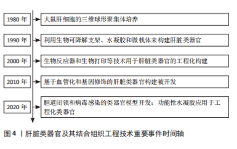
2.1 肝脏类器官 几十年肝细胞和干细胞移植治疗肝病的探索,极大地推动了肝脏类器官研究的发展。干细胞来源的肝样细胞移植很大程度上克服了原代肝细胞难以突破的体外高质量和规模化培养及伦理限制[20],取得了肝脏损伤修复和改善肝功能的疗效[21],但是肝细胞毕竟不能完全替代复杂的肝脏结构和功能[22],肝管破坏导致的胆汁分泌、转运和代谢障碍,及门脉系统的营养物质、毒物或代谢产物转运等突出问题。因此,利用肝细胞、干细胞或肝样细胞与生物材料及肝细胞再生相关因子等形成的肝脏类器官技术应运而生,成为了探讨肝脏组织发育与形成、肝脏疾病致病机制、肝脏再生、终末期肝脏疾病治疗的新策略[23]。肝脏类器官及其结合组织工程技术的重要事件的时间轴(图4),大鼠肝细胞的三维球形聚集体可能是构建肝脏类器官的开端[24]。20世纪90年代,生物可降解支架、水凝胶和微载体等技术应用于肝脏类器官[25-27]。21世纪以来,生物反应器和三维打印技术逐渐在肝脏类器官领域得到应用[28-29]。接下来,具有血管化的肝脏类器官和基因修饰的类器官相继出现[30-31]。直到近几年,构建模式相对成熟的情况下,肝脏类器官扩展到胆道闭锁、病毒感染的类器官疾病模型[32-33]。"

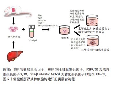
2.1.1 肝脏类器官的构建 (1)肝源组织成体细胞来源的肝脏类器官:虽然不同的种子细胞在肝脏类器官的构建中遵循不同的构建路径,但是它一定是在拥有种子细胞、细胞外基质和相应的信号通路分子3个地基的基础之上,细胞通过模拟肝脏发育过程从而进行自组装并且在空间上重新排列成有序结构[34]。2001年,MICHALOPOULOS等[35]从大鼠肝脏中分离肝细胞,利用胶原包被,辅助肝细胞生长因子和表皮生长因子等简单的细胞因子形成了具有肝结构特征的组织。近年来,利用原代肝细胞作为种子细胞与多种细胞因子和生物材料联合,构建肝类器官也取得了很大的进展[36-37]。研究人员将分离的原代肝细胞悬浮在Matrigel中,辅助以Wnt激动剂(R-spondin1和CHIR99021)、表皮生长因子、成纤维生长因子7、成纤维生长因子10、肝细胞生长因子和转化生长因子β抑制剂等因子进行三维培养,接着分别用优化的肝细胞类器官培养基(人或者鼠)和胆管细胞培养基培养出肝脏类器官(图5)。肝细胞类器官表达肝细胞标志物(白蛋白、肝细胞核因子4α、细胞色素氧化"

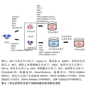
酶Cyp1a2和Cyp3a11)、胎儿肝细胞标志物甲胎蛋白、胆管细胞/祖细胞标记物角蛋白19、祖细胞标志物(Tbx3和Sox9),在一些细胞功能基因(细胞色素P450、糖原代谢、脂质代谢、类固醇代谢、尿素循环和补体激活)出现与原代肝细胞相似的表达谱[37]。此外,肝癌类器官可以直接从手术切除或穿刺活检组织中建立,其方法遵循正常肝脏类器官的流程[38]。 (2) iPSC来源的肝脏类器官:人iPSCs是衍生类器官最常用的种子细胞来源[39]。常用的iPSCs来源肝脏类器官构建方法见图6。"

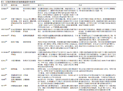
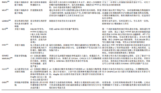
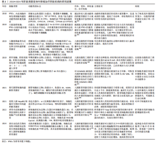
一种是从iPSCs生成成熟肝细胞,随后构建肝脏类器官[40-41]。使用富含激活素 A、骨形态发生蛋白2、成纤维生长因子4、肝细胞生长因子、重组人角质细胞生长因子和抑瘤素M的培养基实现2维成熟肝细胞的分化,然后进行三维培养,加入表皮生长因子、转化生长因子α、成纤维生长因子7、成纤维生长因子10、肝细胞生长因子、转化生长因子β抑制剂A83-01、ROCK抑制剂Y-27632、地塞米松和抑瘤素M因子,最终获得肝脏类器官[40]。 另一种是先建立iPSCs分化早期的球状体,然后进一步诱导为成熟肝脏类器官[42]。使用富含激活素 A、骨形态发生蛋白4、GSK-3抑制剂 CHIR99021和成纤维生长因子4的培养基生成前肠细胞,再将前肠细胞包埋种植于含有成纤维生长因子2、表皮生长因子、血管内皮生长因子、GSK-3 抑制剂 CHIR99021和转化生长因子β抑制剂A83-01等因子的类器官形成培养基进行三维培养,类器官形成后,切换肝脏成熟培养基培养,最终形成肝脏类器官[42]。 虽然大多数方案包括多个阶段,但都是通过激活肝细胞成熟过程中的关键信号通路,进而通过特定的发育标记来验证分化效率[43]。一般来说,通过多个阶段进行更加精确的信号调控诱导细胞分化,可以得到更高的分化效率。总之,肝细胞或干细胞衍生的肝脏类器官具有强大的增殖能力,具有基因组稳定性的优势,且肝脏类器官在一定的微环境下除向肝细胞分化外,还可向胆管细胞分化[32,44]。 2.1.2 肝脏类器官的疾病模型 疾病模型对于研究疾病机制和开创治疗方法是必须的,但是由于动物与人类种属和基因组的差异,动物肝脏疾病模型与人类肝脏疾病致病机制存在不同,人类疾病的组织学复杂性和遗传异质性在许多动物模型中没有得到很好的回馈[45]。目前,利用人iPSCs、胚胎干细胞、肝细胞、肝星状细胞和枯否细胞、肝内胆管细胞和肝外胆管细胞、胆囊细胞、良性胆囊腺瘤组织细胞、成人胆管衍生的双能祖细胞、原发性硬化性胆管炎患者的胆管细胞构建的肝脏类器官已应用于多种肝脏疾病的建模及致病机制研究,见表1。 2.2 肝脏类器官的体内移植 肝脏类器官的终极目标是创造出可移植的肝脏组织替代受损肝脏组织,快速地恢复肝功能[60]。富马酰乙酸水解酶缺乏会导致肝功能衰竭,HUCH等[61]培养的Lgr5+细胞衍生的肝脏类器官具有成熟肝细胞的特征与功能,移植到富马酰乙酸水解酶缺乏小鼠中,肝衰竭小鼠存活率显著增加,并在肝组织中观察到主要由肝细胞核因子4α和角蛋白19阳性的成熟肝细胞组成的富马酰乙酸水解酶阳性结节。同年,TAKEBE课题组[18]将iPSCs生成内胚层细胞通过内皮细胞和间充质干细胞之间的相互作用建立的肝脏类器官移植到肠系膜上探索类器官的功能性和相容性,肝脏类器官移植48 h内与宿主血管系统建立了联系,并执行白蛋白产生和药物代谢功能,成功挽救更昔氯韦诱导的药物性急性肝衰竭,这些是科学家最早探索肝脏类器官体内移植成功治疗肝脏疾病的实验,随后肝脏类器官的体内移植应用得到更多肝脏疾病模型的验证,见表2。此外,采用含有猪肝细胞与人脐静脉内皮细胞球体的改良生物人工肝治疗猴的急性肝衰竭,明显提高了动物的生存率,降低了血氨和炎症因子水平,改善了受损肝脏的再生微环境,很好地恢复了衰竭动物的肝脏功能[62]。"

"

"

"

| [1] DEVARBHAVI H, ASRANI SK, ARAB JP, et al. Global burden of liver disease: 2023 update. J Hepatol. 2023;79(2):516-537. [2] IANSANTE V, MITRY RR, FILIPPI C, et al. Human hepatocyte transplantation for liver disease: current status and future perspectives. Pediatr Res. 2018;83(1-2):232-240. [3] LIU J, YUAN Z, WANG Q. Pluripotent Stem Cell-derived Strategies to Treat Acute Liver Failure: Current Status and Future Directions. J Clin Transl Hepatol. 2022;10(4):692-699. [4] MAN S, DENG Y, MA Y, et al. Prevalence of Liver Steatosis and Fibrosis in the General Population and Various High-Risk Populations: A Nationwide Study With 5.7 Million Adults in China. Gastroenterology. 2023;165(4):1025-1040. [5] YIP TC, FAN JG, WONG VW. China’s Fatty Liver Crisis: A Looming Public Health Emergency. Gastroenterology. 2023;165(4):825-827. [6] LI Y, LU L, CAI X. Liver Regeneration and Cell Transplantation for End-Stage Liver Disease. Biomolecules. 2021;11(12):1907. [7] ZHANG S, YANG Y, FAN L, et al. The clinical application of mesenchymal stem cells in liver disease: the current situation and potential future. Ann Transl Med. 2020;8(8):565. [8] JIN Y, SHI R, QI T, et al. Adipose-derived stem cells show hepatic differentiation potential and therapeutic effect in rats with acute liver failure. Acta Biochim Biophys Sin. 2023;55(4):601-612. [9] YU H, FENG Y, DU W, et al. Off-the-shelf GMP-grade UC-MSCs as therapeutic drugs for the amelioration of CCl4-induced acute-on-chronic liver failure in NOD-SCID mice. Int Immunopharmacol. 2022; 113(Pt A):109408. [10] SCHACHER FC, MARTINS PEZZI DA SILVA A, SILLA L, et al. Bone Marrow Mesenchymal Stem Cells in Acute-on-Chronic Liver Failure Grades 2 and 3: A Phase I-II Randomized Clinical Trial. Can J Gastroenterol Hepatol. 2021;2021:3662776. [11] ANDERSON TN, ZARRINPAR A. Hepatocyte transplantation: past efforts, current technology, and future expansion of therapeutic potential. J Surg Res. 2018;226:48-55. [12] FINKBEINER SR, FREEMAN JJ, WIECK MM, et al. Generation of tissue-engineered small intestine using embryonic stem cell-derived human intestinal organoids. Biol Open. 2015;4(11):1462-1472. [13] 徐灿丽,何文星,汪磊,等. 肝脏类器官研究的文献计量学分析[J].中国组织工程研究,2024,28(7):1099-1104. [14] OGAWA M, OGAWA S, BEAR CE, et al. Directed differentiation of cholangiocytes from human pluripotent stem cells. Nat Biotechnol. 2015;33(8):853-861. [15] SUN L, WANG Y, CEN J, et al. Modelling liver cancer initiation with organoids derived from directly reprogrammed human hepatocytes. Nat Cell Biol. 2019;21(8):1015-1026. [16] SAMPAZIOTIS F, DE BRITO M C, MADRIGAL P, et al. Cholangiocytes derived from human induced pluripotent stem cells for disease modeling and drug validation. Nat Biotechnol. 2015;33(8):845-852. [17] WU F, WU D, REN Y, et al. Generation of hepatobiliary organoids from human induced pluripotent stem cells. J Hepatology. 2019;70(6): 1145-1158. [18] TAKEBE T, SEKINE K, ENOMURA M, et al. Vascularized and functional human liver from an iPSC-derived organ bud transplant. Nature. 2013; 499(7459):481-484. [19] TEN DAM MJM, FREDERIX GWJ, TEN HAM RMT, et al. Toward Transplantation of Liver Organoids: From Biology and Ethics to Cost-effective Therapy. Transplantation. 2023;107(8):1706-1717. [20] YUAN Y, COTTON K, SAMARASEKERA D, et al. Engineered Platforms for Maturing Pluripotent Stem Cell-Derived Liver Cells for Disease Modeling. Cell Mol Gastroenterol Hepatology. 2023;15(5):1147-1160. [21] BAI Y, YANG Z, XU X, et al. Direct chemical induction of hepatocyte-like cells with capacity for liver repopulation. Hepatology. 2023;77(5): 1550-1565. 22] GANDHI N, WILLS L, AKERS K, et al. Comparative transcriptomic and phenotypic analysis of induced pluripotent stem cell hepatocyte-like cells and primary human hepatocytes. Cell Tissue Res. 2024;396(1): 119-139. [23] PRIOR N, INACIO P, HUCH M. Liver organoids: from basic research to therapeutic applications. Gut. 2019;68(12):2228-2237. [24] LANDRY J, BERNIER D, OUELLET C, et al. Spheroidal aggregate culture of rat liver cells: histotypic reorganization, biomatrix deposition, and maintenance of functional activities. J Cell Biol. 1985;101(3):914-923. [25] MIKOS AG, SARAKINOS G, LYMAN MD, et al. Prevascularization of porous biodegradable polymers. Biotechnol Bioeng. 1993;42(6): 716-723. [26] TAKEZAWA T, YAMAZAKI M, MORI Y, et al. Morphological and immuno-cytochemical characterization of a hetero-spheroid composed of fibroblasts and hepatocytes. J Cell Sci. 1992;101(Pt3):495-501. [27] DIXIT V, PISKIN E, ARTHUR M, et al. Hepatocyte immobilization on PHEMA microcarriers and its biologically modified forms. Cell Transplant. 1992;1(6):391-399. [28] MATSUURA T. Bioreactors for 3-dimensional high-density culture of human cells. Hum Cell. 2006;19(1):11-16. [29] YAN Y, WANG X, PAN Y, et al. Fabrication of viable tissue-engineered constructs with 3D cell-assembly technique. Biomaterials. 2005;26(29): 5864-5871. [30] BAPTISTA PM, SIDDIQUI MM, LOZIER G, et al. The use of whole organ decellularization for the generation of a vascularized liver organoid. Hepatology. 2011;53(2):604-617. [31] BROUTIER L, ANDERSSON-ROLF A, HINDLEY CJ, et al. Culture and establishment of self-renewing human and mouse adult liver and pancreas 3D organoids and their genetic manipulation. Nat Protoc. 2016;11(9):1724-1743. [32] HUCH M, GEHART H, VAN BOXTEL R, et al. Long-term culture of genome-stable bipotent stem cells from adult human liver. Cell. 2015; 160(1-2):299-312. [33] DE CRIGNIS E, HOSSAIN T, ROMAL S, et al. Application of human liver organoids as a patient-derived primary model for HBV infection and related hepatocellular carcinoma. Elife. 2021;10:e60747 [34] ZHU X, ZHANG B, HE Y, et al. Liver Organoids: Formation Strategies and Biomedical Applications. Tissue Eng Regen Med. 2021;18(4):573-585. [35] MICHALOPOULOS GK, BOWEN WC, MULÈ K, et al. Histological organization in hepatocyte organoid cultures. Am J Pathol. 2001;159(5): 1877-1887. [36] PENG WC, LOGAN CY, FISH M, et al. Inflammatory Cytokine TNFα Promotes the Long-Term Expansion of Primary Hepatocytes in 3D Culture. Cell. 2018;175(6):1607-1619.e15. [37] HU H, GEHART H, ARTEGIANI B, et al. Long-Term Expansion of Functional Mouse and Human Hepatocytes as 3D Organoids. Cell. 2018;175(6):1591-1606.e19. [38] NUCIFORO S, HEIM MH. Organoids to model liver disease. JHEP Rep. 2021;3(1):100198. [39] YAMANAKA S. Pluripotent Stem Cell-Based Cell Therapy-Promise and Challenges. Cell Stem Cell. 2020;27(4):523-531. [40] GUO J, DUAN L, HE X, et al. A Combined Model of Human iPSC-Derived Liver Organoids and Hepatocytes Reveals Ferroptosis in DGUOK Mutant mtDNA Depletion Syndrome. Adv Sci(Weinh). 2021;8(10): 2004680. [41] MUN SJ, RYU JS, LEE MO, et al. Generation of expandable human pluripotent stem cell-derived hepatocyte-like liver organoids. J Hepatol. 2019;71(5):970-985. [42] SHINOZAWA T, KIMURA M, CAI Y, et al. High-Fidelity Drug-Induced Liver Injury Screen Using Human Pluripotent Stem Cell-Derived Organoids. Gastroenterology. 2021;160(3):831-846.e10. [43] LI Y, YANG X, PLUMMER R, et al. Human Pluripotent Stem Cell-Derived Hepatocyte-Like Cells and Organoids for Liver Disease and Therapy. Int J Mol Sci. 2021;22(19):10471 [44] WANG S, WANG X, TAN Z, et al. Human ESC-derived expandable hepatic organoids enable therapeutic liver repopulation and pathophysiological modeling of alcoholic liver injury. Cell Res. 2019;29(12):1009-1026. [45] ZENG F, ZHANG Y, HAN X, et al. Liver Buds and Liver Organoids: New Tools for Liver Development, Disease and Medical Application. Stem Cell Rev Rep. 2019;15(6):774-784. [46] SOROKA CJ, ASSIS DN, ALRABADI LS, et al. Bile-Derived Organoids From Patients With Primary Sclerosing Cholangitis Recapitulate Their Inflammatory Immune Profile. Hepatology. 2019;70(3):871-882. [47] OUCHI R, TOGO S, KIMURA M, et al. Modeling Steatohepatitis in Humans with Pluripotent Stem Cell-Derived Organoids. Cell Metab. 2019;30(2):374-384.e6. [48] VERSTEGEN MMA, ROOS FJM, BURKA K, et al. Human extrahepatic and intrahepatic cholangiocyte organoids show region-specific differentiation potential and model cystic fibrosis-related bile duct disease. Sci Rep. 2020;10(1):21900. [49] HENDRIKS D, BROUWERS JF, HAMER K, et al. Engineered human hepatocyte organoids enable CRISPR-based target discovery and drug screening for steatosis. Nat Biotechnol. 2023;41(11):1567-1581. [50] SCANU T, SPAAPEN RM, BAKKER JM, et al. Salmonella Manipulation of Host Signaling Pathways Provokes Cellular Transformation Associated with Gallbladder Carcinoma. Cell Host Microbe. 2015;17(6):763-774. [51] GUAN Y, ENEJDER A, WANG M, et al. A human multi-lineage hepatic organoid model for liver fibrosis. Nat Commun. 2021;12(1):6138. [52] MAIER CF, ZHU L, NANDURI LK, et al. Patient-Derived Organoids of Cholangiocarcinoma. Intl J Molecular Sci. 2021;22(16):8675. [53] YUAN B, ZHAO X, WANG X, et al. Patient-derived organoids for personalized gallbladder cancer modelling and drug screening. Clin Transl Med. 2022;12(1):e678. [54] SAITO Y, MURAMATSU T, KANAI Y, et al. Establishment of Patient-Derived Organoids and Drug Screening for Biliary Tract Carcinoma. Cell Rep. 2019;27(4):1265-1276.e4. [55] RAMLI MNB, LIM YS, KOE CT, et al. Human Pluripotent Stem Cell-Derived Organoids as Models of Liver Disease. Gastroenterology. 2020; 159(4):1471-1486.e12. [56] LOARCA L, DE ASSUNCAO TM, JALAN-SAKRIKAR N, et al. Development and characterization of cholangioids from normal and diseased human cholangiocytes as an in vitro model to study primary sclerosing cholangitis. Lab Invest. 2017;97(11):1385-1396. [57] CHOI YJ, KIM MS, RHOADES JH, et al. Patient-Induced Pluripotent Stem Cell-Derived Hepatostellate Organoids Establish a Basis for Liver Pathologies in Telomeropathies. Cell Mol Gastroenterol Hepatol. 2023; 16(3):451-472. [58] GÓMEZ-MARIANO G, MATAMALA N, MARTíNEZ S, et al. Liver organoids reproduce alpha-1 antitrypsin deficiency-related liver disease. Hepatol Int. 2020;14(1):127-137.
[59] CHEN S, LI P, WANG Y, et al. Rotavirus Infection and Cytopathogenesis in Human Biliary Organoids Potentially Recapitulate Biliary Atresia Development. mBio. 2020;11(4):e01968-20. [60] LI J, CHU J, LUI VCH, et al. Bioengineering Liver Organoids for Diseases Modelling and Transplantation. Bioengineering (Basel). 2022;9(12): 796. [61] HUCH M, DORRELL C, BOJ SF, et al. In vitro expansion of single Lgr5+ liver stem cells induced by Wnt-driven regeneration. Nature. 2013; 494(7436):247-250. [62] LI Y, WU Q, WANG Y, et al. Novel spheroid reservoir bioartificial liver improves survival of nonhuman primates in a toxin-induced model of acute liver failure. Theranostics. 2018;8(20):5562-574. [63] NIE YZ, ZHENG YW, OGAWA M, et al. Human liver organoids generated with single donor-derived multiple cells rescue mice from acute liver failure. Stem Cell Res Ther. 2018;9(1):5. [64] TSUCHIDA T, MURATA S, MATSUKI K, et al. The Regenerative Effect of Portal Vein Injection of Liver Organoids by Retrorsine/Partial Hepatectomy in Rats. Int J Mol Sci. 2019;21(1):178. [65] PETTINATO G, LEHOUX S, RAMANATHAN R, et al. Generation of fully functional hepatocyte-like organoids from human induced pluripotent stem cells mixed with Endothelial Cells. Sci Rep. 2019;9(1):8920. [66] LI J, XING F, CHEN F, et al. Functional 3D Human Liver Bud Assembled from MSC-Derived Multiple Liver Cell Lineages. Cell Transplant. 2019; 28(5):510-521. [67] KRUITWAGEN HS, OOSTERHOFF LA, VAN WOLFEREN ME, et al. Long-Term Survival of Transplanted Autologous Canine Liver Organoids in a COMMD1-Deficient Dog Model of Metabolic Liver Disease. Cells. 2020; 9(2):410. [68] SAMPAZIOTIS F, MURARO D, TYSOE OC, et al. Cholangiocyte organoids can repair bile ducts after transplantation in the human liver. Science. 2021;371(6531):839-846. [69] YANG H, SUN L, PANG Y, et al. Three-dimensional bioprinted hepatorganoids prolong survival of mice with liver failure. Gut. 2021; 70(3):567-574. [70] NAM D, PARK MR, LEE H, et al. Induced Endothelial Cell-Integrated Liver Assembloids Promote Hepatic Maturation and Therapeutic Effect on Cholestatic Liver Fibrosis. Cells. 2022;11(14):2242. [71] YUAN X, WU J, SUN Z, et al. Preclinical efficacy and safety of encapsulated proliferating human hepatocyte organoids in treating liver failure. Cell Stem Cell. 2024;31(4):484-498.e5. [72] FIOROTTO R, AMENDUNI M, MARIOTTI V, et al. Liver diseases in the dish: iPSC and organoids as a new approach to modeling liver diseases. Biochim Biophysica Acta Mol Basis Dis. 2019;1865(5):920-928. [73] WILLEMSE J, VAN TIENDEREN G, VAN HENGEL E, et al. Hydrogels derived from decellularized liver tissue support the growth and differentiation of cholangiocyte organoids. Biomaterials. 2022;284: 121473. [74] KIM D H, KIM M J, KWAK S Y, et al. Bioengineered liver crosslinked with nano-graphene oxide enables efficient liver regeneration via MMP suppression and immunomodulation. Nat Commun. 2023;14(1):801. [75] TAMAI M, ADACHI E, KAWASE M, et al. Syngeneic implantation of mouse hepatic progenitor cell-derived three-dimensional liver tissue with dense collagen fibrils. World J Gastroenterol. 2022;28(14): 1444-1454. [76] MATSUMOTO K, YOSHITOMI H, ROSSANT J, et al. Liver organogenesis promoted by endothelial cells prior to vascular function. Science. 2001;294(5542):559-563. [77] YOON Y, GONG SC, KIM MY, et al. Generation of Fibrotic Liver Organoids Using Hepatocytes, Primary Liver Sinusoidal Endothelial Cells, Hepatic Stellate Cells, and Macrophages. Cells. 2023;12(21):2514. [78] RENNERT K, STEINBORN S, GRöGER M, et al. A microfluidically perfused three dimensional human liver model. Biomaterials. 2015;71:119-131. [79] BONANINI F, KUREK D, PREVIDI S, et al. In vitro grafting of hepatic spheroids and organoids on a microfluidic vascular bed. Angiogenesis. 2022;25(4):455-470. [80] SKARDAL A, ALEMAN J, FORSYTHE S, et al. Drug compound screening in single and integrated multi-organoid body-on-a-chip systems. Biofabrication. 2020;12(2):025017. [81] RAJAN SAP, ALEMAN J, WAN M, et al. Probing prodrug metabolism and reciprocal toxicity with an integrated and humanized multi-tissue organ-on-a-chip platform. Acta Biomater. 2020;106:124-135. [82] NGUYEN VVT, YE S, GKOUZIOTI V, et al. A human kidney and liver organoid-based multi-organ-on-a-chip model to study the therapeutic effects and biodistribution of mesenchymal stromal cell-derived extracellular vesicles. J Extracell Vesicles. 2022;11(11):e12280. [83] AKBARI S, ARSLAN N, SENTURK S, et al. Next-Generation Liver Medicine Using Organoid Models. Front Cell Dev Biol. 2019;7:345. [84] LIANG J, WEI J, CAO J, et al. In-organoid single-cell CRISPR screening reveals determinants of hepatocyte differentiation and maturation. Genome Biol. 2023;24(1):251. [85] LAM YK, YU J, HUANG H, et al. TP53 R249S mutation in hepatic organoids captures the predisposing cancer risk. Hepatology. 2023;78(3):727-740. [86] RAMAKRISHNA G, BABU PE, SINGH R, et al. Application of CRISPR-Cas9 based gene editing to study the pathogenesis of colon and liver cancer using organoids. Hepatol Int. 2021;15(6):1309-1317. [87] ZHU S, REZVANI M, HARBELL J, et al. Mouse liver repopulation with hepatocytes generated from human fibroblasts. Nature. 2014;508(7494):93-97. [88] ZAHMATKESH E, KHOSHDEL RAD N, HOSSEIN-KHANNAZER N, et al. Cell and cell-derivative-based therapy for liver diseases: current approaches and future promises. Expert Rev Gastroenterol Hepatol. 2023;17(3): 237-249. [89] JIANG S, XU F, JIN M, et al. Development of a high-throughput micropatterned agarose scaffold for consistent and reproducible hPSC-derived liver organoids. Biofabrication. 2022;15(1). doi: 10.1088/1758-5090/ac933c. [90] YAP KK, GERRAND YW, DINGLE AM, et al. Liver sinusoidal endothelial cells promote the differentiation and survival of mouse vascularised hepatobiliary organoids. Biomaterials. 2020;251:120091. [91] KIM HJ, KIM G, CHI KY, et al. Generation of multilineage liver organoids with luminal vasculature and bile ducts from human pluripotent stem cells via modulation of Notch signaling. Stem Cell Res Ther. 2023;14(1): 19. [92] 刘明昱, 范文娟. 血管化类器官的构建策略[J].中国组织工程研究,2025. https://doi.org/10.12307/2025.052 [93] VELAZQUEZ JJ, LEGRAW R, MOGHADAM F, et al. Gene Regulatory Network Analysis and Engineering Directs Development and Vascularization of Multilineage Human Liver Organoids. Cell Syst. 2021; 12(1):41-55.e11. [94] CROCE S, PELOSO A, ZORO T, et al. A Hepatic Scaffold from Decellularized Liver Tissue: Food for Thought. Biomolecules. 2019; 9(12):813. |
| [1] | Wu Yanting, Li Yu, Liao Jinfeng. Magnesium oxide nanoparticles regulate osteogenesis- and angiogenesis-related gene expressions to promote bone defect healing [J]. Chinese Journal of Tissue Engineering Research, 2026, 30(8): 1885-1895. |
| [2] | Jiang Xinghai, Song Yulin, Li Dejin, Shao Jianmin, Xu Junzhi, Liu Huakai, Wu Yingguo, Shen Yuehui, Feng Sicheng. Vascular endothelial growth factor 165 genes transfected into bone marrow mesenchymal stem cells to construct a vascularized amphiphilic peptide gel module [J]. Chinese Journal of Tissue Engineering Research, 2026, 30(8): 1903-1911. |
| [3] | Liu Xinyue, Li Chunnian, Li Yizhuo, Xu Shifang. Regeneration and repair of oral alveolar bone defects [J]. Chinese Journal of Tissue Engineering Research, 2026, 30(5): 1247-1259. |
| [4] | Yu Shiyu, Yu Sutong, Xu Yang, Zhen Xiangyan, Han Fengxuan. Advances in research and application of tissue engineering therapeutic strategies in oral submucous fibrosis [J]. Chinese Journal of Tissue Engineering Research, 2026, 30(4): 936-948. |
| [5] | Zhou Zixiang, Zhao Baoxiang. Research progress in the relationship between nontraumatic necrosis of the femoral head and lipid metabolism and its treatment [J]. Chinese Journal of Tissue Engineering Research, 2026, 30(3): 680-690. |
| [6] | Zhang Tingting, Li Yalong, Yue Haodi, Li Yanjun, Geng Xiwen, Zhang Yuwei, Liu Xiaozhuan. Protection of exosomes derived from bone marrow mesenchymal stem cells of different mouse ages on radiation-induced lung injury [J]. Chinese Journal of Tissue Engineering Research, 2026, 30(1): 1-9. |
| [7] | Liu Ziwei, Nijati·Tursun, Yin Rui, Li Shuhui, Zhou Jing. Effect of cannabinoid type I receptors on neuronal differentiation of human apical papilla stem cells [J]. Chinese Journal of Tissue Engineering Research, 2026, 30(1): 93-100. |
| [8] | Wu Zhijing, Li Jiali, Zhang Jiaxin, Wang Tangrong, Zheng Yuzhou, Sun Zixuan. Alpha-ketoglutarate engineered small extracellular vesicles delay skin aging [J]. Chinese Journal of Tissue Engineering Research, 2026, 30(1): 120-129. |
| [9] | Li Yiwen, Liu Feixiang, Zhang Yunke. Regulation of lysosome function by stem cells in treatment of lysosomal storage diseases [J]. Chinese Journal of Tissue Engineering Research, 2026, 30(1): 145-152. |
| [10] | Xu Haichao, Luo Lihua, Pan Yihuai. Application and progress of dental pulp stem cells and their derivatives in dental pulp regeneration [J]. Chinese Journal of Tissue Engineering Research, 2026, 30(1): 153-162. |
| [11] | Zhang Zhaowei, Chen Ouzile, Bai Mingru, Wang Chenglin. Therapeutic potential of bioactive substances secreted by dental mesenchymal stem cells for bone repair [J]. Chinese Journal of Tissue Engineering Research, 2026, 30(1): 163-174. |
| [12] | Liu Nian, Dong Xinyue, Wang Songpeng, Xu Yingjiang, Zhang Xiaoming. Stem cell exosomes and biomaterial-assisted exosomes in bone defect repair [J]. Chinese Journal of Tissue Engineering Research, 2026, 30(1): 175-183. |
| [13] | Liu Yu, Gong Senyi, Yang Lihua, Li Weifeng, Hu Yuwen, Yan Qinbiao, Guo Meijin. Isolation, identification, and application of exosomes derived from mesenchymal stem cells [J]. Chinese Journal of Tissue Engineering Research, 2026, 30(1): 194-203. |
| [14] | Zhao Qianwei, Sun Guangyuan . Intestinal organoids: a bibliometric analysis of the latest trends in tissue/organ biology, disease modeling, and clinical applications [J]. Chinese Journal of Tissue Engineering Research, 2026, 30(1): 238-247. |
| [15] | Sun Huiwen, Guo Qiangqiang, Wang Wei, Wu Jie, Xi Kun, Gu Yong. Engineered stem cell bionic periosteum coordinates immune inflammation and vascularization to promote bone regeneration [J]. Chinese Journal of Tissue Engineering Research, 2026, 30(1): 21-33. |
| Viewed | ||||||
|
Full text |
|
|||||
|
Abstract |
|
|||||