Chinese Journal of Tissue Engineering Research ›› 2013, Vol. 17 ›› Issue (28): 5101-5107.doi: 10.3969/j.issn.2095-4344.2013.28.001
Three-dimensional structure and morphology of the mandible in type 1 diabetes mellitus mice
Zhang Jun1, Jiang Li-ting1, Wang Jin-shen2, Zhou Qi2, Gao Yi-ming1
- 1Department of Stomatology, Ruijin Hospital, Shanghai Jiao Tong University School of Medicine, Shanghai 200025, China;
2Shanghai Institute of Traumatology and Orthopedics, Shanghai 200025, China
-
Online:2013-07-09Published:2013-07-29 -
Contact:Gao Yi-ming, M.D., Department of Stomatology, Ruijin Hospital, Shanghai Jiao Tong University School of Medicine, Shanghai 200025, China drgaoym@yahoo.com.cn -
About author:Zhang Jun, Attending physician, Department of Stomatology, Ruijin Hospital, Shanghai Jiao Tong University School of Medicine, Shanghai 200025, China zjjz11071@gmail.com -
Supported by:Medical Guiding Project of Shanghai Science and Technology Committee, No. 114119a3500*

CLC Number:
Cite this article
Zhang Jun, Jiang Li-ting, Wang Jin-shen, Zhou Qi, Gao Yi-ming1. Three-dimensional structure and morphology of the mandible in type 1 diabetes mellitus mice[J]. Chinese Journal of Tissue Engineering Research, 2013, 17(28): 5101-5107.
share this article
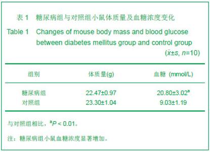
2.1 实验动物数量分析 纳入小鼠共20只,全部进入结果分析,无死亡和感染,无脱落。 2.2 1型糖尿病小鼠体质量和随机血糖浓度检查结果见表1。"
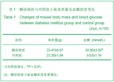
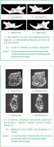
注射链脲佐菌素3周后,与对照组相比,糖尿病组小鼠体质量变化差异无显著性意义,但血糖浓度显著增加(P < 0.01),均大于15 mmol/L,提示诱导1型糖尿病模型成功。 2.3 1型糖尿病小鼠下颌骨松质骨显微结构变化 注射链脲佐菌素3周后,实验选取小鼠第一磨牙牙槽间隔区域的松质骨为感兴趣区,如图1中黑色实线框所示,图2箭头所示糖尿病组较对照组,骨小梁体积变小,数量减少"

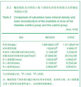
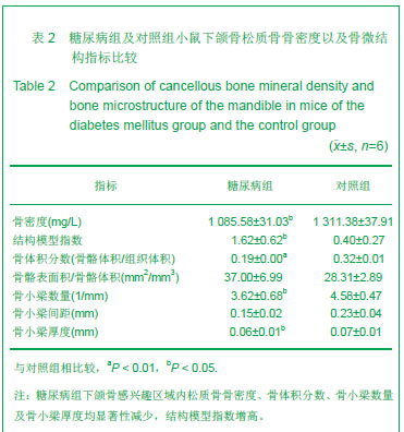
注射链脲佐菌素3周,与对照组比较,糖尿病组下颌骨感兴趣区域内松质骨骨密度、骨体积分数、骨小梁数量及骨小梁厚度均显著性减少(P < 0.01或P < 0.05)。结构模型指数可反映骨小梁结构的改变,结构模型指数为0指骨小梁呈平行板状,3指骨小梁呈杆状改变[13],糖尿病组结构模型指数高于对照组(P < 0.05)。见表2。"
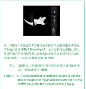
2.4 显微CT测定小鼠下颌骨皮质骨计量学数据 显微CT测定结果显示,注射链脲佐菌素3周后,糖尿病组前牙区松质骨含量少,图3显示颏孔前缘区域几乎没有松质骨,仅见厚实的皮质骨包绕。"
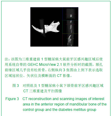
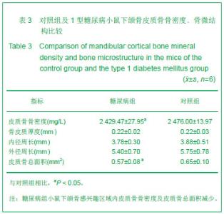
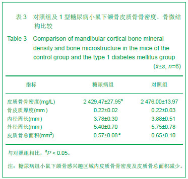
表3数据显示,与对照组相比,糖尿病组小鼠下颌骨感兴趣区域内皮质骨皮质骨骨密度、皮质骨总面积减少(P < 0.05),其他参数两组间比较差异无显著性意义。"
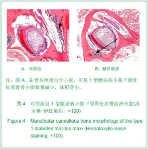
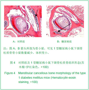
2.5 两组小鼠下颌骨松质骨组织形态 见图4。 注射链脲佐菌素3周后,苏木精-伊红染色可见,与对照组相比,糖尿病组下颌骨松质骨骨小梁数量减少,体积变小。"
| [1]Motyl K, McCabe LR. Streptozotocin, type I diabetes severity and bone. Biol Proced Online. 2009;11:296-315. [2]Motyl KJ, Botolin S, Irwin R, et al. Bone inflammation and altered gene expression with type I diabetes early onset. J Cell Physiol. 2009;218(3):575-583.[3]Shyng YC, Devlin H, Sloan P. The effect of streptozotocin- induced experimental diabetes mellitus on calvarial defect healing and bone turnover in the rat. Int J Oral Maxillofac Surg. 2001;30(1):70-74.[4]Pepelassi E, Xynogala I, Perrea D, et al. Histometric assessment of the effect of diabetes mellitus on experimentally induced periodontitis in rats. J Int Acad Periodontol. 2012;14(2):35-41. [5]Gomes DA, Spolidorio DM, Pepato MT, et al. Effect of induced diabetes mellitus on alveolar bone loss after 30 days of ligature-induced periodontal disease. J Int Acad Periodontol. 2009;11(2):188-192.[6]Okamoto M, Sato T, Shirai H, et al. A histomorphometric analysis on bone dynamics in denture supporting tissue under continuous pressure in streptozotocin-induced diabetic rat. J Oral Rehabil. 2001;28(6):553-559.[7]Zou C, Wang J, Wang S, et al. Characterizing the induction of diabetes in juvenile cynomolgus monkeys with different doses of streptozotocin. Sci China Life Sci. 2012;55(3): 210-218.[8]Chatzigeorgiou A, Halapas A, Kalafatakis K, et al. The use of animal models in the study of diabetes mellitus. In Vivo. 2009; 23(2):245-258.[9]Giglio MJ, Lama MA. Effect of experimental diabetes on mandible growth in rats. Eur J Oral Sci. 2001;109(3):193-197.[10]Duarte VM, Ramos AM, Rezende LA, et al. Osteopenia: a bone disorder associated with diabetes mellitus. J Bone Miner Metab. 2005;23(1):58-68.[11]Thrailkill KM, Liu L, Wahl EC, et al. Bone formation is impaired in a model of type 1 diabetes. Diabetes. 2005;54(10): 2875-2881.[12]Bellenger J, Bellenger S, Bataille A, et al. High pancreatic n-3 fatty acids prevent STZ-induced diabetes in fat-1 mice: inflammatory pathway inhibition. Diabetes. 2011;60(4): 1090-1099. [13]Bouxsein ML, Boyd SK, Christiansen BA, et al. Guidelines for assessment of bone microstructure in rodents using micro-computed tomography. J Bone Miner Res. 2010;25(7): 1468-1486. [14]Singh M, Detamore MS. Biomechanical properties of the mandibular condylar cartilage and their relevance to the TMJ disc. J Biomech. 2009;42(4):405-417. [15]Kuroda S, Tanimoto K, Izawa T, et al. Biomechanical and biochemical characteristics of the mandibular condylar cartilage. Osteoarthritis Cartilage. 2009;17(11):1408-1815.[16]Leidig-Bruckner G, Ziegler R. Diabetes mellitus a risk for osteoporosis? Exp Clin Endocrinol Diabetes. 2001;109 Suppl 2:S493-514.[17]Hamann C, Kirschner S, Günther KP, et al. Bone, sweet bone--osteoporotic fractures in diabetes mellitus. Nat Rev Endocrinol. 2012;8(5):297-305.[18]Unnanuntana A, Rebolledo BJ, Khair MM, et al. Diseases affecting bone quality: beyond osteoporosis. Clin Orthop Relat Res. 2011;469(8):2194-206. [19]DiGirolamo DJ, Clemens TL, Kousteni S. The skeleton as an endocrine organ. Nat Rev Rheumatol. 2012;8(11):674-683.[20]Mishima N, Sahara N, Shirakawa M, et al. Effect of streptozotocin-induced diabetes mellitus on alveolar bone deposition in the rat. Arch Oral Biol. 2002;47(12):843-849.[21]Kunii R, Yamaguchi M, Aoki Y, et al. Effects of experimental occlusal hypofunction, and its recovery, on mandibular bone mineral density in rats. Eur J Orthod. 2008;30(1):52-56.[22]Shimomoto Y, Chung CJ, Iwasaki-Hayashi Y, et al. Effects of occlusal stimuli on alveolar/jaw bone formation. J Dent Res. 2007;86(1):47-51.[23]Verdelis K, Lukashova L, Atti E, et al. MicroCT morphometry analysis of mouse cancellous bone: intra- and inter-system reproducibility. Bone. 2011;49(3):580-587.[24]Layton MW, Goldstein SA, Goulet RW, et al. Examination of subchondral bone architecture in experimental osteoarthritis by microscopic computed axial tomography. Arthritis Rheum. 1988;31(11):1400-1405.[25]Nishiyama KK, Campbell GM, Klinck RJ, et al. Reproducibility of bone micro-architecture measurements in rodents by in vivo micro-computed tomography is maximized with three-dimensional image registration. Bone. 2010;46(1): 155-161.[26]Jiang Y, Zhao J, Liao EY, et al. Application of micro-CT assessment of 3-D bone microstructure in preclinical and clinical studies. J Bone Miner Metab. 2005;23 Suppl:122-131.[27]Umoh JU, Sampaio AV, Welch I, et al. In vivo micro-CT analysis of bone remodeling in a rat calvarial defect model. Phys Med Biol. 2009;54(7):2147-2161.[28]Benhamou CL. Effects of osteoporosis medications on bone quality. Joint Bone Spine. 2007;74(1):39-47.[29]Abbassy MA, Watari I, Soma K. Effect of experimental diabetes on craniofacial growth in rats. Arch Oral Biol. 2008; 53(9):819-825.[30]Ogawa N, Yamaguchi T, Yano S, et al. The combination of high glucose and advanced glycation end-products (AGEs) inhibits the mineralization of osteoblastic MC3T3-E1 cells through glucose-induced increase in the receptor for AGEs. Horm Metab Res. 2007;39(12):871-875. [31]Okazaki K, Yamaguchi T, Tanaka K,et al. Advanced glycation end products (AGEs), but not high glucose, inhibit the osteoblastic differentiation of mouse stromal ST2 cells through the suppression of osterix expression, and inhibit cell growth and increasing cell apoptosis. Calcif Tissue Int. 2012; 91(4):286-296. [32]Holzhausen M, Garcia DF, Pepato MT, et al. The influence of short-term diabetes mellitus and insulin therapy on alveolar bone loss in rats. J Periodontal Res. 2004;39(3):188-193.[33]Coe LM, Zhang J, McCabe LR. Both spontaneous Ins2(+/-) and streptozotocin-induced type I diabetes cause bone loss in young mice. J Cell Physiol. 2013;228(4):689-695. [34]Mc Cabe LR. Understanding the pathology and mechanisms of type I diabetic bone loss. J Cell Biochem. 2007;102(6): 1343-1357.[35]Botolin S, McCabe LR. Bone loss and increased bone adiposity in spontaneous and pharmacologically induced diabetic mice. Endocrinology. 2007;148(1):198-205.[36]Abbassy MA, Watari I, Soma K. The effect of diabetes mellitus on rat mandibular bone formation and microarchitecture. Eur J Oral Sci. 2010;118(4):364-369. [37]Gradin P, Hollberg K, Cassady AI, et al. Transgenic overexpression of tartrate-resistant acid phosphatase is associated with induction of osteoblast gene expression and increased cortical bone mineral content and density. Cells Tissues Organs. 2012;196(1):68-81.[38]Sheng MH, Wergedal JE, Mohan S, et al. Targeted overexpression of osteoactivin in cells of osteoclastic lineage promotes osteoclastic resorption and bone loss in mice. PLoS One. 2012;7(4):e35280.[39]Phan TC, Xu J, Zheng MH. Interaction between osteoblast and osteoclast: impact in bone disease. Histol Histopathol. 2004;19(4):1325-1344.[40]Verhaeghe J, Thomsen JS, van Bree R, et al. Effects of exercise and disuse on bone remodeling, bone mass, and biomechanical competence in spontaneously diabetic female rats. Bone. 2000;27(2):249-256. |
| [1] | Cheng Qisheng, Julaiti·Maitirouzi, Xiao Yang, Zhang Chenwei, Paerhati·Rexiti. Finite element analysis of novel variable-diameter screws in modified cortical bone trajectory of lumbar vertebrae [J]. Chinese Journal of Tissue Engineering Research, 2026, 30(9): 2162-2171. |
| [2] | Li Zhifei, Han Bin, Liu Qiuli, Zhang Zhanming, Wei Haokai, Zuo Kuangshi, Zhang Yisheng. Cervical motion characteristics in patients with cervical spondylotic radiculopathy based on motion capture technology [J]. Chinese Journal of Tissue Engineering Research, 2026, 30(9): 2286-2293. |
| [3] | Chen Ju, Zheng Jinchang, Liang Zhen, Huang Chengshuo, Lin Hao, Zeng Li. Effect and mechanism of beta-caryophyllene in mice with osteoarthritis [J]. Chinese Journal of Tissue Engineering Research, 2026, 30(6): 1341-1347. |
| [4] | Wen Guangwei, Zhen Yinghao, Zheng Taikeng, Zhou Shuyi, Mo Guoye, Zhou Tengpeng, Li Haishan, Lai Yiyi. Effects and mechanisms of isoginkgetin on osteoclastogenesis [J]. Chinese Journal of Tissue Engineering Research, 2026, 30(6): 1348-1358. |
| [5] | Li Linzhen, Jiao Hongzhuo, Chen Weinan, Zhang Mingzhe, Wang Jianlong, Zhang Juntao. Effect of icariin-containing serum on lipopolysaccharide-induced inflammatory damage in human chondrocytes [J]. Chinese Journal of Tissue Engineering Research, 2026, 30(6): 1368-1374. |
| [6] | Pan Hongfei, Zhuang Zhenbing, Xu Baiyun, Yang Zhangyang, Lin Kairui, Zhan Bingqing, Lan Jinghan, Gao Heng, Zhang Nanbo, Lin Jiayu. Inhibitory effects of different concentrations of auranofin on M1 macrophage function and its therapeutic potential in diabetic wound healing [J]. Chinese Journal of Tissue Engineering Research, 2026, 30(6): 1390-1397. |
| [7] | Peng Zhiwei, Chen Lei, Tong Lei. Luteolin promotes wound healing in diabetic mice: roles and mechanisms [J]. Chinese Journal of Tissue Engineering Research, 2026, 30(6): 1398-1406. |
| [8] | Lyu Xiaofan, Huang Yi, Ding Liucheng . Mitochondrial mechanism and intervention therapy in diabetic cystopathy [J]. Chinese Journal of Tissue Engineering Research, 2026, 30(6): 1508-1515. |
| [9] | Cao Xinyan, Yu Zifu, Leng Xiaoxuan, Gao Shiai, Chen Jinhui, Liu Xihua. Effect of repetitive transcranial magnetic stimulation and transcranial direct current stimulation on motor function and gait in children with cerebral palsy: a network meta-analysis [J]. Chinese Journal of Tissue Engineering Research, 2026, 30(6): 1539-1548. |
| [10] | Guo Ying, Tian Feng, Wang Chunfang. Potential drug targets for the treatment of rheumatoid arthritis: large sample analysis from European databases [J]. Chinese Journal of Tissue Engineering Research, 2026, 30(6): 1549-1557. |
| [11] | Yang Zhijie, Zhao Rui, Yang Haolin, Li Xiaoyun, Li Yangbo, Huang Jiachun, Lin Yanping, Wan Lei, HuangHongxing. Postmenopausal osteoporosis: predictive values of muscle mass, grip strength, and appendicular skeletal muscle index [J]. Chinese Journal of Tissue Engineering Research, 2026, 30(5): 1073-1080. |
| [12] | Yin Yongcheng, Zhao Xiangrui, Yang Zhijie, Li Zheng, Li Fang, Ning Bin. Effect and mechanism of peroxiredoxin 1 in microglial inflammation after spinal cord injury [J]. Chinese Journal of Tissue Engineering Research, 2026, 30(5): 1106-1113. |
| [13] | Zhang Jiuxuan, Zhang Jinnan, Sui Xiaofan, Pei Xiaxia, Wei Jianhong, Su Qiang, Li Tian. Effects of ammonia poisoning on cognitive behavior and hippocampal synaptic damage in mice [J]. Chinese Journal of Tissue Engineering Research, 2026, 30(5): 1122-1128. |
| [14] | Sun Yajie, Zhao Xinchen, Bo Shuangling. Spatiotemporal expression of bone morphologic protein 7 in mouse kidney development [J]. Chinese Journal of Tissue Engineering Research, 2026, 30(5): 1156-1161. |
| [15] | Li Haojing, Wang Xin, Song Chenglin, Zhang Shengnan, Chen Yunxin. Therapeutic efficacy of extracorporeal shock wave therapy in the upper trapezius muscle area combined with exercise control training in patients with chronic non-specific neck pain [J]. Chinese Journal of Tissue Engineering Research, 2026, 30(5): 1162-1170. |
| Viewed | ||||||
|
Full text |
|
|||||
|
Abstract |
|
|||||