Chinese Journal of Tissue Engineering Research ›› 2022, Vol. 26 ›› Issue (2): 211-217.doi: 10.12307/2022.035
Previous Articles Next Articles
Asperosaponin VI therapy for Achilles tendinopathy in rabbits
Wang Kun1, He Benxiang2
- 1Institute of Sports Medicine and Health, Chengdu Sport University, Chengdu 610000, Sichuan Province, China; 2Affiliated Sports Hospital, Chengdu Sport University, Chengdu 610000, Sichuan Province, China
-
Received:2020-09-27Revised:2020-09-28Accepted:2020-10-30Online:2022-01-18Published:2021-10-27 -
Contact:He Benxiang, MD, Professor, Affiliated Sports Hospital, Chengdu Sport University, Chengdu 610000, Sichuan Province, China -
About author:Wang Kun, MD candidate, Assistant experimentalist, Institute of Sports Medicine and Health, Chengdu Sport University, Chengdu 610000, Sichuan Province, China -
Supported by:the National Key Research and Development Plan of China, No. 2019YFF0301704 (to HBX)
CLC Number:
Cite this article
Wang Kun, He Benxiang. Asperosaponin VI therapy for Achilles tendinopathy in rabbits[J]. Chinese Journal of Tissue Engineering Research, 2022, 26(2): 211-217.
share this article
Add to citation manager EndNote|Reference Manager|ProCite|BibTeX|RefWorks
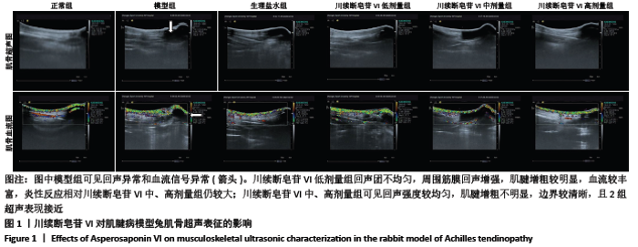
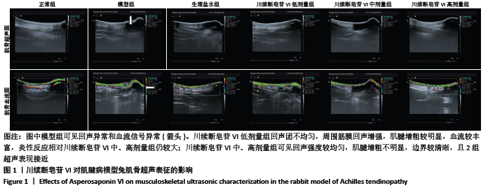
2.1 实验动物数量分析 各组实验动物均顺利完成实验周期,未出现体质量异常、跟腱红肿及流脓等情况或其他疾病表现。 2.2 川续断皂苷VI对肌腱病模型兔肌骨超声表征的影响 正常组肌腱回声均匀连续,边界清晰,未见异常血流信号。模型组可见肌腱明显增粗,回声异常,跟骨上2 cm局部可见多处不均匀回声团,边界不清,周围筋膜回声团信号增强,血流较为丰富。生理盐水组回声基本均匀,未见异常血流信号。川续断皂苷VI低剂量组回声团强度不等,周围筋膜回声增强,血流信号仍较丰富,肌腱较明显增粗,炎性反应较强;川续断皂苷VI中、高剂量组可见回声强度较均匀,肌腱增粗不明显,边界较清晰,且两组超声表现接近,见图1。"
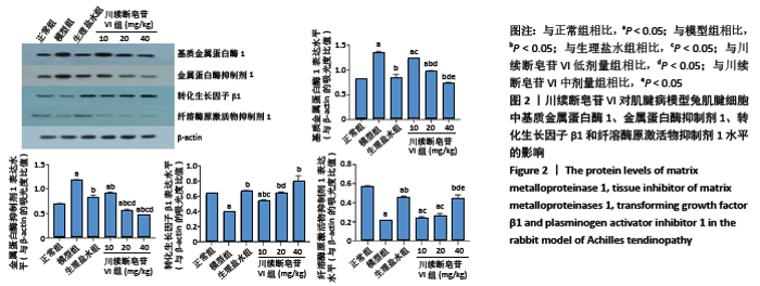
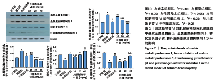
2.3 川续断皂苷VI对肌腱病模型兔肌腱细胞中基质金属蛋白酶1、金属蛋白酶抑制剂1、转化生长因子β1及纤溶酶原激活物抑制剂1表达的影响 与正常组相比,模型组基质金属蛋白酶1表达水平较高;而与模型组相比,川续断皂苷VI中、高剂量组基质金属蛋白酶1表达下调(P < 0.05)。模型组金属蛋白酶抑制剂1水平高于正常组,而在川续断皂苷VI低、中、高剂量组均可降低金属蛋白酶抑制剂1的表达水平(P < 0.05)。与模型组相比,川续断皂苷VI低、中、高剂量转化生长因子β1表达水平上调(P < 0.05)。与模型组相比,川续断皂苷VI高剂量纤溶酶原激活物抑制剂1表达水平升高(P < 0.05),见图2。"
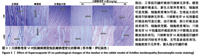
2.4 川续断皂苷VI对肌腱病模型兔肌腱病理变化的影响 模型组肌腱纤维紊乱,排列呈明显波浪形改变,结构不完整,出现新生血管异常增生,炎性细胞浸润,细胞核明显变圆,细胞密度增加明显等现象。川续断皂苷VI低剂量组仍可见较为纤维结构明显松散、些许断裂,部分纤维呈波浪形排列,细胞核仍有变圆趋势,细胞密度增加;川续断皂苷VI中剂量组可见纤维组织有少许断裂及轻微波浪形表现;而川续断皂苷VI高剂量组肌腱纤维组织排列较有序完整,无明显波浪形表现,细胞密度亦无异常,见图3。"

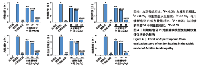
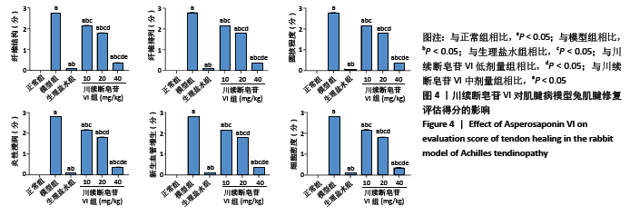
与模型组相比,川续断皂苷VI低、中、高剂量组肌腱修复评估得分依次递减(P < 0.05),见图4。"
| [1] NOURISSAT G, BERENBAUM F, DUPREZ D. Tendon injury: from biology to tendon repair. Nat Rev Rheumatol. 2015;11(4):223-233. [2] RADOVANOVIĆ G, WOLFARTH B, LEGERLOTZ K. Interleukin‐6 levels drop after a 12 week long physiotherapeutic intervention in patients with Achilles tendinopathy—a pilot study. Transl Sports Med. 2019;2: 233-239. [3] WANG Y, HE G, GUO Y, et al. Exosomes from tendon stem cells promote injury tendon healing through balancing synthesis and degradation of the tendon extracellular matrix. J Cell Mol Med. 2019;23(8):5475-5485. [4] KHAN MR, SMITH RK, DAVID F, et al. Evaluation of the Effects of Synovial Multipotent Cells on Deep Digital Flexor Tendon Repair in a Large Animal Model of Intra-Synovial Tendinopathy. J Orthop Res. 2020; 38(1):128-138. [5] WANG Y, HE G, TANG H, et al. Aspirin inhibits inflammation and scar formation in the injury tendon healing through regulating JNK/STAT-3 signalling pathway. Cell Prolif. 2019;52(4):e12650. [6] ALVES C, MENDES D, MARQUES FB. Fluoroquinolones and the risk of tendon injury: a systematic review and meta-analysis. Eur J Clin Pharmacol. 2019;75(10):1431-1443. [7] TEIXEIRA PAG, JAQUET P, BAKOUR O, et al. CT arthrography of the intra-articular long head of biceps tendon: Diagnostic performance outside the labral-bicipital complex. Diagn Interv Imaging. 2019;100(7-8): 437-444. [8] ZAMZAM M, EL YASAKI A, EL GARABAWY N, et al. Shockwave therapy versus local steroid injection in chronic supraspinatus tendinopathy. Egypt Rheumatol Rehabil. 2019;46:141-147. [9] DAN MJ, WALSH WR, CROSS MJ, et al. Treatment of patella tendinopathy by distalising tibial tubercle osteotomy. BMJ Case Rep. 2019;12(7):e229209. [10] BAAR K. Stress Relaxation and Targeted Nutrition to Treat Patellar Tendinopathy. Int J Sport Nutr Exerc Metab. 2019;29(4):453-457. [11] NIE G, WEN X, LIANG X, et al. Additional evidence supports association of common genetic variants in MMP3 and TIMP2 with increased risk of chronic Achilles tendinopathy susceptibility. J Sci Med Sport. 2019;22(10):1074-1078. [12] LEE SY, KIM W, LIM C, et al. Treatment of Lateral Epicondylosis by Using Allogeneic Adipose-Derived Mesenchymal Stem Cells: A Pilot Study. Stem Cells. 2015;33(10):2995-3005. [13] RUI YF, LUI PP, LI G, et al. Isolation and characterization of multipotent rat tendon-derived stem cells. Tissue Eng Part A. 2010;16(5):1549-1558. [14] ARNSDORF EJ, TUMMALA P, JACOBS CR. Non-canonical Wnt signaling and N-cadherin related beta-catenin signaling play a role in mechanically induced osteogenic cell fate. PLoS One. 2009;4(4):e5388. [15] GURTNER GC, WERNER S, BARRANDON Y, et al. Wound repair and regeneration. Nature. 2008;453(7193):314-321. [16] LIU ZG, ZHANG R, LI C, et al. The osteoprotective effect of Radix Dipsaci extract in ovariectomized rats. J Ethnopharmacol. 2009;123(1):74-81. [17] JUNG HW, JUNG JK, SON KH, et al. Inhibitory effects of the root extract of Dipsacus asperoides C.Y. Cheng et al T.M.Ai on collagen-induced arthritis in mice. J Ethnopharmacol. 2012;139(1):98-103. [18] Wong RW, Rabie AB, Hägg EU. The effect of crude extract from Radix Dipsaci on bone in mice. Phytother Res. 2007;21(6):596-598. [19] HUNG TM, NA M, THUONG PT, et al. Antioxidant activity of caffeoyl quinic acid derivatives from the roots of Dipsacus asper Wall. J Ethnopharmacol. 2006;108(2):188-192. [20] KE K, LI Q, YANG X, et al. Asperosaponin VI promotes bone marrow stromal cell osteogenic differentiation through the PI3K/AKT signaling pathway in an osteoporosis model. Sci Rep. 2016;6:35233. [21] LOISELLE AE, YUKATA K, GEARY MB, et al. Development of antisense oligonucleotide (ASO) technology against Tgf-β signaling to prevent scarring during flexor tendon repair. J Orthop Res. 2015;33(6):859-866. [22] QIN W, LIN ZM, DENG R, et al. p38a MAPK is involved in BMP-2-induced odontoblastic differentiation of human dental pulp cells. Int Endod J. 2012;45(3):224-233. [23] 秦川.实验动物学[M].北京:人民卫生出版社,2010. [24] KHAN MH, LI Z, WANG JH. Repeated exposure of tendon to prostaglandin-E2 leads to localized tendon degeneration. Clin J Sport Med. 2005;15(1):27-33. [25] 陈磊.富血小板血浆诱导肌键干细胞增殖分化对肌键微损伤修复的作用[D].重庆:第三军医大学,2012. [26] TSUZAKI M, GUYTON G, GARRETT W, et al. IL-1 beta induces COX2, MMP-1, -3 and -13, ADAMTS-4, IL-1 beta and IL-6 in human tendon cells. J Orthop Res. 2003;21(2):256-264. [27] 史晓伟,龙丽娟,沈勇伟,等,末端病大鼠跟腱病超微结构、胶原、MMP-1及TIMP1的研究[J].中国运动医学杂志,2014,33(9):902-906. [28] YANG S, ZHANG W, XUAN LL, et al. Akebia Saponin D inhibits the formation of atherosclerosis in ApoE-/- mice by attenuating oxidative stress-induced apoptosis in endothelial cells. Atherosclerosis. 2019; 285:23-30. [29] TSUJIMOTO Y. Cell death regulation by the Bcl-2 protein family in the mitochondria. J Cell Physiol. 2003;195(2):158-167. [30] WANG CG, LOU YT, TONG MJ, et al. Asperosaponin VI promotes angiogenesis and accelerates wound healing in rats via up-regulating HIF-1α/VEGF signaling. Acta Pharmacol Sin. 2018;39(3):393-404. [31] KE K, LI Q, YANG X, et al. Asperosaponin VI promotes bone marrow stromal cell osteogenic differentiation through the PI3K/AKT signaling pathway in an osteoporosis model. Sci Rep. 2016;6:35233. [32] NIU Y, LI Y, HUANG H, et al. Asperosaponin VI, a saponin component from Dipsacus asper wall, induces osteoblast differentiation through bone morphogenetic protein-2/p38 and extracellular signal-regulated kinase 1/2 pathway. Phytother Res. 2011;25(11):1700-1706. [33] BI F, SHI Z, JIANG S, et al. Intermittently administered parathyroid hormone [1-34] promotes tendon-bone healing in a rat model. Int J Mol Sci. 2014;15(10):17366-17379. [34] PARK JY, PARK SD, KOH YJ, et al. Aqueous extract of Dipsacus asperoides suppresses lipopolysaccharide-stimulated inflammatory responses by inhibiting the ERK1/2 signaling pathway in RAW 264.7 macrophages. J Ethnopharmacol. 2019;231:253-261. [35] MAJEWSKI M, PORTER RM, BETZ OB, et al. Improvement of tendon repair using muscle grafts transduced with TGF-β1 cDNA. Eur Cell Mater. 2012;23:94-102. [36] OKAMOTO S, TOHYAMA H, KONDO E, et al. Ex vivo supplementation of TGF-beta1 enhances the fibrous tissue regeneration effect of synovium-derived fibroblast transplantation in a tendon defect: a biomechanical study. Knee Surg Sports Traumatol Arthrosc. 2008;16(3):333-339. [37] YAMAZAKI S, YASUDA K, TOMITA F, et al. The effect of transforming growth factor-beta1 on intraosseous healing of flexor tendon autograft replacement of anterior cruciate ligament in dogs. Arthroscopy. 2005; 21(9):1034-1041. [38] PREMDAS J, TANG JB, WARNER JP, et al. The presence of smooth muscle actin in fibroblasts in the torn human rotator cuff. J Orthop Res. 2001;19(2):221-228. [39] 王尉,何恢绪,彭心昭,等.转化生长因子-β1对肌腱细胞DNA及胶原合成的影响[J].中华创伤杂志,2001,17(10):618-619. [40] MICHEL K, ROTH S, TRAUTWEIN C, et al. Analysis of the expression pattern of the latent transforming growth factor beta binding protein isoforms in normal and diseased human liver reveals a new splice variant missing the proteinase-sensitive hinge region. Hepatology. 1998;27(6):1592-1599. [41] 耿震,王震,周海洋.富血小板血浆对肌健愈合影响的实验研究[J].中国修复重建外科杂志,2011,25(3):344-348. [42 ] 史晓伟,周学兰.TGF-β1多效性与肌腱修复研究进展[J].中国运动医学杂志,2016,35(6):588-591. [43] FAVATA M, BEREDJIKLIAN PK, ZGONIS MH, et al. Regenerative properties of fetal sheep tendon are not adversely affected by transplantation into an adult environment. J Orthop Res. 2006;24(11):2124-2132. [44] CHEN Q, LU H, YANG H. Chitosan inhibits fibroblasts growth in Achilles tendon via TGF-β1/Smad3 pathway by miR-29b. Int J Clin Exp Pathol. 2014;7(12):8462-8470. [45] FU SC, WONG YP, CHEUK YC, et al. TGF-beta1 reverses the effects of matrix anchorage on the gene expression of decorin and procollagen type I in tendon fibroblasts. Clin Orthop Relat Res. 2005;(431):226-232. [46] MAUVIEL A, CHUNG KY, AGARWAL A, et al. Cell-specific induction of distinct oncogenes of the Jun family is responsible for differential regulation of collagenase gene expression by transforming growth factor-beta in fibroblasts and keratinocytes. J Biol Chem. 1996;271(18): 10917-10923. [47] MARTIN J, YUNG S, ROBSON RL, et al. Production and regulation of matrix metalloproteinases and their inhibitors by human peritoneal mesothelial cells. Perit Dial Int. 2000;20(5):524-533. [48] MA C, CHEGINI N. Regulation of matrix metalloproteinases (MMPs) and their tissue inhibitors in human myometrial smooth muscle cells by TGF-beta1. Mol Hum Reprod. 1999;5(10):950-954. [49] FARHAT YM, AL-MALIKI AA, CHEN T, et al. Gene expression analysis of the pleiotropic effects of TGF-β1 in an in vitro model of flexor tendon healing. PLoS One. 2012;7(12):e51411. [50] FARHAT YM, AL-MALIKI AA, EASA A, et al. TGF-β1 Suppresses Plasmin and MMP Activity in Flexor Tendon Cells via PAI-1: Implications for Scarless Flexor Tendon Repair. J Cell Physiol. 2015;230(2):318-326. [51] YANG Y, YANG S, CHEN M, et al. Compound Astragalus and Salvia miltiorrhiza Extract exerts anti-fibrosis by mediating TGF-beta/Smad signaling in myofibroblasts. J Ethnopharmacol. 2008;118(2):264-270. [52] TUAN TL, WU H, HUANG EY, et al. Increased plasminogen activator inhibitor-1 in keloid fibroblasts may account for their elevated collagen accumulation in fibrin gel cultures. Am J Pathol. 2003;162(5): 1579-1589. [53] TITAN AL, LONGAKER MT. A fine balance in tendon healing. Nat Cell Biol. 2019;21(12):1466-1467. [54] SPIESZ EM, THORPE CT, CHAUDHRY S, et al. Tendon extracellular matrix damage, degradation and inflammation in response to in vitro overload exercise. J Orthop Res. 2015;33(6):889-897. |
| [1] | Zhang Haobo, Zhao Yunan, Yang Xuejun. Role and therapeutic implications of pyroptosis in intervertebral disc degeneration [J]. Chinese Journal of Tissue Engineering Research, 2022, 26(9): 1445-1451. |
| [2] | Li Rui, Shi Wen, Yang Shicai, Lü Linwei, Zhang Chunqiu. Effect of splintage and Shenxiaosan cataplasm on fracture healing in rabbits with radial fracture model [J]. Chinese Journal of Tissue Engineering Research, 2022, 26(9): 1329-1333. |
| [3] | Chen Xianghe, Liu Bo, Yang Kang, Lu Pengcheng, Yu Huilin. Treadmill exercise improves the myocardial fibrosis of spontaneous type 2 diabetic mice: an exploration on the functional pathway [J]. Chinese Journal of Tissue Engineering Research, 2022, 26(8): 1210-1215. |
| [4] | Xiang Xinjian, Liu Fang, Wu Liangliang, Jia Daping, Tao Yue, Zhao Zhengnan, Zhao Yu. High-dose vitamin C promotes the survival of autologous fat transplantation in rats [J]. Chinese Journal of Tissue Engineering Research, 2022, 26(8): 1242-1246. |
| [5] | Wu Bingshuang, Wang Zhi, Tang Yi, Tang Xiaoyu, Li Qi. Anterior cruciate ligament reconstruction: from enthesis to tendon-to-bone healing [J]. Chinese Journal of Tissue Engineering Research, 2022, 26(8): 1293-1298. |
| [6] | Wang Jifang, Bao Zhen, Qiao Yahong. miR-206 regulates EVI1 gene expression and cell biological behavior in stem cells of small cell lung cancer [J]. Chinese Journal of Tissue Engineering Research, 2022, 26(7): 1027-1031. |
| [7] | Zhu Bingbing, Deng Jianghua, Chen Jingjing, Mu Xiaoling. Interleukin-8 receptor enhances the migration and adhesion of umbilical cord mesenchymal stem cells to injured endothelium [J]. Chinese Journal of Tissue Engineering Research, 2022, 26(7): 1045-1050. |
| [8] | Zhang Yujie, Yang Jiandong, Cai Jun, Zhu Shoulei, Tian Yuan. Mechanism by which allicin inhibits proliferation and promotes apoptosis of rat vascular endothelial cells [J]. Chinese Journal of Tissue Engineering Research, 2022, 26(7): 1080-1084. |
| [9] | Zhang Jinglin, Leng Min, Zhu Boheng, Wang Hong. Mechanism and application of stem cell-derived exosomes in promoting diabetic wound healing [J]. Chinese Journal of Tissue Engineering Research, 2022, 26(7): 1113-1118. |
| [10] | Yin Tingting, Du Dayong, Jiang Zhixin, Liu Yang, Liu Qilin, Li Yuntian. Granulocyte colony-stimulating factors improve myocardial fibrosis in rats with myocardial infarction [J]. Chinese Journal of Tissue Engineering Research, 2022, 26(5): 730-735. |
| [11] | Xu Jing, Yan Yongmin, Cai Mengjie . miR-373 inhibits hepatic stellate cell activation by downregulating transforming growth factor beta type II receptor [J]. Chinese Journal of Tissue Engineering Research, 2022, 26(5): 756-761. |
| [12] | Li Weiming, Xu Qingwen, Li Yijun, Sun Yanbo, Cui Jin, Xu Pengyuan . Deep seawater promotes wound healing in diabetic mice by activating PI3K/Akt pathway [J]. Chinese Journal of Tissue Engineering Research, 2022, 26(5): 724-729. |
| [13] | Yang Sidi, Wang Qian, Xu Nuo, Wang Ronghan, Jin Chuanqi, Lu Ying, Dong Ming. Biodentine enhances the proliferation and differentiation of osteoblasts through upregulating bone morphogenetic protein-2 [J]. Chinese Journal of Tissue Engineering Research, 2022, 26(4): 516-520. |
| [14] | Chen Xiaoxu, Luo Yaxin, Bi Haoran, Yang Kun. Preparation and application of acellular scaffold in tissue engineering and regenerative medicine [J]. Chinese Journal of Tissue Engineering Research, 2022, 26(4): 591-596. |
| [15] | Shen Jiahua, Fu Yong. Application of graphene-based nanomaterials in stem cells [J]. Chinese Journal of Tissue Engineering Research, 2022, 26(4): 604-609. |
| Viewed | ||||||
|
Full text |
|
|||||
|
Abstract |
|
|||||