Chinese Journal of Tissue Engineering Research ›› 2017, Vol. 21 ›› Issue (23): 3767-3772.doi: 10.3969/j.issn.2095-4344.2017.23.027
A meta-analysis of the efficacy of three-dimensional printing-assisted surgery for tibial plateau fractures
Shao Jia-shen, Chang Heng-rui, Zheng Zhan-le, Chen Wei, Zhang Ying-ze
- the Third Hospital of Hebei Medical University, Shijiazhuang 050051, Hebei Province, China
-
Online:2017-08-18Published:2017-09-01 -
Contact:Zhang Ying-ze, Chief physician, the Third Hospital of Hebei Medical University, Shijiazhuang 050051, Hebei Province, China -
About author:Shao Jia-shen, Studying for master’s degree, the Third Hospital of Hebei Medical University, Shijiazhuang 050051, Hebei Province, China -
Supported by:the National Natural Science Foundation for the Youth of China, No. 81401789
CLC Number:
Cite this article
Shao Jia-shen, Chang Heng-rui, Zheng Zhan-le, Chen Wei, Zhang Ying-ze . A meta-analysis of the efficacy of three-dimensional printing-assisted surgery for tibial plateau fractures [J]. Chinese Journal of Tissue Engineering Research, 2017, 21(23): 3767-3772.
share this article

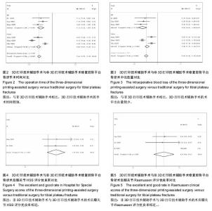
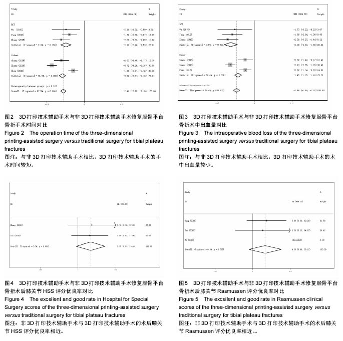
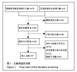
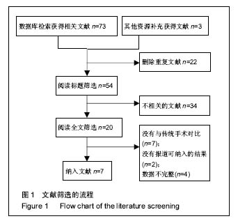
2.2 纳入研究的质量评价 纳入Meta分析的研究类型为随机对照试验时使用Jadad量表评价其研究质量,根据以下8条项目进行评价:随机序列的产生、盲法、撤出和退出、纳入和排除标准、不利影响和统计分析。≤3分为低质量研究,≥4分为高质量研究。其中一项研究为4分[19],两项研究为5分[16,20],一项研究为6分[18]。纳入文章的研究类型为队列研究时使用Newcastle-Ottawa(NOS)量表评价其研究质量,其中两项研究为4分[13,17],一项研究为5分[15]。 2.3 Meta分析结果 2.3.1 手术时间 一共有6篇研究纳入手术时间(min)的分析[13,15,17-20]。各研究间存在较大异质性(P < 0.05,I2=87.5%),为进一步明确异质性来源,根据研究类型不同分为了随机对照试验组(RCT)和前瞻性队列研究试验组(Cohort)两个亚组。Meta分析显示,两种不同的方法在手术时间上的差异有显著性意义(SMD=-2.411,95%CI为-2.718至-2.104,P=0.00),3D打印组的手术时间短于传统手术组。其中,RCT组(SMD=-2.123,95% CI为-2.716至-1.531,P=0.00),异质性分析I2=2.6%;Cohort组(SMD=-2.516,95% CI为-2.875至-2.157,P=0.00),异质性分析I2=94.5%。总的合并结果异质性较大的原因可能为本研究纳入的3篇前瞻性队列研究的异质性较大引起,异质性来源可能由测量方法的差异或手术技术上的差异导致。结果见图2。 2.3.2 术中出血量 一共有6篇研究纳入术中出血量(mL)的分析[13,15,17-20]。各研究间存在异质性(P < 0.05,I2=78.4%),其异质性来源可能为测量方法不同及手术操作者技能水平不同引起。Meta分析显示,两种不同方法在术中出血量上的差异有显著性意义(SMD=-1.579,95% CI为-1.842至-1.316,P=0.00),3D打印组的术中出血量比传统手术组少。其中,RCT组(SMD=-2.084,95%CI为-2.670至-1.499,P=0.00),异质性分析I2= 0.0%;Cohort组(SMD=-1.451,95%CI为-1.745至-1.156,P=0.00),异质性分析I2=89.4%。总的合并结果异质性较大的原因可能为本研究纳入的3篇前瞻性队列研究的异质性较大引起,异质性来源可能由测量方法的差异或手术技术上的差异导致。结果见图3。 2.3.3 术后膝关节HSS评分优良率 一共有2个研究讨论了术后膝关节HSS评分的优良率[15-16],各研究间无异质性(P > 0.05,I2=0%)。Meta分析显示,两种方法术后膝关节功能的HSS 评分差异无显著性意义(OR=3.35,95%CI为0.83-13.48,P > 0.05)。敏感性分析发现分别去除每一项研究,合并效应量仍然无显著性意义并且森林图的方向及异质性未发生改变。结果见图4。 2.3.4 术后膝关节Rasmussen评分优良率 一共有3个研究讨论了术后膝关节Rasmussen评分的优良率[16,19-20],如图5各研究间无异质性(P > 0.05, I2=0%)。Meta分析显示,两种方法术后膝关节功能的Rasmussen评分差异无显著性意义(P > 0.05),2组术后膝关节功能Rasmussen评分差异无显著性意义。敏感性分析发现分别去除每一项研究,合并效应量仍然无显著性意义并且森林图的方向及异质性未发生改变。"

| [1] 张英泽. 临床创伤骨科流行病学[M]. 北京:人民卫生出版社, 2009.[2] Vasanad GH, Antin SM, Akkimaradi RC, et al. "Surgical management of tibial plateau fractures - a clinical study". J Clin Diagn Res. 2013;7:3128-3130.[3] Wasserstein D, Henry P, Paterson JM, et al. Risk of total knee arthroplasty after operatively treated tibial plateau fracture: a matched-population-based cohort study. J Bone Joint Surg Am. 2014;96:144-150.[4] Ch T, Athanasiov A, Wullschleger M, et al. Current concepts in tibial plateau fractures. Acta Chir Orthop Traumatol Cech. 2009;76:363-373.[5] Gardner MJ, Schmidt AH. Tibial plateau fractures. J Knee Surg. 2014;27(1):3-4.[6] Wang SQ, Gao YS, Wang JQ, et al. Surgical approach for high-energy posterior tibial plateau fractures. Indian J Orthop. 2011;45:125-131.[7] Kamineni S. Tibial plateau fracture. Orthopedics. 2002;25: 858-859.[8] Luo CF, Sun H, Zhang B, et al. Three-column fixation for complex tibial plateau fractures. J Orthop Trauma. 2010; 24:683-692.[9] Persiani P, Gurzì MD, Di DM, et al. Risk analysis in tibial plateau fractures: association between severity, treatment and clinical outcome. Musculoskelet Surg. 2013;97:131-136.[10] Lin S, Mauffrey C, Hammerberg EM, et al. Surgical site infection after open reduction and internal fixation of tibial plateau fractures. Eur J Orthop Surg Traumatol. 2014;24: 797-803.[11] 吴云峰,尹勇,黄斐,等. 3D打印技术在复杂胫骨平台骨折临床诊治中的应用[J]. 中华解剖与临床杂志, 2015, 20(4):347-349. [12] Huang H, Hsieh MF, Zhang G, et al. Improved accuracy of 3D-printed navigational template during complicated tibial plateau fracture surgery. Australas Phys Eng Sci Med. 2015; 38:109-117.[13] 章莹,夏远军,万磊,等.计算机三维仿真技术与常规手术治疗胫骨平台骨折的疗效对比分析[J]. 中国骨与关节损伤杂志, 2010, 25(8):686-688.[14] Oremus M, Wolfson C, Perrault A, et al. Interrater reliability of the modified Jadad quality scale for systematic reviews of Alzheimer's disease drug trials. Dementia & Geriatric Cognitive Disorders, 2001,12:232-236.[15] 张计超,宁宇,王向前,等. 3D打印技术在复杂胫骨平台骨折中的应用[J]. 生物骨科材料与临床研究, 2015,12(6):26-28.[16] 左睿,孙永建,吴毅, 等. 3D打印与虚拟手术设计在复杂胫骨平台骨折手术治疗中的应用[J]. 中国骨与关节损伤杂志, 2016,31(4): 369-372.[17] 陈宣煌, 张国栋, 林海滨,等.基于3D打印的胫骨近端骨折标准件库钢板数字化内固定[J]. 中国修复重建外科杂志, 2015,29(6): 704-711.[18] Zhang H, Li Z, Xu Q, et al. Analysis for clinical effect of virtual windowing and poking reduction treatment for Schatzker III tibial plateau fracture based on 3D CT data. Biome Res Int. 2015;2015:231820.[19] 吴云峰,尹勇,黄斐,等. 3D打印技术在复杂胫骨平台骨折临床诊治中的应用[J].中华解剖与临床杂志, 2015,20(4):347-349.[20] 杨龙,王建吉, 孙琦,等 胫骨平台骨折植入物内固定修复中3D打印技术的辅助应用[J]. 中国组织工程研究, 2016,20(13): 1904-1910.[21] Dodd A, Paolucci EO, Korley R. The effect of three-dimensional computed tomography reconstructions on preoperative planning of tibial plateau fractures: a case–control series. BMC Musculoskelet Disord. 2015;16:1-6.[22] Pilson HT, Jr RR, Mutty CE, et al. The long lost art of preoperative planning--resurrected? Orthopedics. 2008;31: 1238-1238.[23] Citak M, Gardner MJ, Kendoff D, et al. Virtual 3D planning of acetabular fracture reduction. J Orthop Res. 2008;26: 547-552.[24] Jr RR, Webb LX. Computer-assisted preoperative planning in the surgical treatment of acetabular fractures. J Surg Orthop Adv. 2007;16:138-143.[25] Bagaria V, Deshpande S, Rasalkar DD, et al. Use of rapid prototyping and three-dimensional reconstruction modeling in the management of complex fractures. Eur J Radiol. 2011;80: 814-820.[26] 张贵林,荣国威. 胫骨平台骨折手术复位效果不佳的原因分析[J]. 中华骨科杂志, 2000,20(4): 219-221.[27] Atesok K, Schemitsch EH. Computer-assisted trauma surgery. J Am Acad Orthop Surg. 2010;18:247-258.[28] Jackson DW, Simon TM. History of computer-assisted orthopedic surgery (CAOS) in sports medicine. Sports Med Arthrosc. 2008,16:62-66.[29] Gray ML, Pearle AD. Advanced imaging and computer-assisted surgery of the knee and hip. Introduction. J Bone Joint Surg Am. 2009;91 Suppl 1:1-2.[30] Lattermann C, Jaramaz B. Section II: Basic science issues in advanced musculoskeletal imaging and computer-assisted surgery. J Bone Joint Surg. 2009;91 Suppl 1:22.[31] Cho WS, Park SS, Kim DH, et al. The Reliability of HSS Knee Rating System. 2000.[32] 赵星,祝少博,李景峰,等. LISS钢板和解剖钢板治疗复杂胫骨平台骨折的Meta分析[J]. 中国矫形外科杂志, 2015,23(14): 1275-1281.[33] Suero EM, Hüfner T, Stübig T, et al. Use of a virtual 3D software for planning of tibial plateau fracture reconstruction. Injury. 2010;41:589-591.[34] Yang P, Du D, Zhou Z, et al. 3D printing-assisted osteotomy treatment for the malunion of lateral tibial plateau fracture. Injury. 2016;47(12):2816-2821. [35] Giannetti S, Bizzotto N, Stancati A, et al. Minimally invasive fixation in tibial plateau fractures using an pre-operative and intra-operative real size 3D printing. Injury. 2017;48(3): 784-788.[36] Qiao F, Li D, Jin Z, et al. A novel combination of computer-assisted reduction technique and three dimensional printed patient-specific external fixator for treatment of tibial fractures. Int Orthop. 2016;40(4):835-841. [37] Chung KJ, Huang B, Choi CH, et al. Utility of 3D Printing for Complex Distal Tibial Fractures and Malleolar Avulsion Fractures: Technical Tip. Foot Ankle Int. 2015;36(12): 1504-1510. [38] Lou Y, Cai L, Wang C, et al. Comparison of traditional surgery and surgery assisted by three dimensional printing technology in the treatment of tibial plateau fractures. Int Orthop. 2017. doi: 10.1007/s00264-017-3445-y.[39] Chen X, Zhang G, Lin H, et al. [Digital design of standard parts database for proximal tibia fractures treated with plating via three-dimensional printing]. Zhongguo Xiu Fu Chong Jian Wai Ke Za Zhi. 2015;29(6):704-711. [40] Huang H, Zhang G, Ouyang H, et al. [Internal fixation surgery planning for complex tibial plateau fracture based on digital design and 3D printing]. Nan Fang Yi Ke Da Xue Xue Bao. 2015;35(2):218-222.[41] Castilho M, Dias M, Gbureck U, et al. Fabrication of computationally designed scaffolds by low temperature 3D printing. Biofabrication. 2013;5(3):035012. |
| [1] | Yao Xiaoling, Peng Jiancheng, Xu Yuerong, Yang Zhidong, Zhang Shuncong. Variable-angle zero-notch anterior interbody fusion system in the treatment of cervical spondylotic myelopathy: 30-month follow-up [J]. Chinese Journal of Tissue Engineering Research, 2022, 26(9): 1377-1382. |
| [2] | An Weizheng, He Xiao, Ren Shuai, Liu Jianyu. Potential of muscle-derived stem cells in peripheral nerve regeneration [J]. Chinese Journal of Tissue Engineering Research, 2022, 26(7): 1130-1136. |
| [3] | Zhang Jinglin, Leng Min, Zhu Boheng, Wang Hong. Mechanism and application of stem cell-derived exosomes in promoting diabetic wound healing [J]. Chinese Journal of Tissue Engineering Research, 2022, 26(7): 1113-1118. |
| [4] | He Yunying, Li Lingjie, Zhang Shuqi, Li Yuzhou, Yang Sheng, Ji Ping. Method of constructing cell spheroids based on agarose and polyacrylic molds [J]. Chinese Journal of Tissue Engineering Research, 2022, 26(4): 553-559. |
| [5] | He Guanyu, Xu Baoshan, Du Lilong, Zhang Tongxing, Huo Zhenxin, Shen Li. Biomimetic orientated microchannel annulus fibrosus scaffold constructed by silk fibroin [J]. Chinese Journal of Tissue Engineering Research, 2022, 26(4): 560-566. |
| [6] | Chen Xiaoxu, Luo Yaxin, Bi Haoran, Yang Kun. Preparation and application of acellular scaffold in tissue engineering and regenerative medicine [J]. Chinese Journal of Tissue Engineering Research, 2022, 26(4): 591-596. |
| [7] | Kang Kunlong, Wang Xintao. Research hotspot of biological scaffold materials promoting osteogenic differentiation of bone marrow mesenchymal stem cells [J]. Chinese Journal of Tissue Engineering Research, 2022, 26(4): 597-603. |
| [8] | Shen Jiahua, Fu Yong. Application of graphene-based nanomaterials in stem cells [J]. Chinese Journal of Tissue Engineering Research, 2022, 26(4): 604-609. |
| [9] | Zhang Tong, Cai Jinchi, Yuan Zhifa, Zhao Haiyan, Han Xingwen, Wang Wenji. Hyaluronic acid-based composite hydrogel in cartilage injury caused by osteoarthritis: application and mechanism [J]. Chinese Journal of Tissue Engineering Research, 2022, 26(4): 617-625. |
| [10] | Li Hui, Chen Lianglong. Application and characteristics of bone graft materials in the treatment of spinal tuberculosis [J]. Chinese Journal of Tissue Engineering Research, 2022, 26(4): 626-630. |
| [11] | Gao Cangjian, Yang Zhen, Liu Shuyun, Li Hao, Fu Liwei, Zhao Tianyuan, Chen Wei, Liao Zhiyao, Li Pinxue, Sui Xiang, Guo Quanyi. Electrospinning for rotator cuff repair [J]. Chinese Journal of Tissue Engineering Research, 2022, 26(4): 637-642. |
| [12] | Guan Jian, Jia Yanfei, Zhang Baoxin , Zhao Guozhong. Application of 4D bioprinting in tissue engineering [J]. Chinese Journal of Tissue Engineering Research, 2022, 26(3): 446-455. |
| [13] | Liu Zemin, Lü Xin. Application of intramedullary nailing in the treatment of long tubular bone fractures of the extremities: reaming and non-reaming [J]. Chinese Journal of Tissue Engineering Research, 2022, 26(3): 461-467. |
| [14] | Yang Ruijia, Jiang Lingkai, Dong Zhengquan, Wang Yunfei, Ma Zhou, Cong Linlin, Guo Yanjing, Gao Yangyang, Li Pengcui. Open reduction and internal fixation versus circular external fixation for tibial plateau fractures: a meta-analysis [J]. Chinese Journal of Tissue Engineering Research, 2022, 26(3): 480-486. |
| [15] | Huang Bo, Chen Mingxue, Peng Liqing, Luo Xujiang, Li Huo, Wang Hao, Tian Qinyu, Lu Xiaobo, Liu Shuyun, Guo Quanyi . Fabrication and biocompatibility of injectable gelatin-methacryloyl/cartilage-derived matrix particles composite hydrogel scaffold [J]. Chinese Journal of Tissue Engineering Research, 2022, 10(16): 2600-2606. |
| Viewed | ||||||
|
Full text |
|
|||||
|
Abstract |
|
|||||


