Chinese Journal of Tissue Engineering Research ›› 2017, Vol. 21 ›› Issue (19): 3102-3107.doi: 10.3969/j.issn.2095-4344.2017.19.023
Previous Articles Next Articles
Pedicle screw fixation for thoracic and lumbar diseases: a comparative analysis and prospects
Liu Zheng, Li Hong-wei, Wang Shuang
- Department of Orthopedics, No. 202 Hospital of Chinese PLA, Shenyang 110003, Liaoning Province, China
-
Online:2017-07-08Published:2017-08-10 -
Contact:Li Hong-wei, Chief physician, Department of Orthopedics, No. 202 Hospital of Chinese PLA, Shenyang 110003, Liaoning Province, China -
About author:Liu Zheng, Studying for master’s degree, Physician, Department of Orthopedics, No. 202 Hospital of Chinese PLA, Shenyang 110003, Liaoning Province, China -
Supported by:the Foundation of Science and Technology Department of Liaoning Province, No. 201602746
CLC Number:
Cite this article
Liu Zheng, Li Hong-wei, Wang Shuang. Pedicle screw fixation for thoracic and lumbar diseases: a comparative analysis and prospects [J]. Chinese Journal of Tissue Engineering Research, 2017, 21(19): 3102-3107.
share this article
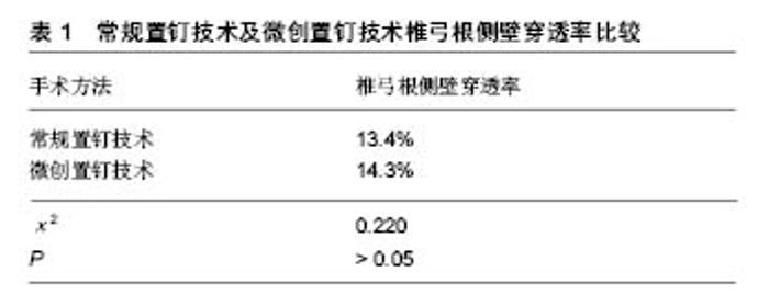
2.1 徒手置钉技术 如人字嵴顶点法、Weinstein法、Magerl法等。虽然国内外众多学者根据自己的经验与研究提出了多种进钉法[5-6]。但目前临床最常用的仍是以上3种方法。为了使椎弓根螺钉准确置入,术中需使用C形臂X射线机等影像设备进行辅助,使手术人员及患者暴露于辐射危险中。 2.1.1 人字嵴法 解剖学研究发现在腰椎峡部有一隆起的纵嵴,在上关节突根部的后外侧也有一隆起的纵嵴,两嵴汇合形成了“人”字结构,故称人字嵴,人字嵴结构比较稳定,有报道其出现率为94.5%,其解剖结构变异较少,只有少数人字嵴在干燥标本上较浅和不明显,但在活体中一般较为容易辨认,可作为理想的定位点。 临床应用时,将腰背肌剥离至关节突关节外缘即可充分显露人字嵴,因椎弓根与矢状面有一定的夹角,所以进钉时需向外倾斜5°-10°。尤其在T12-L4节段采用人字嵴定位的准确程度较高。如因小关节增生肥大,导致人字嵴结构难以辨认,则需要结合横突判断入钉点。以人字嵴作为进钉定位点,不需切开关节突关节的关节囊,对关节突关节的稳定性影响较小,且该点易于显露,手术操作较容易。另外,有研究者认为,组成人字嵴的两条纵嵴上无肌肉附着,不易发生退变,故出现解剖变异的概率较低,是理想的椎弓根定位方法。 2.1.2 Weinstein法 采用固定椎的上关节突外缘垂直延长线与横突中轴水平线的交点,即预植入椎的上关节突的颈背部,该处椎板外缘有一典型的骨嵴,定点标志也相当于紧靠骨嵴外上方的凹陷处。Weinstein法的椎弓根定点标志相当于椎弓根轴线在椎弓根后方的投影点,此法也较常用。 2.1.3 Magerl法 需显露上关节突和横突,以上关节突外缘切线及横突中线的交点为进钉点,常规置钉,螺钉平行于椎体终板,内倾约15°。以横突中线进行定位,但椎弓根中点不一定在横突中线上。有学者通过对干燥骨标本研究发现L1、L2、L3的椎弓根中心点分别位于横突中轴线上方3.3 mm、2.5 mm和1.8 mm处,L4椎弓根中心点接近中轴线,位于横突中轴线下0.3 mm处,L5椎弓根中心点则位于横突中轴线下方1.2 mm处。所以有一定的置钉失误率。 以上各方法在术中应用时,都会出现一些需要二次置钉的情况,由于椎弓根毗邻结构的复杂性以及个体化差异,徒手置钉的失误率可高达20%-40%。传统的椎弓根钉置钉技术要点为螺钉型号、进钉点、进钉方向、进钉深度。术者依据书本中对其技术的描述,依靠患者X射线片或CT数据在脑海中进行术前的手术模拟,以确定手术方案。这种手术方案质量的高低,取决于医生的临床经验与技能,且需要整个手术组成员精确领会制定手术方案人员头脑中模拟的手术方案。患者的个体差异、解剖变异、脊柱退变等因素都可能导致术者不能对进钉点产生个性化的准确认知,最终可能造成错误判断,引起螺钉误置,甚至导致严重的并发症,如脊髓及神经损伤、大血管损伤、硬膜撕裂及脑脊液漏等。虽然随着手术医生经验的积累,置钉准确率有所提高,但对于颈椎、上胸椎或者解剖结构畸变的病例,也仍有一定的置钉失败率。 2.2 经皮椎弓根螺钉固定技术 目前也被广泛应用于胸腰椎伤病的治疗中[7]。这一微创治疗方式使手术切口最小化、减少椎旁肌肉剥离和出血量,但其准确度仍是脊柱外科医师关心的问题。 Raley等[8]回顾分析88例患者(424枚微创螺钉)的CT扫描结果发现螺钉误置率达9.7%,其中穿透椎弓根侧壁的仅占4%,仅1例出现术后L4神经根损伤。Oh等[9]对比558枚常规椎弓根螺钉和498枚微创螺钉的CT影像资料,常规螺钉组与微创螺钉组椎弓根侧壁穿透率分别为13.4%和14.3%,差异无显著性意义(表1)。2组误置螺钉多数为轻度穿透;常规椎弓根螺钉置入技术的外侧壁穿透率(66.7%)高于微创螺钉置入组(43.7%);而经皮组椎弓根内侧壁、上壁和下壁穿透率略高于开放组,但两者差异均无显著性意义。原因可能是:①常规组螺钉置入时,周围肌肉组织厚或肌肉张力过大,常导致置钉角度偏小,容易使螺钉穿透椎弓根外侧壁。有时即使椎弓根螺钉钉道制备完毕探针探查没有问题,拧入螺钉时也容易因为周围软组织挤压,导致螺钉内倾角度变小并穿透外侧壁;②常规组术中很少常规采用后前位透视明确螺钉位置,术中很难及时发现椎弓根螺钉穿透内外侧皮质的情况。而经皮组全程在X射线透视下操作,安全性高。"


2.3 计算机辅助手术导航系统(computer-assisted surgery navigation system,CASNS) 是以影像技术为基础,通过立体定位和虚拟成像技术显示局部解剖结构与手术器械的空间位置关系,以帮助术者提高椎弓根螺钉置入的准确率[10]。 自20世纪90年代Steinmann等将计算机辅助手术导航系统应用于脊柱外科手术以来,其已成为推动脊柱外科向精准化、数字化发展的重要技术之一。 传统手术中出现的并发症,如螺钉穿透椎体前缘主要与螺钉置入角度以及螺钉长度选择不当有关,精确控制螺钉置入角度和螺钉长度对降低并发症起重要作用。传统手术主要依据术者的临床经验和术中X射线影像估计螺钉角度和长度,一旦失误则可能导致螺钉穿透椎体前缘。而导航技术中O-arm导航系统提供了清晰的三维图像,可以准确地控制螺钉的置入角度和深度,其中螺钉置入角度与导航图像中虚拟钉道角度具有高度的一致性。 此外,通过导航图像可以使虚拟钉道深度测量的较为准确,选择合适长度的螺钉,从而实现对螺钉置入角度的精确控制和置钉深度的最大化,降低螺钉穿透椎体前缘发生血管及内脏损伤的风险[11]。 目前,导航技术仍处于发展阶段,有学者观察到导航辅助下置入椎弓根螺钉时存在手术对象的解剖位置在三维导航图像中出现相对移位的“漂移”现象[12],可导致置钉失败[13]。对于此种现象,目前尚无有效对策,仍需借助术者的经验和术中的严密观察及时发现并予以校正。据统计,计算机导航技术辅助置钉的成功率为95%-98%,但也存在一定误差,如进钉点的确定等。该技术目前的主要不足在于准备阶段耗时长,延长了手术时间,增加了出血量和术后感染等并发症的发生率。此外,计算机导航设备价格昂贵,全面普及仍存在较大困难。 2.4 3D打印技术 实质是“激光快速成型技术”,也被称为“增量技术”、“增材技术”,是20世纪80年代末兴起的一门新技术[14]。3D打印的基础是数字模型文件,通过可以“打印”出真实物体的3D打印机,采用分层加工、迭加成形的方式逐层增加材料以生成3D实体,其最突出的优点是无需机械加工或模具,就能直接从计算机图形数据中生成任何形状的物体。该技术以往常在模具制造、工业设计等领域被用于制造模型,现在也逐渐被应用于数字骨科领域,为骨科手术尤其是脊柱手术精确性的提高提供了新的方法。 近年来国内一些学者报道应用3D打印技术制备个体化导航模板以辅助椎弓根螺钉置入手术,可有效提高置钉准确性,能有效减少手术并发症[15],具有较好的临床应用前景[16]。"

| [1] Kasten MD, Rao LA, Priest B. Long-term results of iliac wing fixation below extensive fusions in ambulatory adult patients with spinal disorders. J Spinal Disord Tech. 2010;23(7):e37-42.[2] Siewe J, Bredow J,Oppermann J,et al. Evaluation of efficacy of a new hybrid fusion device : a randomized ,two-centre controlled trial. BMC Musculoskelet Disord. 2014;15:249. [3] 龚全,孔清泉,李涛,等.后路复位技术在重度腰椎滑脱症治疗中的应用[J].中国修复重建外科杂志,2013,27(4):393-398.[4] Zebala LP, Bridwell KH, Baldus C, et al. Minimum 5-year radiographic results of long scoliosis fusion in juvenile spinal muscular atrophy patients: major curve progression after instrumented fusion. J Pediatr Orthop. 2011;31(5):480-488.[5] 刘军,项良碧,陈语,等.“触摸法”经皮椎弓根钉内固定治疗不稳定胸腰椎骨折[J]. 颈腰痛杂志,2010,31(5):330-334.[6] 李永成,丁元霞,单玉云,等.直视下胸腰椎椎弓根螺钉旋转置人法临床应用[J]. 颈腰痛杂志,2009,30(5):430-432.[7] Lefhnc M,Peltier J,Fichten A,et al.Dual, minimally invasive fixation in acute,double,thoracic spine fmcture.Minim Invasive Neumsurg. 201l;54(5-6):253-256.[8] Raley DA, Mobbs RJ. Restrospective computed tomography scan analysis of percutaneously inserted pediclescrews for posteriortrans pedicular stabilization of the thoracic and lumbar spine:accuracy and complication rates.Spine(Phila Pa 1976). 2012;37(12):1092-1100.[9] Oh HS,Kim JS,Lee SH,et al.Comparison between the accuracy of percutaneous and open pedicle screw fixations in lumbosacral fusion.Spine J. 2013;13(12) :1751-1757.[10] Sonntag VK.Pedicle screw fixation made safer by the use of computed tomography-guided intraoperative navigation. World Neurosurg. 2013;79(2):286-287.[11] Parker SL,Amin AG,Santiago-Dieppa D,et al.Incidence and clinical significance of vascular encroachment resulting from free-hand placement of pedicle screws in the thoracic and lumbar spine:analysis of 68 16 consecutive screws.Spine. 2014;39(8):683-687.[12] Rivkin MA.Yocom SS.Thoracolumbar instrumentation with CT-guided navigation(0-arm)in 270 consecutive patients: accuracy rates and lessons learned.Neurosurg Focus. 2014;36(3):E7.[13] 周东生.实用骨科导航技术[M].2版. 北京:人民军医出版社, 2012:75.[14] 周伟民,闵国全,李小丽.3D打印医学[J].组织工程与重建外科杂志,2014,10(1):1-7.[15] 周锦.3D打印技术在骨科临床中的应用[J].中华骨科杂志, 2014, 34(10):1078.[16] 庞骄阳,赵岩,肖宇龙,等.3D打印技术在脊柱外科的应用[J].中国组织工程研究, 2016,20(4):577-581.[17] Kurtz SM, Lanman TH, Higgs G, et al. Retrieval analysis of PEEK rods for posterior fusion and motion preservation. Eur Spine J. 2013;22(12): 2752-2759.[18] 郭昭庆,陈仲强,齐强,等.重度发育不良性腰椎滑脱的手术治疗[J]. 中华外科杂志,2014,52(11):845-850.[19] Okuda S, Oda T, Yamasaki R, et al. Posterior lumbar interbody fusion with total facetectomy for low-dysplastic isthmic spondylolisthesis: effects of slip reduction on surgical outcomes: Clinical article. J Neurosurg Spine. 2014;21(2): 171-178.[20] Zang L, Du P, Hai Y, et al. Device related complications of the Coflex interspinous process implant for the lumbar spine.Chin Med J (Engl). 2013;126(13):2517-2522.[21] Paik H, Kang DG, Lehman RA Jr, et al. The biomechanical consequences of rod reduction on pedicle screws: should it be avoided? Spine J. 2013;13(11):1617-1626.[22] Joglekar SB, Mehbod AA. Surgeon's view of pedicle screw implantation for the monitoring neurophysiologist. J Clin Neurophysiol. 2012;29(6):482-488.[23] Kosmopoulos V, Schizas C. Pedicle screw placement accuracy: a meta-analysis. Spine (PhilaPa 1976). 2007; 32(3):111-120. [24] Olerud S, Sjostrom L, Karlstrom G, et al. Spontaneous effect of increased stability of the lower lumbar spine in cases of severe chronic back pain. Clin Orthop Relat Res.2012;20(3): 67-74. [25] Soini J, Slatis P, Kannisto M, et al. External transpedicular fixation test of the lumbar spine correlates with the outcome of subsequent lumbar fusion. Clin Orthop Relat Res. 2013;293: 89-96.[26] Lefhnc M, Peltier J, Fichten A, et al. Dual,minimally invasive fixation in acute, double,thoracic spine fmcture. Minim Invasive Neumsurg. 201l;54(5-6):253-256.[27] Ni WF, Huang YX, Chi YL, et al. Percutaneous pedicle screw fixation for neurologic intact thoracolumbar burst fractures. J Spinal Disord Tech. 2010;23(8): 530-537.[28] 刘洪涛,镇万新,朱杰诚,等. 经皮椎弓根内固定术在胸腰椎骨折中的应用[J]中国矫形外科杂志, 2005, 13(16):1227-1229[29] Kim MC, Chung HT, Cho JL, et al. Factors affecting the accurate placement of percutaneous pedicle screws during minimally invasive transfomminal lumbar interbody fusion.Eur Spine J. 2011;20(10);1635-1643.[30] 周伟民,闵国全,李小丽. 3D 打印医学[J].组织工程与重建外科杂志,2014, 10(1):1-7. [31] Hu Y,Yuan ZS,Spiker WR,et al .Deviation analysis of C2 translaminar screw placement assisted by a novel rapid prototyping drill template: acadaveric study. Eur Spine J. 2013;22(12):2770-2776.[32] 戎帅,滕勇,乌日开西•艾依提,等.基于3D打印技术的腰椎多节段峡部裂个性化手术治疗[J].中国矫形外科杂志, 2013,21(21): 2222-2226.[33] Putzier M,Strube P,Cecchinato R,et al.A new navigational tool for pedicle screw placement in patients with severe scoliosis:a pilot study to prove feasibility,accuracy,and identify operative challenges. Clin Spine Surg. 2017;30(4):E430-E439.[34] Kaneyama S,Sugawara T,Sumi M,et al.A novel screw guiding method with a screw guide template system for posterior C-2 fixation:clinical article. J Neurosurg Spine. 2014;21(2): 231-238.[35] Hu Y,Yuan ZS,Kepler CK,et al.Deviation analysis of atlantoaxial pedicle screws assisted by a drill template. Orthopedics. 2014;37(5):e420-427. [36] Hu Y,Yuan ZS,Kepler CK,et al.Deviation analysis of C1-C2 transartieular screw placement assisted by a novel rapid prototyping drill template:a cadaveric study. J Spinal Disord Tech. 2014;27(5):E181-186. [37] Merc M,Drstvensek I,Vogrin M,et al.Error rate of multi-level rapid prototyping trajeetories for pedicle screw placement in lumbar and sacral spine.Chin J Traumatol. 2014;17(5): 261-266.[38] Yang JC,Ma XY,Xia H,et al.Clinical application of computer aided design-rapid prototyping in C1--C2 operation techniques for complex atlantoaxial instability.J Spinal Disord Tech. 2014;27 (4):E143-150.[39] Merc M,Drstvensek I,Vogrin M,et al.A multi-level rapid prototyping drill guide template reduces the perforation risk of pedicle screw placement in the lumbar and sacral spine.Arch Orthop Trauma Surg. 2013;133(7):893-899.[40] Ferrari V,Parchi P,Condino S,et al. An optimal design for patient-specific templates for pedicle spine screws placement. Int J Med Robot. 2013;9(3):298-304.[41] 胡勇,袁振山,谢辉,等.快速成型导向模板技术在枢椎椎板交叉螺钉置钉中的应用[J].中华骨科杂志,2013,33(6):640-648.[42] 陈国平,陆声,徐永清,等.计算机辅助导航模板辅助颈椎椎弓根置钉可行性研究[J]. 解剖与临床,2010,15(3):164-167.[43] 尹庆水,万磊,夏虹,等.计算机辅助设计寰枢椎椎弓根内固定数字化导向模板精确置钉[J].中华骨科杂志,2009,29(12):1089-1092.[44] 陆声,徐永清,张元智,等.快速成形个体化导航模板辅助枢椎椎板螺钉的植入[J]. 中华骨科杂志,2009,29(8):739-743.[45] 刘亚军,田伟,靳培浩,等.导航微创与传统切开经椎间孔入路椎间植骨融合术治疗成人腰椎滑脱症的对照研究[J].中华创伤骨科杂志,2014,16(3):194-198.[46] Tian W , Xu YF, Liu B, et al. Computer-assisted Minimally Invasive Transforaminal Lumbar Interbody Fusion May Be Better than Open surgery for Treating Degenerative Lumbar Disease. J Spinal Disord Tech. 2014.[47] Larson AN, Santos ER, Polly DW Jr, et al. Pediatric pedicle screw placement using intraoperative computed tomography and 3-dimensional image-guided navigation. Spine. 2012; 37(3):E188-E194.[48] Kantelhardt SR, Martinez R, Baerwinkel S, et al. Perioperative course and accuracy of screw positioning in conventional, open robotic-guided and percutaneous robotic-guided, pedicle screw placement. Eur Spine J. 2011;20(6):860-868.[49] Eck JC, Lange J, Street J, et al. Accuracy of intraoperative computed tomography-based navigation for placement of percutaneous pedicle screws. Global Spine J. 2013;3(2): 103-108.[50] Faizan, A, Kiapour A, Kiapour AM, et al. Biomechanical analysis of various footprints of transforaminal lumbar interbody fusion devices. J Spinal Disord Tech. 2014;27(4): E118-127.[51] 吴静,茅金宝,孔祥云,等.导航与普通透视对手术室医务人员放射量的对比分析[J].医学影像学杂志, 2013, 23(10):1631-1634.[52] Yang M, Li C, Li Y, et al. Application of 3D rapid prototyping technology in posterior corrective surgery for Lenke 1 adolescent idiopathic scoliosis patients. Medicine(Baltimore). 2015;94(8):e582.[53] 韩今华,马德春,白淼,等.腰椎椎弓根螺钉进钉点的个体化定位[J].中国组织工程研究与临床康复,2011,15(48):9066-9069.[54] 夏刚,万军,江汉.强化椎弓根螺钉的研究及应用前景[J].实用骨科杂志,2004,10(2):621-623.[55] Wang L, Huang H, Zhang Z, et al. Biomechanical Evaluation of a Novel Autogenous Bone Interbody Fusion Cage for Posterior Lumbar Interbody Fusion in a Cadaveric Model. Spine (Phila Pa 1976). 2014.[56] 袁强,张贵林,吴静晔,等. 3D 计算机导航下经椎弓根骨水泥增强螺钉的应用[J].中华创伤骨科杂志,2014,16(3):199-203.[57] Bourgeois AC, Faulkner AR , Bradley YC, et al. Improved Accuracy of Minimally Invasive Transpedicular Screw Placement in the Lumbar Spine with Three-dimensional Stereotactic Image Guidance: A Comparative Meta-analysis. J Spinal Disord Tech. 2015;28(9):324-329. [58] Silbermann J, Riese F, Allam Y, et al. Computer tomography assessment of pedicle screw placement in lumbar and sacral spine: comparison between free-hand and O-arm based navigation techniques. Eur Spine J. 2011;20(6): 875-881.[59] Wang S, Zhang J, Qiu G, et al. Posterior hemivertebra resection with bisegmental fusion for congenital scoliosis: more than 3 year outcomes and analysis of unanticipated surgeries. Eur Spine J. 2013;22(2):387-393.[60] 赵江涛,李洪涛,宫云昭.计算机导航辅助下椎弓根螺钉内固定疗效观察[J] .医学综述,2009,15(11):1746-1748. |
| [1] | Yao Xiaoling, Peng Jiancheng, Xu Yuerong, Yang Zhidong, Zhang Shuncong. Variable-angle zero-notch anterior interbody fusion system in the treatment of cervical spondylotic myelopathy: 30-month follow-up [J]. Chinese Journal of Tissue Engineering Research, 2022, 26(9): 1377-1382. |
| [2] | Jiang Huanchang, Zhang Zhaofei, Liang De, Jiang Xiaobing, Yang Xiaodong, Liu Zhixiang. Comparison of advantages between unilateral multidirectional curved and straight vertebroplasty in the treatment of thoracolumbar osteoporotic vertebral compression fracture [J]. Chinese Journal of Tissue Engineering Research, 2022, 26(9): 1407-1411. |
| [3] | An Weizheng, He Xiao, Ren Shuai, Liu Jianyu. Potential of muscle-derived stem cells in peripheral nerve regeneration [J]. Chinese Journal of Tissue Engineering Research, 2022, 26(7): 1130-1136. |
| [4] | Zhang Jinglin, Leng Min, Zhu Boheng, Wang Hong. Mechanism and application of stem cell-derived exosomes in promoting diabetic wound healing [J]. Chinese Journal of Tissue Engineering Research, 2022, 26(7): 1113-1118. |
| [5] | Song Jiawei, Yang Yongdong, Yu Xing, Yang Jizhou, Wang Fengxian, Qu Yi, Bi Lianyong. Mid-term effect of Isobar EVO non-fusion dynamic fixation in the treatment of adjacent segment disease after lumbar fusion [J]. Chinese Journal of Tissue Engineering Research, 2022, 26(6): 908-913. |
| [6] | He Yunying, Li Lingjie, Zhang Shuqi, Li Yuzhou, Yang Sheng, Ji Ping. Method of constructing cell spheroids based on agarose and polyacrylic molds [J]. Chinese Journal of Tissue Engineering Research, 2022, 26(4): 553-559. |
| [7] | He Guanyu, Xu Baoshan, Du Lilong, Zhang Tongxing, Huo Zhenxin, Shen Li. Biomimetic orientated microchannel annulus fibrosus scaffold constructed by silk fibroin [J]. Chinese Journal of Tissue Engineering Research, 2022, 26(4): 560-566. |
| [8] | Chen Xiaoxu, Luo Yaxin, Bi Haoran, Yang Kun. Preparation and application of acellular scaffold in tissue engineering and regenerative medicine [J]. Chinese Journal of Tissue Engineering Research, 2022, 26(4): 591-596. |
| [9] | Kang Kunlong, Wang Xintao. Research hotspot of biological scaffold materials promoting osteogenic differentiation of bone marrow mesenchymal stem cells [J]. Chinese Journal of Tissue Engineering Research, 2022, 26(4): 597-603. |
| [10] | Shen Jiahua, Fu Yong. Application of graphene-based nanomaterials in stem cells [J]. Chinese Journal of Tissue Engineering Research, 2022, 26(4): 604-609. |
| [11] | Zhang Tong, Cai Jinchi, Yuan Zhifa, Zhao Haiyan, Han Xingwen, Wang Wenji. Hyaluronic acid-based composite hydrogel in cartilage injury caused by osteoarthritis: application and mechanism [J]. Chinese Journal of Tissue Engineering Research, 2022, 26(4): 617-625. |
| [12] | Li Hui, Chen Lianglong. Application and characteristics of bone graft materials in the treatment of spinal tuberculosis [J]. Chinese Journal of Tissue Engineering Research, 2022, 26(4): 626-630. |
| [13] | Gao Cangjian, Yang Zhen, Liu Shuyun, Li Hao, Fu Liwei, Zhao Tianyuan, Chen Wei, Liao Zhiyao, Li Pinxue, Sui Xiang, Guo Quanyi. Electrospinning for rotator cuff repair [J]. Chinese Journal of Tissue Engineering Research, 2022, 26(4): 637-642. |
| [14] | Guan Jian, Jia Yanfei, Zhang Baoxin , Zhao Guozhong. Application of 4D bioprinting in tissue engineering [J]. Chinese Journal of Tissue Engineering Research, 2022, 26(3): 446-455. |
| [15] | Huang Bo, Chen Mingxue, Peng Liqing, Luo Xujiang, Li Huo, Wang Hao, Tian Qinyu, Lu Xiaobo, Liu Shuyun, Guo Quanyi . Fabrication and biocompatibility of injectable gelatin-methacryloyl/cartilage-derived matrix particles composite hydrogel scaffold [J]. Chinese Journal of Tissue Engineering Research, 2022, 10(16): 2600-2606. |
| Viewed | ||||||
|
Full text |
|
|||||
|
Abstract |
|
|||||