Chinese Journal of Tissue Engineering Research ›› 2015, Vol. 19 ›› Issue (7): 996-1002.doi: 10.3969/j.issn.2095-4344.2015.07.003
Previous Articles Next Articles
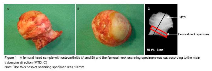
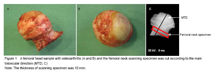
Three-dimensional microarchitecture of the proximal femur in osteoarthritis and rheumatoid arthritis
- 1 Department of Joint Surgery, China-Japan Friendship Hospital, Beijing 100029, China2 The Orthopaedic Research Laboratory, Department of Orthopaedic Surgery, Odense University Hospital, University of Southern Denmark, Odense, Denmark3 Department of Orthopaedic Surgery, Odense University Hospital, and Clinical Institute, University of Southern Denmark, Odense, Denmark
-
Online:2015-02-12Published:2015-02-12 -
Contact:Wang Bai-liang, Department of Joint Surgery, China-Japan Friendship Hospital, Beijing 100029, China -
About author:Wang Bai-liang, M.D., Associate chief physician, Department of Joint Surgery, China-Japan Friendship Hospital, Beijing 100029, China -
Supported by:the Youth Foundation of China-Japan Friendship Hospital, No. 2013-QN-19
CLC Number:
Cite this article
Wang Bai-liang, Ming Ding, Søren Overgaard. Three-dimensional microarchitecture of the proximal femur in osteoarthritis and rheumatoid arthritis[J]. Chinese Journal of Tissue Engineering Research, 2015, 19(7): 996-1002.
share this article
Difference in the three-dimensional microstructure of femoral neck specimens between osteoarthritis and rheumatoid arthritis groups All the data were not distributed normally. Analyses of variation in the microstructural properties of different regions in the two groups are summarized in Table 1. The patients with osteoarthritis were older than those with rheumatoid arthritis, but there was no significant difference. The entire cortical bone of the femoral neck in patients with rheumatoid arthritis had higher BV/TV, Co.Th*, Co.N* and Co.CD, lower Co.Sp, Co.DA and CSA than those with osteoarthritis, but there was no significant difference between the two groups. The entire trabecular bone in the rheumatoid arthritis group had higher BV/TV, Tb.N*, Tb.CD, Tb.DA and CSA, lower Tb.Th*, Tb.Sp than that in the osteoarthritis group, but there was no significant difference between the two groups. Properties of the femoral neck were similar to those of the trabecular bone. This decrease in bone volume in the osteoarthritis group resulted from the decreased amount of cortical and trabecular bone tissues as well as a consequent increase in dispersion degree. The osteoarthritis group was also characterized by a loss of the connectivity, an increase in DA for the cortical bone, but a decrease in DA for the cancellous bone and the entire trabecular bone when compared to the rheumatoid arthritis group. Relations between the microstructural parameters of femoral neck specimens in osteoarthritis and rheumatoid arthritis groups Among all the parameters, BV/TV had the strong correlation with Th*, Sp, SMI, and N* for the entire femoral head, cortical and trabecular bone in both osteoarthritis and rheumatoid arthritis groups. For all the three regions of rheumatoid arthritis and osteoarthritis patients, we found a BV/TV-related increase in Th* and a BV/TV-related decrease in Sp and SMI. But for the correlation between BV/TV and N*, there was no consistent tendency in the three regions of osteoarthritis and rheumatoid arthritis groups. The significant correlations between Sp and N* were also found in the three regions of osteoarthritis and rheumatoid arthritis groups. "
| [1]Sun SS, Ma HL, Liu CL. Difference in femoral head and neck material properties between osteoarthritis and osteoporosis. Clin Biomech. 2008;23:S39-S47.[2]Issever AS, Burghardt A, Patel V, et al. A micro-computed tomography study of the trabecular bone structure in the femoral head. J Musculoskelet Neuronal Interact. 2003; 3(2):176-184. [3]Radin EL, Rose RM. Role of subchondral bone in the initiation and progression of cartilage damage. Clin Orthop. 1986;213:34-40. [4]Li BH, Aspden RM. Mechanical and material properties of the subchondral bone plate from the femoral head of patients with osteoarthritis or osteoporosis. Ann Rheum Dis. 1997;56:247-254. [5]Ding M, Christian Danielsen C, Hvid I. Bone density does not reflect mechanical properties in early-stage arthrosis. Acta Orthop Scand. 2001;72(2):181-185. [6]Dieppe P, Cushnaghan J, Young P, et al. Prediction of the progression of joint space narrowing in osteoarthritis of the knee by bone scintigraphy. Ann Rheum Dis. 1993;52: 557-563. [7]Chappard C, Peyrin F, Bonnassie A, et al. Subchondral bone micro-architectural alterations in osteoarthritis: a synchrotron micro-computed tomography study. Osteoarthritis Cartilage. 2006;14(3):215-223.[8]Cooper C, Cook PL, Osmond C, et al. Osteoarthritis of the hip and osteoporosis of the proximal femur. Ann Rheum Dis. 1991;50:540-542. [9]Dequeker J, Johnell O. Osteoathritis protects against femoral neck fracture: the MEDOS study experience. Bone. 1993;14:S51-S56. [10]Verstraeten A, Van Ermen H, Haghebaert G, et al. Osteoarthrosis retards the development of osteoporosis. Observation of the coexistence of osteoarthrosis and osteoporosis. Clin Orthop. 1991;264:169-177. [11]Gong H, Zhang M, Yeung HY, et al. Regional variations in microstructural properties of vertebral trabeculae with aging. J Bone Miner Metab. 2005;23:174-180. [12]Inaba M, Nagata M, Goto H, et al. Preferential reductions of paraarticular trabecular bone component in ultradistal radius and of calcaneus ultrasonography in early-stage rheumatoid arthritis. Osteoporos Int. 2003;14(8):683-687. [13]Korczowska I, Olewicz-Gawlik A, Trefler J, et al. Does low-dose and short-term glucocorticoids treatment increase the risk of osteoporosis in rheumatoid arthritis female patients? Clin Rheumatol. 2008;27(5):565-572. [14]Böttcher J, Pfeil A, Mentzel H, et al. Peripheral bone status in rheumatoid arthritis evaluated by digital X-ray radiogrammetry and compared with multisite quantitative ultrasound. Calcif Tissue Int. 2006;78(1):25-34. [15]Enokida M, Yamasaki D, Okano T, et al. Bone mass changes of tibial and vertebral bones in young and adult rats with collagen-induced arthritis. Bone. 2001;28(1): 87-93. [16]Le Corroller T, Pithioux M, Chaari F, et al. Bone texture analysis is correlated with three-dimensional microarchitecture and mechanical properties of trabecular bone in osteoporotic femurs. J Bone Miner Metab. 2013; 31(1):82-88. [17]Ollivier M, Le Corroller T, Blanc G, et al. Radiographic bone texture analysis is correlated with 3D microarchitecture in the femoral head, and improves the estimation of the femoral neck fracture risk when combined with bone mineral density. Eur J Radiol. 2013; 82(9):1494-1498. [18]Deodhar AA, Woolf AD. Bone mass measurement and bone metabolism in rheumatoid arthritis: a review. Br J Rheumatol. 1996;35(4):309-322. [19]Homminga J, Van-Rietbergen B, Lochmuller EM, et al. The osteoporotic vertebral structure is well adapted to the loads of daily life, but not to infrequent ‘‘error’’ loads. Bone. 2004; 34: 510-516. [20]Blain H, Chavassieux P, Portero-Muzy N, et al. Cortical and trabecular bone distribution in the femoral neck in osteoporosis and osteoarthritis. Bone. 2008;43(5):862-868. [21]Mayhew PM, Thomas CD, Clement JG, et al. Relation between age, femoral neck cortical stability, and hip fracture risk. Lancet. 2005;366:129-135. [22]Basso T, Klaksvik J, Syversen U, et al. A biomechanical comparison of composite femurs and cadaver femurs used in experiments on operated hip fractures. J Biomech. 2014; 18;47(16):3898-3902. [23]Bouxsein ML, Fajardo RF. Cortical stability of the femoral neck and hip fracture risk. Lancet. 2005;366:1532-1534.[24]Rivadeneira F, Zillikens MC. Femoral neck BMD is a strong predictor of hip fracture susceptibility in elderly men and women because it detects cortical bone instability: the rotterdam study. J Bone Miner Res. 2007;22:1781-1790. [25]Fazzalari NL, Parkinson IH. Femoral trabecular bone of osteoarthritic and normal subjects in an age and sex matched group. Osteoarth Cart. 1998;6:377-382. [26]Fazzalari NL, Moore RJ, Manthey BA, et al. Comparative study of iliac crest and subchondral femoral bone in osteoarthritis patients. Bone. 1992;13:331-335. [27]Hildebrand T, Rüegsegger P. Quantification of bone microarchitecture with the structure model index. Comput Methods Biomech Biomed Engin. 1997;1(1):15-23. [28]Odgaard A. Three-dimensional methods for quantification of cancellous bone architecture. Bone. 1997;20(4):315-328. [29]Tanck E, Bakker AD, Kregting S, et al. Predictive value of femoral head heterogeneity for fracture risk. Bone. 2009; 44(4):590-595. [30]Duque G, Troen BR. Understanding the mechanisms of senile osteoporosis: new facts for a major geriatric syndrome. J Am Geriatr Soc. 2008;56:935-941. [31]Bell KL, Loveridge N, Power J et al. Structure of the femoral neck in hip fracture: Cortical bone loss in the inferoanterior to superoposterior axis. J Bone Miner Res 1999;14: 111-119. [32]Lotz JC, Cheal EJ, Hayes WC. Stress distribution within the proximal femur during fait and falls: implications for osteoporotic fracture. Osteoporos Int. 1995;5:252-261. [33]Bell KL, Loveridge N, Power J, et al. Regional differences in cortical porosity in the fractured femoral neck. Bone. 1999; 24:57-64. [34]Lane NE, Pressman AR, Star VL, et al. Rheumatoid arthritis and bone mineral density in elderly women. The study of osteoporotic fractures research group. J Bone Miner Res. 1995;10(2):257-263. [35]Aguila LA, Lopes MR, Pretti FZ, et al. Clinical and laboratory features of overlap syndromes of idiopathic inflammatory myopathies associated with systemic lupus erythematosus, systemic sclerosis, or rheumatoid arthritis. Clin Rheumatol. 2014;33(8):1093-1098. [36]Heidari B, Hassanjani Roushan MR. Rheumatoid arthritis and osteoporosis. Caspian J Intern Med. 2012;3(3): 445-446. [37]Chen H, Kubo KY. Bone three-dimensional microstructural features of the common osteoporotic fracture sites. World J Orthop. 2014;18;5(4):486-495. [38]Chen H, Zhou X, Fujita H, et al. Age-related changes in trabecular and cortical bone microstructure. Int J Endocrinol. 2013;2013:213234. [39]Courtland HW, Kennedy OD, Wu Y, et al. Low levels of plasma IGF-1 inhibit intracortical bone remodeling during aging. Age (Dordr). 2013;35(5):1691-1703. [40]Giambini H, Wang HJ, Zhao C, et al. Anterior and posterior variations in mechanical properties of human vertebrae measured by nanoindentation. J Biomech. 2013;46(3): 456-461.[41]Chen H, Zhou X, Washimi Y, et al. Three-dimensional microstructure of the bone in a hamster model of senile osteoporosis. Bone 2008;43:494-500. [42]Popp AW, Buffat H, Eberli U, et al. Microstructural parameters of bone evaluated using HR-pQCT correlate with the DXA-derived cortical index and the trabecular bone score in a cohort of randomly selected premenopausal women. PLoS One. 2014;9(2):e88946. [43]Chiba K, Burghardt AJ, Osaki M, et al. Heterogeneity of bone microstructure in the femoral head in patients with osteoporosis: an ex vivo HR-pQCT study. Bone. 2013;56(1): 139-146.[44]Nicks KM, Amin S, Melton LJ 3rd,et al. Three-dimensional structural analysis of the proximal femur in an age-stratified sample of women. Bone. 2013;55(1):179-188. [45]Manske SL, Liu-Ambrose T, Cooper DM, et al. Cortical and trabecular bone in the femoral neck both contribute to proximal femur failure load prediction. Osteoporos Int. 2009; 20(3):445-453.[46]Hordon LD, Peacock M. The architecture of cancellous and cortical bone in femoral neck fracture. Bone Miner. 1990;11: 335-345. [47]Lang TF, Keyak JH, Heitz MW, et al. Volumetric quantitative computed tomography of the proximal femur: precision and relation to bone strength. Bone. 1997;21:101-108. [48]Riggs BL, Melton LJ 3rd, Robb RA, et al. Population-based study of age and sex differences in bone volumetric density, size, geometry, and structure at different skeletal sites. J Bone Miner Res. 2004;19:1945-1954. |
| [1] | Wei Wei, Li Jian, Huang Linhai, Lan Mindong, Lu Xianwei, Huang Shaodong. Factors affecting fall fear in the first movement of elderly patients after total knee or hip arthroplasty [J]. Chinese Journal of Tissue Engineering Research, 2021, 25(9): 1351-1355. |
| [2] | Peng Zhihao, Feng Zongquan, Zou Yonggen, Niu Guoqing, Wu Feng. Relationship of lower limb force line and the progression of lateral compartment arthritis after unicompartmental knee arthroplasty with mobile bearing [J]. Chinese Journal of Tissue Engineering Research, 2021, 25(9): 1368-1374. |
| [3] | Zhang Chong, Liu Zhiang, Yao Shuaihui, Gao Junsheng, Jiang Yan, Zhang Lu. Safety and effectiveness of topical application of tranexamic acid to reduce drainage of elderly femoral neck fractures after total hip arthroplasty [J]. Chinese Journal of Tissue Engineering Research, 2021, 25(9): 1381-1386. |
| [4] | Chen Junming, Yue Chen, He Peilin, Zhang Juntao, Sun Moyuan, Liu Youwen. Hip arthroplasty versus proximal femoral nail antirotation for intertrochanteric fractures in older adults: a meta-analysis [J]. Chinese Journal of Tissue Engineering Research, 2021, 25(9): 1452-1457. |
| [5] | Huang Dengcheng, Wang Zhike, Cao Xuewei. Comparison of the short-term efficacy of extracorporeal shock wave therapy for middle-aged and elderly knee osteoarthritis: a meta-analysis [J]. Chinese Journal of Tissue Engineering Research, 2021, 25(9): 1471-1476. |
| [6] | Liu Xiangxiang, Huang Yunmei, Chen Wenlie, Lin Ruhui, Lu Xiaodong, Li Zuanfang, Xu Yaye, Huang Meiya, Li Xihai. Ultrastructural changes of the white zone cells of the meniscus in a rat model of early osteoarthritis [J]. Chinese Journal of Tissue Engineering Research, 2021, 25(8): 1237-1242. |
| [7] | Liu Lihua, Sun Wei, Wang Yunting, Gao Fuqiang, Cheng Liming, Li Zirong, Wang Jiangning. Type L1 steroid-induced osteonecrosis of the femoral head through femoral head and neck junction decompression by fenestration: a single-center prospective clinical study [J]. Chinese Journal of Tissue Engineering Research, 2021, 25(6): 906-911. |
| [8] | Liu Xin, Yan Feihua, Hong Kunhao. Delaying cartilage degeneration by regulating the expression of aquaporins in rats with knee osteoarthritis [J]. Chinese Journal of Tissue Engineering Research, 2021, 25(5): 668-673. |
| [9] | Ma Zetao, Zeng Hui, Wang Deli, Weng Jian, Feng Song. MicroRNA-138-5p regulates chondrocyte proliferation and autophagy [J]. Chinese Journal of Tissue Engineering Research, 2021, 25(5): 674-678. |
| [10] | Cao Xuhan, Bai Zixing, Sun Chengyi, Yang Yanjun, Sun Weidong. Mechanism of “Ruxiang-Moyao” herbal pair in the treatment of knee osteoarthritis based on network pharmacology [J]. Chinese Journal of Tissue Engineering Research, 2021, 25(5): 746-753. |
| [11] | Li Yonghua, Feng Qiang, Tan Renting, Huang Shifu, Qiu Jinlong, Yin Heng. Molecular mechanism of Eucommia ulmoides active ingredients treating synovitis of knee osteoarthritis: an analysis based on network pharmacology [J]. Chinese Journal of Tissue Engineering Research, 2021, 25(5): 765-771. |
| [12] | Song Shan, Hu Fangyuan, Qiao Jun, Wang Jia, Zhang Shengxiao, Li Xiaofeng. An insight into biomarkers of osteoarthritis synovium based on bioinformatics [J]. Chinese Journal of Tissue Engineering Research, 2021, 25(5): 785-790. |
| [13] | Deng Zhenhan, Huang Yong, Xiao Lulu, Chen Yulin, Zhu Weimin, Lu Wei, Wang Daping. Role and application of bone morphogenetic proteins in articular cartilage regeneration [J]. Chinese Journal of Tissue Engineering Research, 2021, 25(5): 798-806. |
| [14] | Zhao Xiang, Wei Cuilan, Zhang Yeting. Neurogenesis and neuroinflammation under exercise: alteration and regulation [J]. Chinese Journal of Tissue Engineering Research, 2021, 25(5): 813-820. |
| [15] | Zhang Yu, Feng Shuo, Yang Zhi, Zhang Ye, Sun Jianning, An Lun, Chen Xiangyang. Three-dimensional gait of patients with developmental dysplasia of hip undergoing total hip arthroplasty with high hip center [J]. Chinese Journal of Tissue Engineering Research, 2021, 25(3): 350-355. |
| Viewed | ||||||
|
Full text |
|
|||||
|
Abstract |
|
|||||