Chinese Journal of Tissue Engineering Research ›› 2015, Vol. 19 ›› Issue (46): 7419-7424.doi: 10.3969/j.issn.2095-4344.2015.46.008
Previous Articles Next Articles
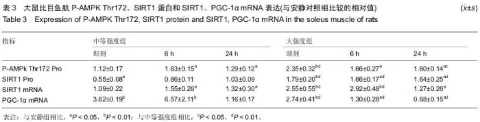
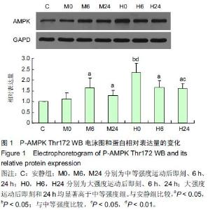
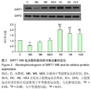
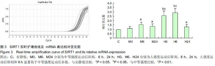
High-intensity endurance exercise inhibits mitochondrial biogenesis in the skeletal muscle
Zhang Guo-hua, Chen Shu-zhuang, Li Su-ping
- Sports Institute of Hanshan Normal University, Chaozhou 521041, Guangdong Province, China
-
Received:2015-08-22Online:2015-11-12Published:2015-11-12 -
About author:Zhang Guo-hua, Ph.D., Professor, Sports Institute of Hanshan Normal University, Chaozhou 521041, Guangdong Province, China -
Supported by:the Natural Science Foundation of Guangdong Province, No. S2012010010767; the Science Team Project of Hanshan Normal University, No. LT201203
Cite this article
Zhang Guo-hua, Chen Shu-zhuang, Li Su-ping. High-intensity endurance exercise inhibits mitochondrial biogenesis in the skeletal muscle[J]. Chinese Journal of Tissue Engineering Research, 2015, 19(46): 7419-7424.
share this article
"
"
"
"
"

| [1] Geng T, Li P, Okutsu M,et al.PGC-1 plays a functional role in exercise-induced mitochondrial biogenesis and angiogenesis but not fiber-type transformation in mouse skeletal muscle.Am J Physiol Cell Physiol.2010;298(3):572-579. [2] Uguccioni G, Hood DA. The importance of PGC-1 in contractile activity-induced mitochondrial adaptation.Am J Physiol Endocrinol Metab.2011;300(2): 361-371.
[3] Bartlett JD,Hwa Joo C,Jeong TS, et al. Matched work high-intensity interval and continuous running induce similar increases in PGC-1α mRNA, AMPK, p38, and p53 phosphorylation in human skeletal muscle. Warren GAppl Physiol.2012;112(7): 1135-1143.
[4] Frier BC, Hancock CR, Little JP,et al.Reductions in RIP140 are not required for exercise- and AICAR-mediated increases in skeletal muscle mitochondrial content.J Appl Physiol.2011; 111(3): 688-695.
[5] Steiner JL, Murphy EA, McClellan JL,et al. McClellan,et al.Exercise training increases mitochondrial biogenesis in the brain. J Appl Physiol.2011;111(4): 1066-1071.
[6] Bedford TG,Tipton CM,Wilson NC,et al.Maximum oxygen consumption of rats and its changes with various ex-perimental procedures. J Appl Physiol.1979;47(6):1278 -1283.
[7] Wisloff U,Helgerud J,Kemi OJ,et al.Intensity-con-trolled treadmill running in rats: VO2max and cardiac hy-pertrophy. Am J Physiol Heart Circ Physiol.2001;280: 1301-1310.
[8] Cassano P,Sciancalepore AG,Pesce V,et al. Acetyl-L-carnitine feeding to unloaded rats triggers in soleus muscle the coordinated expression of genes involved in mitochondrial biogenesis. Biochimica et Biophysica Acta ( BBA) Bioener -getics.2006;1757:1421-1428.
[9] Wagatsuma A,Kotake N,Kawachi T,et al. Mitochondrial adaptations in skeletal muscle to hindlimb unloading. Molecular and cellular biochemistry.2010;350(1-2):1-11.
[10] Liu J,Peng YH,Cui ZW,et al. Depressed mitochondrial biogenesis and dynamic remodeling in mouse tibialis anterior and gastrocnemius induced by 4-week hindlimb unloading. IUBMB life.2012;64(11):901-910.
[11] Wu Z,Puigserver P,Andersson U,et al.Mechanisms controlling mitochondrial biogenesis and respiration through the thermogenic coactivator PGC-1. Cell.1999;98(1):115-124.
[12] Fujimoto E,Yamaguchi W,Terada S,et al.Change inPGC-1α Expression in Rat Skeletal Muscle after Low-intensity Prolonged Swimming Exercise.J Physiol Anthropol. 2011; 30(1): 23-27.
[13] Li L, Chen Y, Gibson SB.Starvation-induced autophagy is regulated by mitochondrial reactive oxygen species leading to AMPK activation. Cell Signal. 2013;25(1):50-65.
[14] Mackenzie RM, Salt IP, Miller WH,et al. Mitochondrial reactive oxygen species enhance AMP-activated protein kinase activation in the endothelium of patients with coronary artery disease and diabetes. Clin Sci (Lond).2013;124(6): 403-411.
[15] Davis JM, Murphy EA, Carmichael MD,et al.Quercetin increases brain and muscle mitochondrial biogenesis and exercise tolerance.Am J Physiol Regulatory Integrative Comp Physiol.2009;296(4): 1071-1077.
[16] Canto C, Jiang LQ, Deshmukh AS,et al.Interdependence of AMPK and SIRT1 for metabolic adaptation to fasting and exercise in skeletal muscle. Cell Metab.2010; 11(3): 213-219.
[17] Funk JA, Odejinmi S, Schnellmann RG.Schnellmann. SRT1720 induces mitochondrial biogenesis and rescues mitochondrial function after oxidant injury in renal proximal tubule cells. J Pharmacol Exp Ther.2010; 333(2): 593-601.
[18] Khalil HS, Tummala H, DeCaris L, et al.Pharmacological inhibition of ATM results in mitochondrial biogenesis in AMPK independent manner. Cancer Res. 2013; 73(4): 1697.
[19] Menzies KJ, Singh K, Saleem A, et al.Sirtuin 1-mediated effects of exercise and resveratrol on mitochondrial biogenesis. J Biol Chem.2013;288: 6968-6979.
[20] Chabi B, Adhihetty PJ, O'Leary MF, et al.Relationship between Sirt1 expression and mitochondrial proteins during conditions of chronic muscle use and disuse. J Appl Physiol. 2009;107(6): 1730-1735.
[21] Gurd BJ, Yoshida Y, McFarlan JT,et al.Nuclear SIRT1 activity, but not protein content, regulates mitochondrial biogenesis in rat and human skeletal muscle. Am J Physiol Regulatory Integrative Comp Physiol.2011; 301(1): 67-75. |
| [1] | Bai Shengchao, Gao Yang, Wang Bo, Li Junping, Wang Ruiyuan. Dynamic changes of mitochondrial function of the skeletal muscle after acupuncture intervention in rats with heavy load exercise-induced injury [J]. Chinese Journal of Tissue Engineering Research, 2021, 25(23): 3648-3653. |
| [2] | Chen Tiangui, Gao Lei, Li Tianbo, Wang Jiangning. Effects of Mailuoning Injection on mitochondrial autophagy and PINK1/Parkin pathway in a pig model of crush injury syndrome [J]. Chinese Journal of Tissue Engineering Research, 2021, 25(17): 2676-2680. |
| [3] | Wang Yue, Wang Xinjun, Yuan Yinpeng, Wang Yuze. Mechanism of DAIa2GIP inhibiting mitochondrial apoptosis in chondrocytes [J]. Chinese Journal of Tissue Engineering Research, 2021, 25(11): 1652-1657. |
| [4] |
Peng Xu, Yang Jibin, You Qi, Jin Ying, Zhang Jun, Ge Zhen, Zou Gang, Jiang Kongjun, Liu Yi.
Effect of Psammosilene gavage on the expression of
apoptosis-related proteins in chondrocytes of a rabbit osteoarthritis model |
| [5] | Fang Wen, Li Ze, Liu Zhaoming, Feng Hong. Changes of mitochondrial respiratory function and PHB1 expression in rat skeletal muscle after moderate-intensity training [J]. Chinese Journal of Tissue Engineering Research, 2020, 24(8): 1207-1212. |
| [6] | Wang Zhangling, Yu Limei, Zhao Chunhua. Tissue repair using mesenchymal stem cells via mitochondrial transfer [J]. Chinese Journal of Tissue Engineering Research, 2020, 24(7): 1123-1129. |
| [7] | Li Yuanqi, Lin Hai, Luo Hongrong, Zhang Xingdong. Relationship between mitochondrial autophagy and chondrogenesis of bone marrow mesenchymal stem cells [J]. Chinese Journal of Tissue Engineering Research, 2020, 24(31): 4954-4960. |
| [8] | Zhou Wenming, Lin Yifeng, Zhang Zhen, Chi Liye. Effect of Bushen Zhuangdu Fang serum on mitochondrial apoptotic pathway of nucleus pulposus cells [J]. Chinese Journal of Tissue Engineering Research, 2020, 24(23): 3643-3648. |
| [9] | Yin Lian, Zhao Jin, Lei Xuemei, Li Miaomiao, Wang Kun, Zhang Tingran, Luo Jiong. Effect of exercise-induced irisin on myocardial fibrosis [J]. Chinese Journal of Tissue Engineering Research, 2020, 24(23): 3730-3736. |
| [10] | Liu Jing, Sun Zhilu, Zhou Jun, Liao Yuan, Sun Guanghua, Wu Qi, Zhou Guijuan, Zhong Peirui, Cheng Guo, Xiao Hao, Li Lan, Liao Ying. Effect of ibandronate on mitogen-activated protein kinase signaling pathway in rat models of osteoarthritis [J]. Chinese Journal of Tissue Engineering Research, 2020, 24(2): 181-186. |
| [11] | Zhong Ping, Cui Xing. Mechanism of angelica polysaccharide regulating mitochondrial apoptosis for improving bone marrow failure [J]. Chinese Journal of Tissue Engineering Research, 2020, 24(13): 2074-2079. |
| [12] | Ye Liang, Yuan Miao, Xiao Wenfeng. Role of Pink1/Parkin-mediated mitochondrial autophagy in a rat model of brain injury [J]. Chinese Journal of Tissue Engineering Research, 2020, 24(11): 1695-1700. |
| [13] | Cao Haixin, Wang Xiaomei. Rhodiola intervention improves mitochondrial autophagy and fusion-division in skeletal muscle cells of mice with high intensity exercise [J]. Chinese Journal of Tissue Engineering Research, 2020, 24(1): 136-140. |
| [14] | Zhao Min, Chen Yao, Li Xuguang, Li Wenhua. Protein acetylation in hypoxic environment: research advance and the existing problems [J]. Chinese Journal of Tissue Engineering Research, 2019, 23(34): 5570-5576. |
| [15] | Li Xiang, Meng Zhichao, Jiao Yang, Yu Bingxiao, Talatibaike•Maimaitijuma, Cao Yongping . Transmembrane protein 208 affects autophagy and mitochondrial function in chondrocytes [J]. Chinese Journal of Tissue Engineering Research, 2019, 23(23): 3636-3642. |
| Viewed | ||||||
|
Full text |
|
|||||
|
Abstract |
|
|||||