Chinese Journal of Tissue Engineering Research ›› 2015, Vol. 19 ›› Issue (28): 4562-4566.doi: 10.3969/j.issn.2095-4344.2015.28.024

Previous Articles     Next Articles

In vivo homing of bone marrow mesenchymal stem cells transplanted through different ways in rats exposed to silica dust

Huang Ming, Zhou Yong-mei, Yan Ling, Li Bin, Wu Qi-feng, Liang Wei-hui   

  1. Guangdong Province Hospital for Occupational Disease Prevention and Treatment, Guangzhou 510300, Guangdong Province, China
  • Online:2015-07-02 Published:2015-07-02
  • About author:Huang Ming, M.D., Associate chief physician, Guangdong Province Hospital for Occupational Disease Prevention and Treatment, Guangzhou 510300, Guangdong Province, China
  • Supported by:

    the National Natural Science Foundation of China, No. 81302396; the National Science and Technology Support Program of China during the 12th Five-Year Period, No. 2014BAI12B01; the Project of Guangdong Provincial Key Laboratory of Occupational Disease Prevention, No. 2012A061400007; Guangdong Medical Research Foundation in 2015

Abstract:

BACKGROUND: Bone marrow mesenchymal stem cells transplanted into rats exposed to silica dust can home to the injured lung, but the homing effects via different ways are still unclear.
OBJECTIVE: To comparatively observe the distribution of bone marrow mesenchymal stem cells transplanted via different ways into rats exposed to silica dust. 
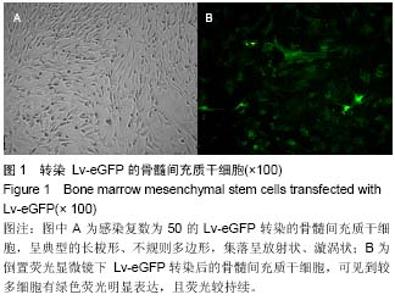
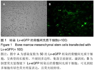
METHODS: Bone marrow mesenchymal stem cells of donor rats were isolated through whole bone marrow adherent method and transfected by Lv-eGFP. Receptor rats were exposed to silica dust through windpipe injection and randomly divided into intravenous injection and intratracheal injection groups. Then, transfected bone marrow mesenchymal stem cells were injected via the vein and trachea into acceptor rats. The acceptor rats were killed at weeks 1, 2, 3, 4 after transplantation to take the lung, heart, liver, spleen, kidney, and brain tissue that were made into frozen sections and observed under fluorescence microscopy. The intensity of green 
fluorescence (absorbance value) was analyzed using image analysis software.
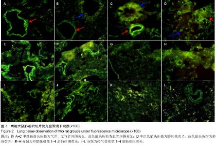
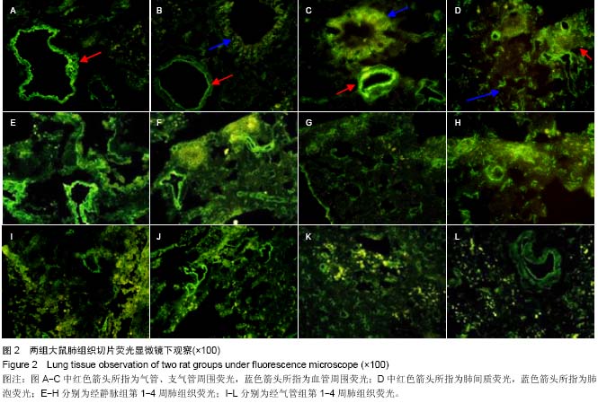
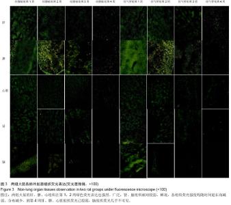
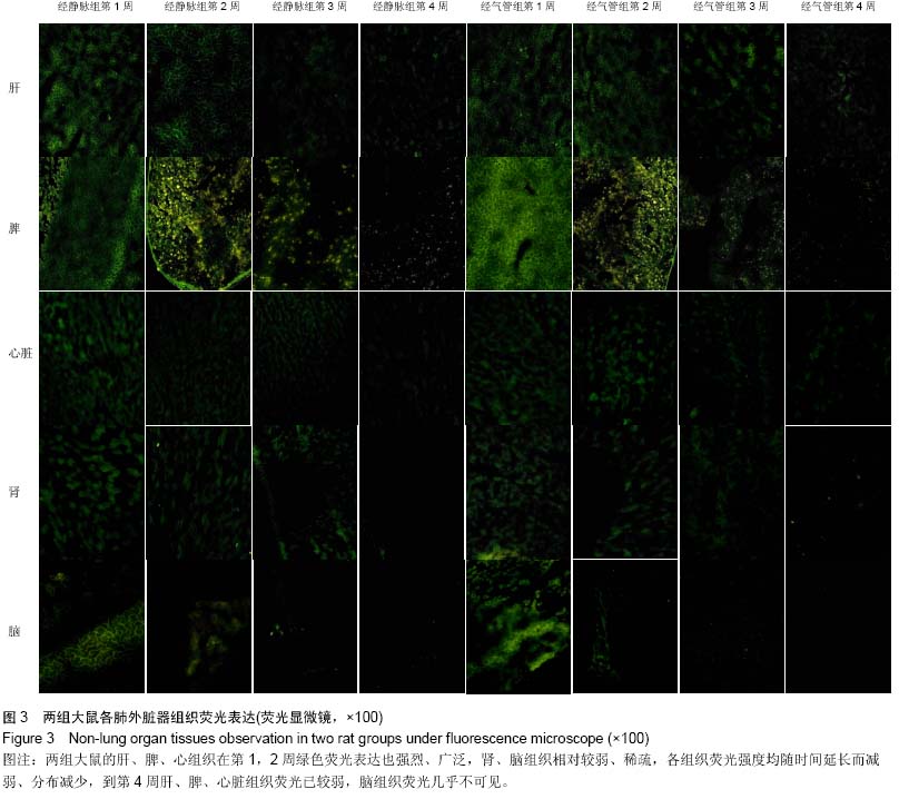
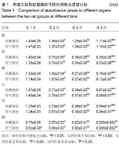
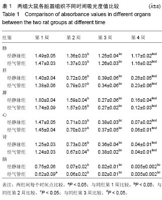
RESULTS AND CONCLUSION: Strong, wide and lasting green fluorescence was both observed in the lung tissue of intravenous injection and intratracheal injection groups, which was especially remarkable around the bronchus and blood vessels. The fluorescence intensities of both two groups were slightly decreased with time, but there was no difference between the two groups (P > 0.05). The fluorescence in the other organs of both two groups was also observed at early stage. It was stronger and wider in the liver, spleen and heart, while fainter and less in the kidney and brain, and reduced with time in all the organs. Fluorescence could be observed few and faint only in the liver and spleen at late stage, and could hardly be seen in the brain. The fluorescence intensities of the liver, spleen, heart, kidney and brain had no significant difference between the two groups at the same time (P > 0.05), but the fluorescence intensity in the brain at the 1st week showed significant difference between the two groups (P < 0.05). These findings indicate that the intravenous injection and intratracheal injection of bone marrow mesenchymal stem cells have similar homing effects in rats exposed to silica dust.

中国组织工程研究杂志出版内容重点:干细胞;骨髓干细胞;造血干细胞;脂肪干细胞;肿瘤干细胞;胚胎干细胞;脐带脐血干细胞;干细胞诱导;干细胞分化;组织工程

Key words: Bone Marrow, Mesenchymal Stem Cell Transplantation, Lung, Drug Administration Routes

CLC Number: