Chinese Journal of Tissue Engineering Research ›› 2015, Vol. 19 ›› Issue (26): 4235-4241.doi: 10.3969/j.issn.2095-4344.2015.26.026
Previous Articles Next Articles
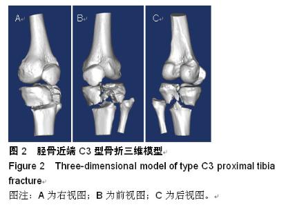
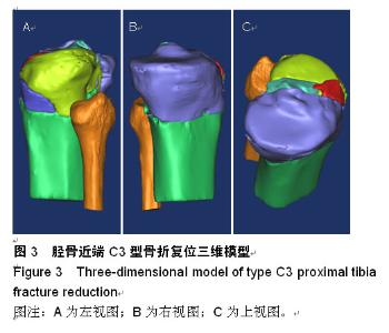
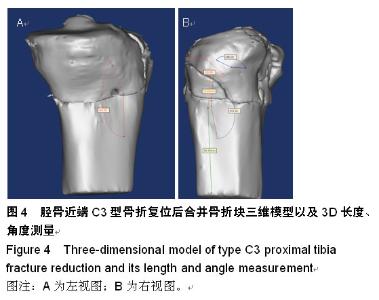
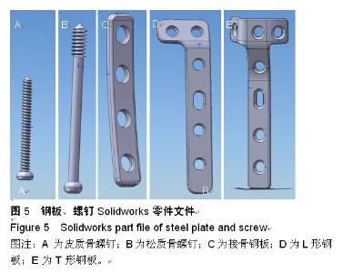
Three-dimensional reconstruction reduction and design of digital plates for proximal tibia fracture
Chen Xuan-huang1, Wu Xian-wei1, Lin Hai-bin1, Wu Chang-fu1, Zheng Feng1, Guo Qing-qing2, Zhang Guo-dong1
- 1Department of Orthopedics, Affiliated Hospital of Putian University (Affiliated Putian Hospital of Southern Medical University), Teaching Hospital, Fujian Medical University, Putian 351100, Fujian Province, China; 2Department of Medical Imaging, Affiliated Hospital of Putian University (Affiliated Putian Hospital of Southern Medical University), Teaching Hospital, Fujian Medical University, Putian 351100, Fujian Province, China)
-
Received:2015-04-12Online:2015-06-25Published:2015-06-25 -
Contact:Zhang Guo-dong, M.D., Department of Orthopedics, Affiliated Hospital of Putian University (Affiliated Putian Hospital of Southern Medical University), Teaching Hospital, Fujian Medical University, Putian 351100, Fujian Province, China -
About author:Chen Xuan-huang, Master, Associate chief physician, Department of Orthopedics, Affiliated Hospital of Putian University (Affiliated Putian Hospital of Southern Medical University), Teaching Hospital, Fujian Medical University, Putian 351100, Fujian Province, China -
Supported by:the Putian City Science and Technology Program of Fujian Province, No. 2013S03(6)
CLC Number:
Cite this article
Chen Xuan-huang, Wu Xian-wei, Lin Hai-bin, Wu Chang-fu, Zheng Feng, Guo Qing-qing,Zhang Guo-dong. Three-dimensional reconstruction reduction and design of digital plates for proximal tibia fracture[J]. Chinese Journal of Tissue Engineering Research, 2015, 19(26): 4235-4241.
share this article
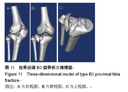
"

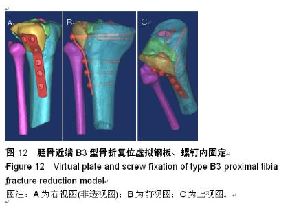
"

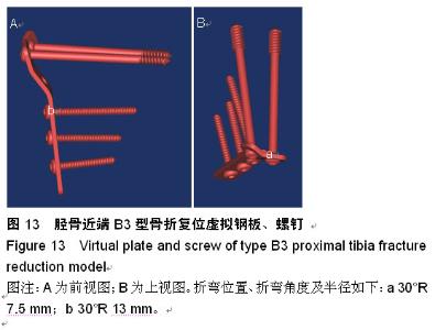
"

"


"

"

"

"

"

"

"

"

| [1] Rüedi TP,Murphy WM. 王满宜,杨庆铭,曾炳芳,译.骨折治疗的AO原则[M]. 2版.上海:上海科学技术出版社,2010:470-473. [2] 郑晓晖,张国栋,黄华军,等.基于三维编辑的四肢骨折快速虚拟复位[J]. 中华临床医师杂志(电子版),2015,(9)4:617- 622. [3] 张国栋,林海滨,陈宣煌,等.基于多平面三维测量的髋臼骨折数字化内固定植入方案[J].中华临床医师杂志:电子版,2012, 6(8): 2010-2015. [4] Gösling T, Klingler K, Geerling J, et al. Improved intra-operative reduction control using a three-dimensional mobile image intensifier--a proximal tibia cadaver study. Knee. 2009;16(1):58-63. [5] Papagelopoulos PJ, Partsinevelos AA, Themistocleous GS,et al. Complications after tibia plateau fracture surgery. Injury. 2006; 37(6):475-484. [6] 蔡伟斌,胡鸿璇,郭新辉. 锁定加压钢板与解剖钢板置入内固定治疗复杂胫骨平台骨折[J].中国组织工程研究,2012,16(52): 9750-9755. [7] Taylor J, Langenbach A, Marcellin-Little DJ. Risk factors for fibular fracture after TPLO. Vet Surg. 2011;40(6):687-693. [8] Yu Z,Zheng L,Zhang Y,et al.Functional and radiological evaluations of high-energy tibial plateau fractures treated with double -buttress plate fixation. Eur J Med Res. 2009;14(5): 200-2005. [9] 刘俊,刘锦波,王生介,等.双钢板治疗胫骨平台粉碎性骨折的临床研究[J]. 实用临床用药杂志,2014,18(7):129-131. [10] Yu B, Han K, Zhan C, et al. Fibular head osteotomy:a new approach for the treatment of lateral or posterolateral tibial plateau fractures. Knee. 2010;17(5):313. [11] Shen F, Chen B, Guo Q, et al. Augmented reality patient-specific reconstruction plate design for pelvic and acetabular fracture surgery.Int J Comput Assist Radiol Surg. 2013;8(2):169-179. [12] Hu YL, Ye FG, Ji AY, et al. Three-dimensional computed tomography imaging increases the reliability of classification systems for tibial plateau fractures. Injury. 2009;40(12): 1282-1285. [13] Hu Y,Li H,Qiao G,et al.Computer-assisted virtual surgical procedure for acetabular fractures based on real CT data. Injury. 2011;42(10):1121-1124. [14] Cimerman M, Kristan A. Preoperative planning in pelvic and acetabular surgery: The value of advanced computerised planning modules. J Care Injured. 2007;38; 442-449. [15] 扈延龄,金丹,苏秀云,等.基于三维CT数据的髋臼骨折计算机辅助虚拟手术设计[J].中华创伤骨科杂志, 2008,10(2):137-139. [16] Doornberg JN, Rademakers MV, van den Bekerom MP, et al.Two-dimensional and three-dimensional computed tomography for the classification and characterisation of tibial plateau fractures. Injury. 2011;42(12):1416-1425. [17] Waschke A, Walter J, Duenisch P, et al. CT-navigation versus fluoroscopy-guided placement of pedicle screws at the thoracolumbar spine: single center experience of 4,500 screws. Eur Spine J. 2013;22(3):654-660. [18] Villard J, Ryang YM, Demetriades AK, et al. Radiation exposure to the surgeon and the patient during posterior lumbar spinal instrumentation: a prospective randomized comparison of navigated versus non-navigated freehand techniques. Spine (Phila Pa 1976). 2014;39(13): 1004-1009. [19] 田伟,郎昭,刘亚军,等.术中即时三维导航辅助下轴性旋转腰椎椎弓根螺钉置入的实验研究[J].中华外科杂志,2010,48(11): 838-841. [20] Tormenti MJ, Kostov DB, Gardner PA, et al. Intraoperative computed tomography image-guided navigation for posterior thoracolumbar spinal instrumentation in spinal deformity surgery. Neurosurg Focus. 2010,28(3): E11. [21] Lee MH, Lin MH, Weng HH, et al. Feasibility of Intra-operative Computed Tomography Navigation System for Pedicle Screw Insertion of the Thoraco-lumbar Spine J Spinal Disord Tech. 2012. [22] Hodges SD, Eck JC, Newton D. Analysis of CT-based navigation system for pedicle screw placement. Orthopedics. 2012;35(8):1221-1224. [23] Tabaraee E, Gibson AG, Karahalios DG, et al. Intraoperative cone beam-computed tomography with navigation (O-ARM) versus conventional fluoroscopy (C-ARM): a cadaveric study comparing accuracy, efficiency, and safety for spinal instrumentation. Spine (Phila Pa 1976). 2013;38(22):1953-1958. [24] Oertel M F, Hobart J, Stein M, et al. Clinical and methodological precision of spinal navigation assisted by 3D intraoperative O-arm radiographic imaging. J Neurosurg Spine. 2011;14(4): 532-536. [25] Silbermann J, Riese F, Allam Y, et al. Computer tomography assessment of pedicle screw placement in lumbar and sacral spine: comparison between free-hand and O-arm based navigation techniques. Eur Spine J. 2011;20(6):875-881. [26] Kantelhardt S R, Martinez R, Baerwinkel S, et al. Perioperative course and accuracy of screw positioning in conventional, open robotic-guided and percutaneous robotic-guided, pedicle screw placement. Eur Spine J. 2011;20(6):860-868. [27] Bourgeois AC, Faulkner AR, Bradley YC, et al. Improved Accuracy of Minimally Invasive Transpedicular Screw Placement in the Lumbar Spine with Three-dimensional Stereotactic Image Guidance: A Comparative Meta-analysis.J Spinal Disord Tech.2014. [28] Lu S, Xu YQ, Zhang YZ, et al. A novel computer-assisted drill guide template for placement of C2 laminar screws. Eur Spine J. 2009;18(9):1379-1385. [29] Merc M, Drstvensek I, Vogrin M, et al. A multi-level rapid prototyping drill guide template reduces the perforation risk of pedicle screw placement in the lumbar and sacral spine. Arch Orthop Trauma Surg. 2013;133(7):893-899. [30] Ma T, Xu YQ, Cheng YB, et al. A novel computer-assisted drill guide template for thoracic pedicle screw placement: a cadaveric study. Arch Orthop Trauma Surg. 2012;132(1):65-72. [31] Khoury A,Liebergall M,Weil Y,et al. Computerized fluoroscopic- based navigation-assisted intramedullary nailing. Am J Orthop. 2007;36(11):582-585. [32] Osti M,Philipp H,Meusburger B,et a1.Analysis of failure following anterior screw fixation of Type II odontoid fractures in geriatric patients. Eur Spine J. 2011;20(11):1915-1920. [33] Chen C,Ruan D,Wu C,et al. CT morphometric analysis to determine the anatomical basis for the use of transpedicular screws during reconstruction and fixations of anterior cervical vertebrae. PLoS One. 2013;8(12):e81159. [34] Messmer P,Brig G,Suhm N,et al.Three-dimensional fracture simulation for preoperative planning and education. Eur J Trauma. 2001;27:171-177. [35] Osti M,Philipp H,Meusburger B,et al.Analysis of failure following anterior screw fixation of Type II odontoid fractures in geriatric patients. Eur Spine J. 2011;20(11):1915-1920. [36] 张国栋,陶圣祥,郑和平,等.基于三维重建技术的股骨远端数字钢板设计[J].实用骨科学杂志,2010,16(1):35-38. [37] 陈宣煌,张国栋,吴长福,等.数字化颈前路螺钉内固定方案设计:Ⅱ型齿状突骨折的临床应用[J].中国组织工程研究,2013,17(26): 4926-4933. [38] 陈宣煌,林海滨,张国栋,等.腰椎后路数字化内固定手术方案及临床应用[J].中华临床医师杂志:电子版,2012,6(17):5249-5251. [39] 陈宣煌,林海滨,张国栋,等.枕颈融合内固定治疗C1 Jefferson骨折的数字化设计及意义[J].中华临床医师杂志:电子版,2013,7(9): 4110-4112. [40] 陈宣煌,张国栋,吴长福.C1侧块骨折Magerl技术内固定的数字化模拟及应用[J].临床骨科杂志,2013,16(6):712-715. |
| [1] | Wang Liang, Huang Zhaozhao, Yu Jiaona, Gu Weidong, Wang Ren, Qian Zhiyi. Biomechanical characteristics of bridge-link type combined internal fixation system with mixed-rod versus double-rod in the treatment of femoral and tibial fractures [J]. Chinese Journal of Tissue Engineering Research, 2020, 24(6): 888-892. |
| [2] | Fang Yi, Zhao Wenzhi, Pan Deyue, Han Xin, Zhang Lu, He Hongtao, Shi Feng, Tian Tingxiao. Acromioclavicular joint dislocation: how to achieve anatomical reduction, sustained stability and micro-motion [J]. Chinese Journal of Tissue Engineering Research, 2020, 24(5): 796-802. |
| [3] | Mu Shuai, Xiao Peng, Zhang Jianliang, Hou Weiguang. Incidence of femoral neck shortening after internal fixation of femoral neck fracture and prognostic factors [J]. Chinese Journal of Tissue Engineering Research, 2020, 24(21): 3304-3309. |
| [4] | Fu Jiaxin, Xiao Lianping, Wang Shusen, Li Xiaodong, Han Liqiang, Wang Tonghao. Therapeutic effects of paraspinal approach combined with internal fixation through pedicle of fractured vertebra versus traditional AF screw-rod system for thoracolumbar fractures [J]. Chinese Journal of Tissue Engineering Research, 2019, 23(8): 1177-1181. |
| [5] | Qiu Zhongpeng, Li Ke, Li Gang, Liu Keyu, Du Xinhui, Meng Defeng, Shi Chenhui, Wang Weishan. Different treatments for two-part and three-part proximal humeral fractures by Neer classification: follow-up results analyzed using clinical economics [J]. Chinese Journal of Tissue Engineering Research, 2019, 23(8): 1188-1195. |
| [6] | Ke Wei, Li Ke, Wang Sibo, Du Xinhui, Qiu Zhongpeng, Kang Zhilin, Wang Weishan, Li Gang . Open reduction and plate fixation versus closed reduction and external fixation for distal radius fractures: scores and linear regression analysis [J]. Chinese Journal of Tissue Engineering Research, 2019, 23(8): 1196-1202. |
| [7] | Fan Zhirong, Peng Jiajie, Zhong Degui, Zhou Lin, Su Haitao, Huang Yongquan, Wu Jianglin, Liang Yihao. Suture anchor combined with open reduction and internal fixation versus open reduction and internal fixation for ankle fracture combined with deltoid ligament injury: a meta-analysis [J]. Chinese Journal of Tissue Engineering Research, 2019, 23(8): 1307-1312. |
| [8] | Zhou Yu, Liu Yuehong, Liu Shuping, Chen Xi, Qin Wei, Li Qifeng. Spinal stability of intervertebral grafting reinforced by five or six augmenting screws versus transvertebral grafting reinforced by four augmenting screws for thoracolumbar vertebral fractures [J]. Chinese Journal of Tissue Engineering Research, 2019, 23(4): 505-511. |
| [9] | Li Xianzhou, Wang Qian, Zhang Cunxin . Lumbar spondylolisthesis: status and prospects of implant treatment [J]. Chinese Journal of Tissue Engineering Research, 2019, 23(4): 621-627. |
| [10] | Yin Hao, Zhou Enchang, Pan Zhengjun, Chen Guang, Jiang Hua. Finite element analysis of the four and three cannulated screws combined with buttress plate fixation for the treatment of Pauwels III femoral neck fractures [J]. Chinese Journal of Tissue Engineering Research, 2019, 23(32): 5133-5137. |
| [11] |
Wang Lei, Li Zilong, Yuan Binbin, Wu Qingwei, Tang Fengming.
Clinical effect of locking plate versus anterograde intramedullary nail in the treatment of adult humeral shaft fractures: a meta-analysis
[J]. Chinese Journal of Tissue Engineering Research, 2019, 23(24): 3924-3930.
|
| [12] | Hao Liang, Zhang Zhonglin, Wang Baodong, Bi Zhenggang. Intertrochanteric fracture of the femur: improvement of internal fixation device, surgical changes and related disputes [J]. Chinese Journal of Tissue Engineering Research, 2019, 23(18): 2927-2935. |
| [13] | Yao Liquan, Ling Qinjie, Li Jiaying, Zhong Letian, Zhou Xingping, Liu Jintao, He Erxing, Yin Zhixun. Antibiotic artificial bone implantation for treating pyogenic spondylodiscitis [J]. Chinese Journal of Tissue Engineering Research, 2019, 23(14): 2133-2139. |
| [14] | Gong Zhibing, Wu Zhaoke, Zhang Huantang, Xu Zhiqing, Zhuang Zhikun, Zhang Qianjin. Allogeneic cortical bone plate combined with locking plate for Vancouver type B1 and C osteoporotic periprosthetic femoral fractures after hip arthroplasty in older adults [J]. Chinese Journal of Tissue Engineering Research, 2019, 23(12): 1812-1817. |
| [15] | Li Xiaofeng, Xie Furong, Zhan Long, Yang Yuan. Design of locking compression plate through transoral approach [J]. Chinese Journal of Tissue Engineering Research, 2019, 23(12): 1824-1828. |
| Viewed | ||||||
|
Full text |
|
|||||
|
Abstract |
|
|||||