Chinese Journal of Tissue Engineering Research ›› 2026, Vol. 30 ›› Issue (6): 1398-1406.doi: 10.12307/2026.558
Previous Articles Next Articles
Luteolin promotes wound healing in diabetic mice: roles and mechanisms
Peng Zhiwei1, 2, Chen Lei3, Tong Lei2
- 1Dalian Medical University, Dalian 116000, Liaoning Province, China; 2Department of Sports Medicine, The Second Affiliated Hospital of Xuzhou Medical University, Xuzhou 221000, Jiangsu Province, China; 3Department of Burn and Plastic Surgery, The First Affiliated Hospital of Wannan Medical College, Wuhu 241000, Anhui Province, China
-
Received:2024-10-31Accepted:2025-01-14Online:2026-02-28Published:2025-07-14 -
Contact:Tong Lei, Chief physician, Master’s supervisor, Department of Sports Medicine, The Second Affiliated Hospital of Xuzhou Medical University, Xuzhou 221000, Jiangsu Province, China -
About author:Peng Zhiwei, PhD candidate, Attending physician, Dalian Medical University, Dalian 116000, Liaoning Province, China; Department of Sports Medicine, The Second Affiliated Hospital of Xuzhou Medical University, Xuzhou 221000, Jiangsu Province, China -
Supported by:Xuzhou Key Research and Development Program (Social Development), No. KC22213 (to TL)
CLC Number:
Cite this article
Peng Zhiwei, Chen Lei, Tong Lei. Luteolin promotes wound healing in diabetic mice: roles and mechanisms[J]. Chinese Journal of Tissue Engineering Research, 2026, 30(6): 1398-1406.
share this article
Add to citation manager EndNote|Reference Manager|ProCite|BibTeX|RefWorks

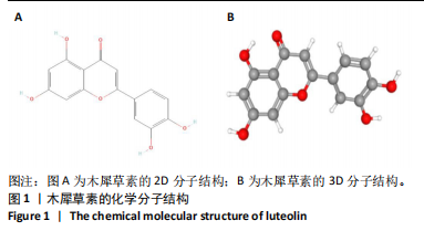
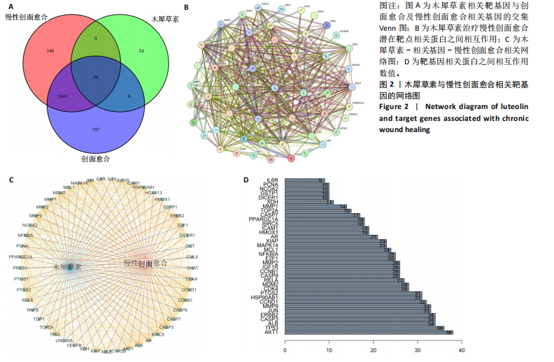
2.1 木犀草素的化学分子式及药理学信息 利用PubChem数据库成功获取了木犀草素的二维(2D)及三维(3D)化学分子结构图(图1)[31]。根据数据库TCMSP提供数据可得木犀草素的相对分子质量为286.25,药物半衰期为15.94 h ,辛醇/水分配系数"
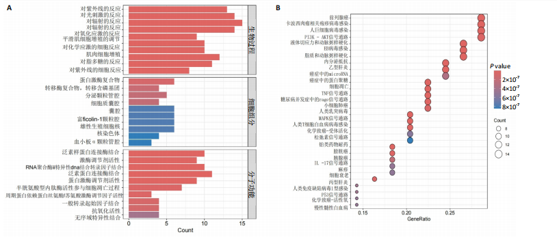
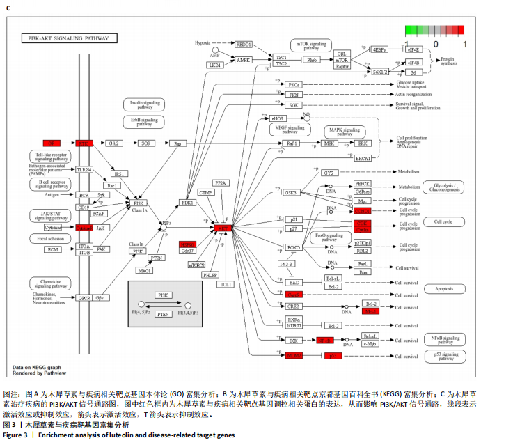
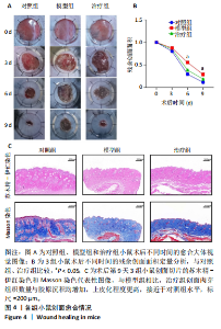
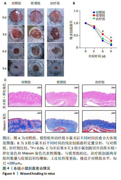
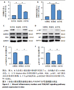
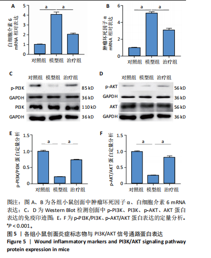
为2.07,氢键供体和受体数目分别为4和6,口服生物利用度为36.16%,渗透性为0.19,药物相似性指数为0.25[32]。 2.2 木犀草素相关的靶点基因 利用TCMSP数据库成功筛选出木犀草素的51个潜在靶点基因,通过PharmMapper数据库识别出33个靶点,对这2个数据库中的靶点基因进行并集运算后,共得到79个潜在靶点基因。进一步地将这些靶点与GeneCards数据库中与创面愈合及慢性创面相关的基因进行交集分析,最终筛选出56个与木犀草素治疗慢性创面相关的潜在靶点基因(图2A)。 2.3 木犀草素治疗创面愈合相关靶点蛋白的相互作用 将与药物相关的疾病靶点基因导入STRING数据库进行蛋白-蛋白相互作用分析,以揭示靶点间的相互作用关系(图2B)。利用Cytoscape软件对分析结果进行可视化展示(图2C),分析结果显示,靶点间联系的丰富程度与基因自由度呈正相关,进而影响其作为潜在治疗靶点的可靠性。通过R语言进行的网络分析揭示,AKT基因与其他靶点基因的相互作用最为紧密(图2D),据此推断,AKT基因可能是木犀草素促进慢性创面愈合过程中的关键靶点。 2.4 木犀草素与疾病靶基因的GO和KEGG分析 GO分析结果显示,在生物过程方面,目标基因显著富集于紫外线反应、光刺激反应、辐射反应、氧化应激反应及平滑肌细胞增殖调节等领域;在细胞组分方面,目标基因主要富集于蛋白激酶复合体、转移酶复合体、细胞质囊腔等结构;在分子功能方面,目标基因富集于泛素样蛋白连接酶结合、激酶调节剂活性、RNA聚合酶Ⅱ特异性DNA结合转录因子结合等分子功能(图3A)。KEGG分析结果显示,木犀草素目标基因主要富集于PI3K/AKT(磷脂酰肌醇3-激酶/蛋白激酶B)信号通路(图3B,C)。在56个目标基因中,有14个基因富集于PI3K/AKT通路,同时,TNF-α(肿瘤坏死因子α)、IL-17(白细胞介素17)、MAPK(丝裂原活化蛋白激酶)、P53等信号通路亦被富集,并与炎症反应及细胞凋亡的调控密切相关。PI3K信号通路激活后,可进一步激活下游的AKT、P53和NF-κB(核因子κB)信号通路,从而促进创面愈合过程。 2.5 体内实验验证木犀草素促进糖尿病小鼠创面愈合 2.5.1 实验动物数量分析 18只小鼠全部进入结果分析。 2.5.2 各组小鼠创面愈合情况 图4A展示了各组小鼠在创面形成后不同时间点的愈合状况。在创面形成后的第3天,治疗组创面表面已形成痂皮,未见红肿现象,创面面积相较于模型组略有缩小;至第6天,对照组与治疗组创面均可见少量新生肉芽组织形成。随着术后时间的延长,3组创面面积逐渐缩小,对照组、治疗组创面形成后第6,9天的残余创面面积均小于模型组(P < 0.05),见图4B。 2.5.3 苏木精-伊红与Masson染色结果 苏木精-伊红染色结果显示,与模型组相比,治疗组创面肉芽组织数量更多、上皮化程度更高,接近于对照组水平(图4C)。Masson染色结果显示,与模型组相比,治疗组创面胶原沉积增加,具有较高的胶原纤维密度,接近于对照组水平(图4C)。由此说明,木犀草素在动物体内具有促进皮肤创面愈合的作用。 2.5.4 木犀草素抑制小鼠创面炎症反应 慢性创面的形成与持续的炎症反应密切相关,而肿瘤坏死因子α和白细胞介素6是创面微环境中的主要促炎因子。qRT-PCR检测结果显示,对照组、治疗组肿瘤坏死因子α和白细胞介素6 mRNA表达均低于模型组(P < 0.001),见图5A,B,表明木犀草素能够有效缓解创面的炎症反应。 2.5.5 木犀草素通过调节PI3K/AKT信号通路促进创面愈合 Western Blot检测结果显示,对照组、治疗组p-PI3K、p-AKT蛋白表达高于模型组(P < 0.001),见图5C-F,表明木犀草素可能通过激活PI3K/AKT信号传导途径促进了糖尿病小鼠创面的愈合过程。"

"

"
"
"
| [1] HARTINGER R, SINGH K, LEVERETT J, et al. Enhancing Cellular Homeostasis: Targeted Botanical Compounds Boost Cellular Health Functions in Normal and Premature Aging Fibroblasts. Biomolecules. 2024;14(10):1310. [2] LIAO Y, ZHANG Z, ZHAO Y, et al. Glucose oxidase: An emerging multidimensional treatment option for diabetic wound healing. Bioact Mater. 2025;44:131-151. [3] SUN R, LIU C, LIU J, et al. Integrated network pharmacology and experimental validation to explore the mechanisms underlying naringenin treatment of chronic wounds. Sci Rep. 2023;13(1):132. [4] O’REILLY S, MARKIEWICZ E, IDOWU OC. Aging, senescence, and cutaneous wound healing-a complex relationship. Front Immunol. 2024;15:1429716. [5] KOMI D, KHOMTCHOUK K, SANTA MP. A Review of the Contribution of Mast Cells in Wound Healing: Involved Molecular and Cellular Mechanisms. Clin Rev Allergy Immunol. 2020;58(3):298-312. [6] HOSEMANN W, WIGAND ME, GÖDE U, et al. Normal wound healing of the paranasal sinuses: clinical and experimental investigations. Eur Arch Otorhinolaryngol. 1991;248(7):390-394. [7] GONG C, XIA C, LIU L. Exosomes derived from epidermal growth factor-like domain protein 6-preconditioned mesenchymal stem cells for diabetic wound healing. Regen Ther. 2024;26:932-940. [8] BLAIR M. Diabetes Mellitus Review. Urol Nurs. 2016;36(1):27-36. [9] YAZDANPANAH L, SHAHBAZIAN H, NAZARI I, et al. Risk factors associated with diabetic foot ulcer-free survival in patients with diabetes. Diabetes Metab Syndr. 2018;12(6):1039-1043. [10] DEN DEKKER A, DAVIS FM, KUNKEL SL, et al. Targeting epigenetic mechanisms in diabetic wound healing. Transl Res. 2019;204:39-50. [11] SCHAPER NC, Van NETTEN JJ, APELQVIST J, et al. Prevention and management of foot problems in diabetes: a Summary Guidance for Daily Practice 2015, based on the IWGDF Guidance Documents. Diabetes Metab Res Rev. 2016;32 Suppl 1:7-15. [12] EZHILARASU H, VISHALLI D, DHEEN ST, et al. Nanoparticle-Based Therapeutic Approach for Diabetic Wound Healing. Nanomaterials (Basel). 2020;10(6):1234. [13] SUN S, DING C, LIU X, et al. Silk protein/polyvinylpyrrolidone nanofiber membranes loaded with puerarin accelerate wound healing in mice by reducing the inflammatory response. Biomater Adv. 2022;135:212734. [14] 谭海燕,吴仁擎.益气活血通阳法治疗糖尿病足临床研究[J].实用中医药杂志,2023,39(5):859-861. [15] 刘振华,张云霞.中药毛稔对伤口愈合的治疗效果与机制预测[J].中国药物应用与监测,2024,21(3):310-314. [16] 宁永玲,沃达,袁志莹,等.绿茶多酚表没食子儿茶素没食子酸酯对高脂高糖饮食诱导肥胖小鼠伤口愈合的影响[J].中国中西医结合杂志,2023,43(3):337-344. [17] HOPKINS AL. Network pharmacology: the next paradigm in drug discovery. Nat Chem Biol. 2008;4(11):682-690. [18] DENG Y, YE X, CHEN Y, et al. Chemical Characteristics of Platycodon grandiflorum and its Mechanism in Lung Cancer Treatment. Front Pharmacol. 2020;11:609825. [19] LUO TT, LU Y, YAN SK, et al. Network Pharmacology in Research of Chinese Medicine Formula: Methodology, Application and Prospective. Chin J Integr Med. 2020;26(1):72-80. [20] SU M, GUO C, LIU M, et al. Therapeutic targets of vitamin C on liver injury and associated biological mechanisms: A study of network pharmacology. Int Immunopharmacol. 2019;66:383-387. [21] EMIG D, IVLIEV A, PUSTOVALOVA O, et al. Drug target prediction and repositioning using an integrated network-based approach. PLoS One. 2013;8(4):e60618. [22] LOTFI SM, GHADIRI N, MOUSAVI SR, et al. A review of network-based approaches to drug repositioning. Brief Bioinform. 2018;19(5):878-892. [23] KOTLYAR M, FORTNEY K, JURISICA I. Network-based characterization of drug-regulated genes, drug targets, and toxicity. Methods. 2012; 57(4):499-507. [24] HOU X, WU W, ZHAO F, et al. Construction of an electrochemical sensor with graphene aerogel doped with ZrO2 nanoparticles and chitosan for the selective detection of luteolin. Mikrochim Acta. 2021;188(3):86. [25] ZHANG X, XU W, LI H, et al. Luteolin prevents cadmium-induced PC12 cell death by suppressing the Akt/mTOR signaling pathway. Medicine (Baltimore). 2024;103(44):e40372. [26] GENG YF, YANG C, ZHANG Y, et al. An innovative role for luteolin as a natural quorum sensing inhibitor in Pseudomonas aeruginosa. Life Sci. 2021;274:119325. [27] ATTIQ A, JALIL J, HUSAIN K, et al. Luteolin and apigenin derived glycosides from Alphonsea elliptica abrogate LPS-induced inflammatory responses in human plasma. J Ethnopharmacol. 2021;275:114120. [28] WU HT, LIN J, LIU YE, et al. Luteolin suppresses androgen receptor-positive triple-negative breast cancer cell proliferation and metastasis by epigenetic regulation of MMP9 expression via the AKT/mTOR signaling pathway. Phytomedicine. 2021;81:153437. [29] YU R, CHEN L, LAN R, et al. Computational screening of antagonists against the SARS-CoV-2 (COVID-19) coronavirus by molecular docking. Int J Antimicrob Agents. 2020;56(2):106012. [30] XIE Y, ZHANG T, CHEN Y, et al. Fabrication of core-shell magnetic covalent organic frameworks composites and their application for highly sensitive detection of luteolin. Talanta. 2020;213:120843. [31] KIM S. Exploring Chemical Information in PubChem. Curr Protoc. 2021; 1(8):e217. [32] RU J, LI P, WANG J, et al. TCMSP: a database of systems pharmacology for drug discovery from herbal medicines. J Cheminform. 2014;6:13. [33] CONSORTIUM TU. UniProt: the universal protein knowledgebase in 2021. Nucleic Acids Res. 2021;49(D1):D480-D489. [34] 鲍丹,郭蕊,侯道荣.角蛋白17基因敲除加剧糖尿病小鼠的创面愈合障碍[J].中国比较医学杂志,2023,33(10):54-60. [35] MA Q, CHEN G, LI Y, et al. The molecular genetics of PI3K/PTEN/AKT/mTOR pathway in the malformations of cortical development. Genes Dis. 2024;11(5):101021. [36] JIN Y, HUANG Y, ZENG G, et al. Advanced glycation end products regulate macrophage apoptosis and influence the healing of diabetic foot wound through miR-361-3p/CSF1R and PI3K/AKT pathway. Heliyon. 2024;10(2):e24598. [37] QIU F, FAN S, DIAO Y, et al. The mechanism of Chebulae Fructus Immaturus promote diabetic wound healing based on network pharmacology and experimental verification. J Ethnopharmacol. 2024;322:117579. [38] MING G, LIU J, WU Y, et al. Strictosamide promotes wound healing through activation of the PI3K/AKT pathway. Heliyon. 2024;10(9):e30169. [39] WANG N, HONG B, ZHAO Y, et al. Dopamine-grafted oxidized hyaluronic acid/gelatin/cordycepin nanofiber membranes modulate the TLR4/NF-kB signaling pathway to promote diabetic wound healing. Int J Biol Macromol. 2024;262:130079. [40] WU S, ZHU L, NI S, et al. Hyaluronic acid-decorated curcumin-based coordination nanomedicine for enhancing the infected diabetic wound healing. Int J Biol Macromol. 2024;263:130249. [41] XIAO J, LIANG Y, SUN T, et al. A functional dual responsive CMC/OHA/SA/TOB hydrogel as wound dressing to enhance wound healing. Sci Rep. 2024;14(1):26854. [42] HUANG C, NG MY, LIN T, et al. Quercetin ameliorates advanced glycation end product-induced wound healing impairment and inflammaging in human gingival fibroblasts. J Dent Sci. 2024;19(1):268-275. [43] GENG X, WANG Y, LI H, et al. Total iridoid glycoside extract of Lamiophlomis rotata (Benth) Kudo accelerates diabetic wound healing by the NRF2/COX2 axis. Chin Med. 2024;19(1):53. [44] SHAH A, AMINI-NIK S. The Role of Phytochemicals in the Inflammatory Phase of Wound Healing. Int J Mol Sci. 2017;18(5):1068. [45] YOUJUN D, HUANG Y, LAI Y, et al. Mechanisms of resveratrol against diabetic wound by network pharmacology and experimental validation. Annals of medicine (Helsinki). 2023;55(2):2280811. [46] FANG C, RONG X, JIANG W, et al. Geniposide promotes wound healing of skin ulcers in diabetic rats through PI3K/Akt pathway. Heliyon. 2023; 9(11):e21331. [47] ZHANG H, CHEN S, YAN X, et al. Egg white-derived peptide KPHAEVVLR promotes wound healing in rat palatal mucosa via PI3K/AKT/mTOR pathway. Peptides. 2023;168:171074. [48] ZHU X, HU D, ZENG Z, et al. XB130 inhibits healing of diabetic skin ulcers through the PI3K/Akt signalling pathway. World J Diabetes. 2023; 14(9):1369-1384. [49] JERE SW, ABRAHAMSE H, HOURELD NN. Interaction of the AKT and β-catenin signalling pathways and the influence of photobiomodulation on cellular signalling proteins in diabetic wound healing. J Biomed Sci. 2023;30(1):81. [50] LI Y, LU Y, ZHAO Y, et al. Deciphering the Wound-Healing Potential of Collagen Peptides and the Molecular Mechanisms: A Review. J Agric Food Chem. 2024;72(47):26007-26026. |
| [1] | Fu Lyupeng, Yu Peng, Liang Guoyan, Chang Yunbing. Electroactive materials applied in spinal surgery [J]. Chinese Journal of Tissue Engineering Research, 2026, 30(8): 2113-2123. |
| [2] | Tan Jing, Li Li, Wang Liangliang, Qin Xiangyu. Bionic functional coating improves the integration of titanium implants and skin tissue interface [J]. Chinese Journal of Tissue Engineering Research, 2026, 30(8): 2014-2022. |
| [3] | Pan Zhiyi, Huang Jiawen, Xue Wenjun, Xu Jianda. Advantages of MXene-based flexible electronic sensors and their application in monitoring diabetic foot wounds [J]. Chinese Journal of Tissue Engineering Research, 2026, 30(8): 2023-2032. |
| [4] | Zhou Sirui, Xu Yukun, Zhao Kewei. Ideas and methods of anti-melanogenesis of Angelica dahurica extracellular vesicles [J]. Chinese Journal of Tissue Engineering Research, 2026, 30(7): 1747-1754. |
| [5] | Li Zhenyu, Zhang Siming, Bai Jiaxiang, Zhu Chen. Osthole improves osteogenic differentiation function of bone marrow mesenchymal stem cells under high-glucose conditions [J]. Chinese Journal of Tissue Engineering Research, 2026, 30(7): 1641-1648. |
| [6] | Chen Yulin, He Yingying, Hu Kai, Chen Zhifan, Nie Sha Meng Yanhui, Li Runzhen, Zhang Xiaoduo , Li Yuxi, Tang Yaoping. Effect and mechanism of exosome-like vesicles derived from Trichosanthes kirilowii Maxim. in preventing and treating atherosclerosis [J]. Chinese Journal of Tissue Engineering Research, 2026, 30(7): 1768-1781. |
| [7] | Pan Hongfei, Zhuang Zhenbing, Xu Baiyun, Yang Zhangyang, Lin Kairui, Zhan Bingqing, Lan Jinghan, Gao Heng, Zhang Nanbo, Lin Jiayu. Inhibitory effects of different concentrations of auranofin on M1 macrophage function and its therapeutic potential in diabetic wound healing [J]. Chinese Journal of Tissue Engineering Research, 2026, 30(6): 1390-1397. |
| [8] | Sun Yajie, Zhao Xinchen, Bo Shuangling. Spatiotemporal expression of bone morphologic protein 7 in mouse kidney development [J]. Chinese Journal of Tissue Engineering Research, 2026, 30(5): 1156-1161. |
| [9] | Yu Huifen, Mo Licun, Cheng Leping. The position and role of 5-hydroxytryptamine in the repair of tissue injury [J]. Chinese Journal of Tissue Engineering Research, 2026, 30(5): 1196-1206. |
| [10] | Wen Fan, Xiang Yang, Zhu Huan, Tuo Yanfang, Li Feng. Exercise improves microvascular function in patients with type 2 diabetes [J]. Chinese Journal of Tissue Engineering Research, 2026, 30(5): 1225-1235. |
| [11] | Wen Xiaolong, Weng Xiquan, Feng Yao, Cao Wenyan, Liu Yuqian, Wang Haitao. Effects of inflammation on serum hepcidin and iron metabolism related parameters in patients with type 2 diabetes mellitus: a meta-analysis [J]. Chinese Journal of Tissue Engineering Research, 2026, 30(5): 1294-1301. |
| [12] | Yang Zeyu, Zhi Liang, Wang Jia, Zhang Jingyi, Zhang Qingfang, Wang Yulong, Long Jianjun. A visualized analysis of research hotspots in high-frequency repetitive transcranial magnetic stimulation from the macroscopic perspective [J]. Chinese Journal of Tissue Engineering Research, 2026, 30(5): 1320-1330. |
| [13] | Wang Mingqi, Feng Shiya, Han Yinhe, Yu Pengxin, Guo Lina, Jia Zixuan, Wang Xiuli. Construction and evaluation of a neuralized intestinal mucosal tissue engineering model in vitro [J]. Chinese Journal of Tissue Engineering Research, 2026, 30(4): 892-900. |
| [14] | Wang Jie, Huang Rui, Zhang Ye, Shou Zhaoxi, Yao Jie, Liu Chenxi, Liao Jian. Role and mechanism of probiotics in peri-implantitis [J]. Chinese Journal of Tissue Engineering Research, 2026, 30(4): 901-907. |
| [15] | Yang Xiao, Bai Yuehui, Zhao Tiantian, Wang Donghao, Zhao Chen, Yuan Shuo. Cartilage degeneration in temporomandibular joint osteoarthritis: mechanisms and regenerative challenges [J]. Chinese Journal of Tissue Engineering Research, 2026, 30(4): 926-935. |
| Viewed | ||||||
|
Full text |
|
|||||
|
Abstract |
|
|||||