Chinese Journal of Tissue Engineering Research ›› 2026, Vol. 30 ›› Issue (36): 9497-9504.doi: 10.12307/2026.800
Previous Articles Next Articles
Magnetic resonance imaging, virtual simulation, and motion capture analyses of biomechanical characteristics of the paracervical muscles in cervical spondylotic myelopathy
Wei Haokai1, Zuo Kuangshi1, Liu Qiuli1, Li Zhifei1, Han Bin1, Zhang Zhanming1, Zhou Jinyan2
- 1Graduate School, Guangxi University of Chinese Medicine, Nanning 530000, Guangxi Zhuang Autonomous Region, China; 2First Affiliated Hospital, Guangxi University of Chinese Medicine, Nanning 530001, Guangxi Zhuang Autonomous Region, China
-
Received:2025-07-18Revised:2025-12-12Online:2026-12-28Published:2026-05-22 -
Contact:Zhou Jinyan, MD, Associate chief physician, First Affiliated Hospital, Guangxi University of Chinese Medicine, Nanning 530001, Guangxi Zhuang Autonomous Region, China -
About author:Wei Haokai, Master candidate, Graduate School, Guangxi University of Chinese Medicine, Nanning 530000, Guangxi Zhuang Autonomous Region, China -
Supported by:Guangxi Natural Science Foundation (General Project), No. 2022JJA140858 (to LZF)

CLC Number:
Cite this article
Wei Haokai, Zuo Kuangshi, Liu Qiuli, Li Zhifei, Han Bin, Zhang Zhanming, Zhou Jinyan. Magnetic resonance imaging, virtual simulation, and motion capture analyses of biomechanical characteristics of the paracervical muscles in cervical spondylotic myelopathy[J]. Chinese Journal of Tissue Engineering Research, 2026, 30(36): 9497-9504.
share this article
Add to citation manager EndNote|Reference Manager|ProCite|BibTeX|RefWorks
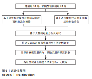
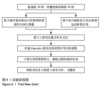
2.1 参与者数量分析 共纳入30例受试者,其中脊髓型颈椎病组20例,健康组10例。所有受试者均完成了试验,未出现样本脱落情况。 2.2 试验流程图 见图6。"
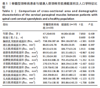
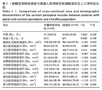
2.3 颈椎旁肌横截面积及人口学特征比较 脊髓型颈椎病与健康人群两组间性别、年龄未见明显差异(P > 0.05)。磁共振定量分析结果表明,健康组的胸锁乳突肌横截面积显著大于脊髓型颈椎病组(t=-2.501,P=0.019);同样,健康组的多裂肌横截面积显著大于脊髓型颈椎病组(t=-2.437,P=0.022);其余肌肉两组间比较差异均无显著性意义(P > 0.05)。与正常人群相比,脊髓型颈椎病患者的颈前肌群和颈侧肌群中有更明显的肌肉萎缩现象,描述性特征详见表1。"
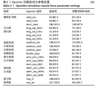
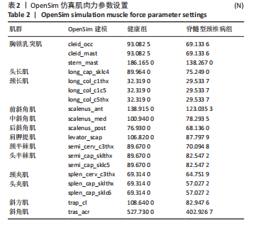
2.4 OpenSim仿真肌肉力参数比较 OpenSim虚拟仿真模型中健康人群与脊髓型颈椎病组椎旁肌肌肉力参数显示,脊髓型颈椎病组颈椎旁肌肌肉力相较于健康人群显著减弱,仿真模型的肌肉力参数描述性特征详见表2。"

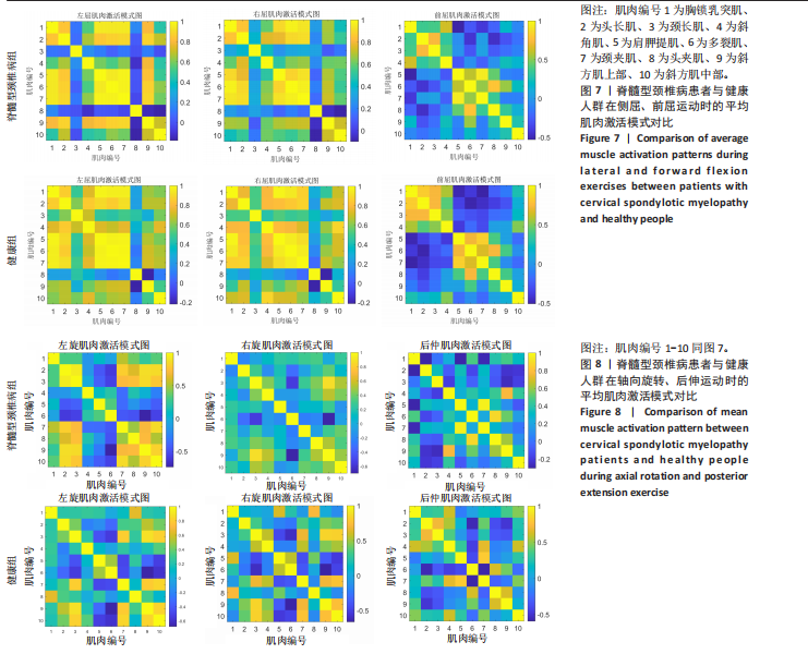
2.5 颈椎旁肌肌肉激活模式比较 6种特定运动的肌肉激活模式图直观地反映了肌肉间协同作用的强度。两块肌肉相交的格子颜色越亮,表明两肌肉之间的关联性越强,协同作用越显著。颜色从蓝色逐渐过渡到黄色,亮度依次增加,黄色代表较高的关联性和协同作用,而蓝色则表示较低的关联性。肌肉激活模式对比显示,脊髓型颈椎病组颈椎旁肌肌肉在前屈、后伸、侧屈及轴向旋转运动下的协同激活显著低于健康组,说明了脊髓型颈椎病患者存在颈部肌肉群的协同失衡,这提示临床上需选择增强颈部肌肉协同模式的治疗方向。肌肉激活描述性特征详见图7,8。 2.6 不良事件 脊髓型颈椎病患者在进行头颈部运动轨迹的动作捕捉时,可能会引起短暂性的头颈部疼痛加重现象。脊髓型颈椎病患者的避痛行为也可能影响运动学参数的真实性。为确保试验的安全性及数据的可靠性,受试者可采用头颈部渐进式运动以提前预知头颈部安全活动范围。"
| [1] SOUSA SS, ANDRADE MJ, FERNANDES CS, et al. Patients with cervical spondylotic myelopathy and dependency in activities of daily living during hospitalization-descriptive and correlational study. Porto Biomed J. 2024;9(3):252. [2] LEE S, LEE YH, CHUNG TS, et al. Accuracy of Diffusion Tensor Imaging for Diagnosing Cervical Spondylotic Myelopathy in Patients Showing Spinal Cord Compression. Korean J Radiol. 2015;16(6):1303-1312. [3] TERASHIMA Y, YURUBE T, SUMI M, et al. Clinical and Radiological Characteristics of Cervical Spondylotic Myelopathy in Young Adults: A Retrospective Case Series of Patients under Age 30. Medicina (Kaunas). 2023;59(3):539. [4] GUADARRAMA-ORTIZ P, RUÍZ-RIVERO CO, CAPI-CASILLAS D, et al. Demographic and Clinical Characteristics of Patients With Cervical Spine Degeneration Reveal Frequent Cervicolumbar Tandem Spinal Stenosis in Mexico. Int J Spine Surg. 2023;17(5):670-677. [5] APAYDIN AS, THAN K. Commentary on “Radiological and Clinical Significance of Cervical Dynamic Magnetic Resonance Imaging for Cervical Spondylotic Myelopathy”. Neurospine. 2024;21(2):455-457. [6] CANT DA, ANDERSEN SBK, HØY K. Cervical spondylotic myelopathy. Ugeskr Laeger. 2024;186(36):V02240149. [7] CHERNYSH AA, LOFTUS DH, ZHENG B, et al. Utility of Diffusion Tensor Imaging for Prognosis and Management of Cervical Spondylotic Myelopathy: A PRISMA Review. World Neurosurg. 2024;190:88-98. [8] LEE A, WU J, LIU C, et al. Using deep learning to enhance reporting efficiency and accuracy in degenerative cervical spine MRI. Spine J. 2025. doi:10.1016/j.spinee.2025.03.009 [9] NI M, LI S, WEN X, et al. A matched case-control study of early cervical spondylotic myelopathy based on diffusion magnetic resonance imaging. Insights Imaging. 2024;15(1):25. [10] DAMMEYER C, NÜESCH C, VISSCHER RMS, et al. Classification of inertial sensor-based gait patterns of orthopaedic conditions using machine learning: A pilot study. J Orthop Res. 2024;42(7):1463-1472. [11] KHAN AF, HAYNES G, MOHAMMADI E, et al. Utility of MRI in Quantifying Tissue Injury in Cervical Spondylotic Myelopathy. J Clin Med. 2023;12(9):3337. [12] EJIDE C, BANDYOPADHYAY S, LAKHOO K. Paediatric Spinal Cord Injury: A Review of Current Management. Cureus. 2024;16(6):e63306. [13] 《现代脊柱外科学》已出版[J]. 脊柱外科杂志,2007(6):353. [14] 梁渊耀. 基于动作捕捉及opensim数字仿真技术探讨神经根型颈椎病患者颈椎运动的内在生物力学特征[D].南宁:广西中医药大学,2024. [15] KEIDAN L, IBRAHIM R, OHAYON E, et al. Multi-Planar Cervical Motion Dataset: IMU Measurements and Goniometer. Sci Data. 2025;12(1):13. [16] JIN C, YU JM, LI R, et al. Regional biomechanical characterization of the spinal cord tissue: dynamic mechanical response. Front Bioeng Biotechnol. 2024;12: 1439323. [17] SUZUKI H, FUNABA M, FUJIMOTO K, et al. Current Concepts of Cervical Spine Alignment, Sagittal Deformity, and Cervical Spine Surgery. J Clin Med. 2024; 13(5):1196. [18] HARA Y, YOSHII Y. Diagnostic Dilemmas in Carpal Tunnel Syndrome and Cervical Spine Disorders: A Comprehensive Review. Diagnostics (Basel). 2025;15(2):122. [19] SCHUPPER AJ, DIDOMENICO J, FARBER SH, et al. Predictors of patient satisfaction in the surgical treatment of cervical spondylotic myelopathy. J Neurosurg Spine. 2024;41(5):611-618. [20] ELFIKY T, BESSADA B, STIENEN MN, et al. Endplate and Facet Joint Changes in Cervical Spondylotic Myelopathy. World Neurosurg. 2023;175:e361-e366. [21] HACHIYA K, KAWABATA S, MICHIKAWA T, et al. Severity of locomotive syndrome in surgical cervical spondylotic myelopathy patients: a cross-sectional study. Fujita Med J. 2023;9(3):246-252. [22] LUO W, BU W, SHAO R, et al. Neuroimaging feature in identifying acute myelopathy etiologies: comparison between neuromyelitis optica spectrum disorder and cervical spondylotic myelopathy. BMC Neurol. 2024;24(1):369. [23] RANGANKAR VP, RAO PM, DESHMUKH SK. Post-Gadolinium Pancake-Like Spinal Cord Enhancement in Spondylotic Cervical Compressive Myelopathy. Neurol India. 2024;72(4):915-916. [24] NGUYEN AN, NIU AV, ONG LF, et al. An Uncommon Presentation of Cervical Myelopathy. Cureus. 2023;15(9):e45609. [25] LI X, KRECKE KN, WEINSHENKER BG, et al. Hemicord White Matter Enhancement and the Croissant Sign in Cervical Spondylotic Myelopathy. Neurology. 2023;101(21):968-969. [26] 李智斐,钟远鸣,张家立,等.动态MRI检测颈椎健康者、颈肩部不适者脊髓型颈椎病未确诊及确诊者的价值[J].中国组织工程研究与临床康复, 2010,14(48):8998-9002. [27] LIU Q, SHAO H, LIU C, et al. Quantitative evaluation of the spinal cord compression in patients with cervical spondylotic myelopathy using synthetic MRI. Front Physiol. 2023;14:1140870. [28] TAO Q, WANG K, WEN B, et al. Assessment of image quality and diagnostic accuracy for cervical spondylosis using T2w-STIR sequence with a deep learning-based reconstruction approach. Eur Spine J. 2024;33(8):2982-2996. [29] WANG H, YE W, XIONG J, et al. Application of Short T1 Inversion Recovery Sequence in Increased Signal Intensity Following Cervical Spondylotic Myelopathy. World Neurosurg. 2024;187:e1097-e1105. [30] NARVEKAR M, DAVE BR, KRISHNAN A, et al. Utility of cervical dynamic magnetic resonance imaging for evaluating patients with cervical myelopathy: a retrospective study. Asian Spine J. 2024;18(5):647-653. [31] HEJRATI N, PEDRO K, ALVI MA, et al. Degenerative cervical myelopathy: Where have we been? Where are we now? Where are we going? Acta Neurochir (Wien). 2023;165(5):1105-1119. [32] ŞERIFOĞLU L, KARAASLANLI A. The Role of Dynamic Cervical Magnetic Resonance Imaging in Determining the Level of Posterior Decompression in Cervical Spondylotic Myelopathy. World Neurosurg. 2025;195:123698. [33] HESNI S, BAXTER D, SAIFUDDIN A. The imaging of cervical spondylotic myeloradiculopathy. Skeletal Radiol. 2023;52(12):2341-2365. [34] PAHUTA M, SARRAJ M, BUSSE J, et al. Nonoperative Care Versus Surgery for Degenerative Cervical Myelopathy: An Application of a Health Economic Technique to Simulate Head-to-Head Comparisons. JB JS Open Access. 2024;9(4):e23.00166. [35] SCHAEFER SD, DAVIES BM, NEWCOMBE VFJ, et al. Could spinal cord oscillation contribute to spinal cord injury in degenerative cervical myelopathy? Brain Spine. 2023;3:101743. [36] KIM MW, PARK YS, KANG CN, et al. Cervical spondylotic myelopathy and radiculopathy: a stepwise approach and comparative analysis of surgical outcomes: a narrative review of recent literature. Asian Spine J. 2025;19(1):121-132. [37] CHAN AK, BISSON EF, BYDON M, et al. Introduction. Toward a contemporaneous understanding of cervical spondylotic myelopathy. Neurosurg Focus. 2023;55(3):E1. [38] PARK C, BHOWMICK DA, SHAFFREY CI, et al. Do class III obese patients achieve similar outcomes and satisfaction to nonobese patients following surgery for cervical myelopathy? A QOD study. J Neurosurg Spine. 2024;41(5):604-610. [39] WANG H, XU H, WANG X, et al. Imbalance of Muscles Around the Cervical Spine in Patients with Degenerative Cervical Spondylotic Kyphosis and Myelopathy. World Neurosurg. 2025;195:123605. [40] LIN BH, ZHONG GQ, MENG XH, et al. Application effect of diversified health-promoting models on rehabilitation exercises for cervical spondylotic myelopathy. Technol Health Care. 2024;32(2):705-718. [41] HU J, LI X, ZHENG P, et al. The impact of neck pain and movement performance on the interarticular compressive force of the cervical spine: a cross-sectional study based on OpenSim. J Neuroeng Rehabil. 2025;22(1):26. [42] MILLS K, HÜBSCHER M, O’LEARY H, et al. Current concepts in joint pain in knee osteoarthritis. Aktuelles zum Gelenkschmerz bei Kniearthrose. Schmerz. 2019;33(1):22-29. [43] SIPKO T, BARCZYK-PAWELEC K, PIKSA M, et al. Impact of Standing and Sitting Postures on Spinal Curvature and Muscle Mechanical Properties in Young Women: A Photogrammetric and MyotonPro Analysis. Med Sci Monit. 2024;30:e944930. [44] XU G, LI C, SHENG Z. Biomechanical effects of altered multifidus muscle morphology on cervical spine tissues. Front Bioeng Biotechnol. 2025;13:1524844. [45] THAKAR S, MOHAN D, FURTADO SV, et al. Paraspinal muscle morphometry in cervical spondylotic myelopathy and its implications in clinicoradiological outcomes following central corpectomy: clinical article. J Neurosurg Spine. 2014;21(2):223-230. [46] NAKAMURA K, TERAGUCHI M, NAKATA T, et al. Associated Factors of Cervical Spinal Cord Injury Without Radiographic Evidence of Trauma: A Retrospective Study. Cureus. 2025;17(3):e80006. [47] 周鑫,马迅,李强.颈椎旁肌退变与颈椎病的发生和手术预后相关性的研究进展[J].中国医药导报,2024,21(6):46-49. [48] DANDUMAHANTI BP, SUBRAMANIYAM M. Biomechanical analysis of cervical spine (C2-C7) at different flexed postures. Int J Artif Organs. 2024;47(3):205-211. [49] CABRAL HV, OXENDALE C, DEVECCHI V, et al. The Effect of Experimentally Induced Pain in the Cervical, Shoulder, or Orofacial Regions on Cervical Neuromuscular and Kinematic Features: A Systematic Review and Meta-analysis. J Pain. 2024;25(12):104660. [50] LI Z, ZHANG B, FANG B, et al. Finite element analysis of a three-dimensional cervical spine model with muscles based on CT scan data. Comput Methods Biomech Biomed Engin. 2025;28(16):2463-2473. [51] YEN LW, CHEN HB, REZAEI A, et al. A comparison of conventional intervention and home-based approach with head-mounted device for cervical spondylotic myelopathy after surgery. Sci Rep. 2025;15(1):8099. [52] XU G, LI C, SHENG Z. Biomechanical effects of altered multifidus muscle morphology on cervical spine tissues. Front Bioeng Biotechnol. 2025;13:1524844. |
| [1] | Zhou Daobin, Wang Kehao, Xie Yang, Ning Rende. Biomechanical characteristics of volar locking plate only versus combined dorsal mini-plate fixation of distal radius fractures with dorsal ulnar fragment [J]. Chinese Journal of Tissue Engineering Research, 2026, 30(9): 2255-2261. |
| [2] | Zheng Wangyang, Fei Ji, Yang Di, Zhao Lang, Wang Lingli, Liu Peng, Li Haiyang. Finite element analysis of the force changes of the supraspinatus tendon and glenohumeral joint during the abduction and flexion of the humerus [J]. Chinese Journal of Tissue Engineering Research, 2026, 30(9): 2199-2207. |
| [3] | Cai Qirui, Dai Xiaowei, Zheng Xiaobin, Jian Sili, Lu Shaoping, Liu Texi, Liu Guoke, Lin Yuanfang. Mechanical effects of Long’s traction orthopedic method on cervical functional units: quantitative analysis of biomechanical model of head and neck [J]. Chinese Journal of Tissue Engineering Research, 2026, 30(9): 2208-2216. |
| [4] | Rao Jingcheng, Li Yuwan, Zheng Hongbing, Xu Zhi, Zhu Aixiang, Shi Ce, Wang Bing, Yang Chun, Kong Xiangru, Zhu Dawei. Biomechanical differences between the new proximal femoral stable intramedullary nail and traditional intramedullary nail#br# [J]. Chinese Journal of Tissue Engineering Research, 2026, 30(9): 2217-2225. |
| [5] | Chen Long, Wang Xiaozhen, Xi Jintao, Lu Qilin. Biomechanical performance of short-segment screw fixation combined with expandable polyetheretherketone vertebral body replacement in osteoporotic vertebrae [J]. Chinese Journal of Tissue Engineering Research, 2026, 30(9): 2226-2235. |
| [6] | Yan Xiangning, Chen Lei, Chen Yonghuan, Wang Chao, Li Xiaosheng. Influence of different depths and loads on knee joint mechanics and peripheral muscle force characteristics during squatting [J]. Chinese Journal of Tissue Engineering Research, 2026, 30(9): 2236-2247. |
| [7] | Zhang Nan, Meng Qinghua, Bao Chunyu. Characteristics and clinical application of ankle joint finite element models [J]. Chinese Journal of Tissue Engineering Research, 2026, 30(9): 2343-2349. |
| [8] | Zeng Xuan, Weng Rui, Ye Shicheng, Tang Jiadong, Mo Ling, Li Wenchao. Two lumbar rotary manipulation techniques in treating lumbar disc herniation: a finite element analysis of biomechanical differences [J]. Chinese Journal of Tissue Engineering Research, 2026, 30(9): 2153-2161. |
| [9] | Wu Hongxu, Liu Xuanyu, Wang Taoyu, Wang Shiyao, Cheng Jingyi, Zhang Mingwen, Zhang Yinxia, Liu Zhihua, Wang Xiaojie. Finite element simulation of scoliosis with muscle unit introduction: verification of correction effect under bidirectional load [J]. Chinese Journal of Tissue Engineering Research, 2026, 30(9): 2172-2181. |
| [10] | Liu Jiafu, Ren Ruxia, Liao Zhouwei, Zhou Xiali, Wu Yihong, Zhang Shaoqun. Three-dimensional finite element analysis of cervical spine biomechanical characteristics in a rat model of cervical vertigo [J]. Chinese Journal of Tissue Engineering Research, 2026, 30(9): 2182-2190. |
| [11] | Zhang Xianxu, Ma Zhong, Liu Xin, Huang Lei, Shen Wenxiang, Luo Zhiqiang . Lumbar fusion combined with unilateral fixation for lumbar degenerative diseases: biomechanics, technical evolution, and clinical applications [J]. Chinese Journal of Tissue Engineering Research, 2026, 30(9): 2334-2342. |
| [12] | Zhong Caihong, Xiao Xiaoge, Li Ming, Lin Jianhong, Hong Jing. Biomechanical mechanism of sports-related patellar tendinitis [J]. Chinese Journal of Tissue Engineering Research, 2026, 30(6): 1417-1423. |
| [13] | Lin Shi, Yang Binbin, Tang Zijia, Dongzhi Zhuoma, Wu Jianjun, Chen Tongying, Wan Lei, Huang Hongxing, Huang Hong. Bushen Jianpi Huoxue Formula inhibits bone loss and improves trabecular biomechanical parameters in ovariectomized rats [J]. Chinese Journal of Tissue Engineering Research, 2026, 30(35): 9128-9134. |
| [14] | Yan Jin, Xu Mengzhen, Bao Jiayi. Spinal injury risk assessment of a double-layer cushion for ejection seats based on ABAQUS [J]. Chinese Journal of Tissue Engineering Research, 2026, 30(32): 8471-8478. |
| [15] | Shang Depeng, Wei Haiyu, Yang Fan. Finite element analysis for three different types of internal screw fixation in treatment of severe lumbar 1 vertebral body fractures [J]. Chinese Journal of Tissue Engineering Research, 2026, 30(3): 537-545. |
| Viewed | ||||||
|
Full text |
|
|||||
|
Abstract |
|
|||||