Chinese Journal of Tissue Engineering Research ›› 2026, Vol. 30 ›› Issue (35): 9165-9173.doi: 10.12307/2026.422
Previous Articles Next Articles
Mechanisms by which voluntary wheel running improves endothelial progenitor cell function in diabetic rats
Pan Dong1, Yang Jialing1, Tian Wei2, Wang Dongji2, Zhu Zheng3, Ma Wenchao3, Liu Na3, Fu Changxi3
- 1School of Physical Education, Lianyungang Normal University, Lianyungang 222006, Jiangsu Province, China; 2Department of Endocrinology, Lianyungang Hospital of Traditional Chinese Medicine, Lianyungang 222100, Jiangsu Province, China; 3Department of Physical Education, Xuzhou University of Technology, Xuzhou 221008, Jiangsu Province, China
-
Received:2025-08-17Revised:2026-01-04Online:2026-12-18Published:2026-04-28 -
Contact:Fu Changxi, PhD, Associate professor, Department of Physical Education, Xuzhou University of Technology, Xuzhou 221008, Jiangsu Province, China -
About author:Pan Dong, MS, Lecturer, School of Physical Education, Lianyungang Normal University, Lianyungang 222006, Jiangsu Province, China -
Supported by:Jiangsu Provincial Industry-Academia-Research Collaboration Project, No. BY20221256 (to FCX); Jiangsu Provincial Social Science Fund Project, No. 22TYD001 (to FCX)
CLC Number:
Cite this article
Pan Dong, Yang Jialing, Tian Wei, Wang Dongji, Zhu Zheng, Ma Wenchao, Liu Na, Fu Changxi. Mechanisms by which voluntary wheel running improves endothelial progenitor cell function in diabetic rats[J]. Chinese Journal of Tissue Engineering Research, 2026, 30(35): 9165-9173.
share this article
Add to citation manager EndNote|Reference Manager|ProCite|BibTeX|RefWorks
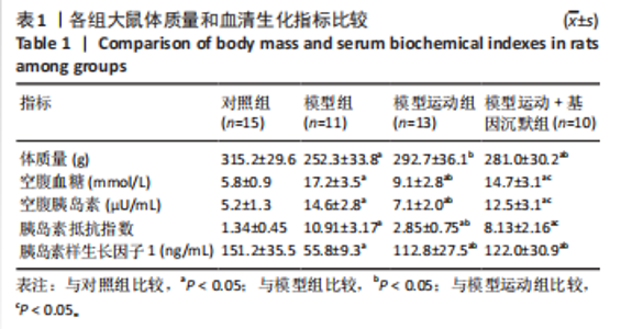
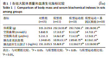
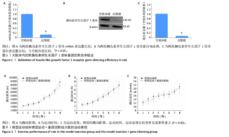
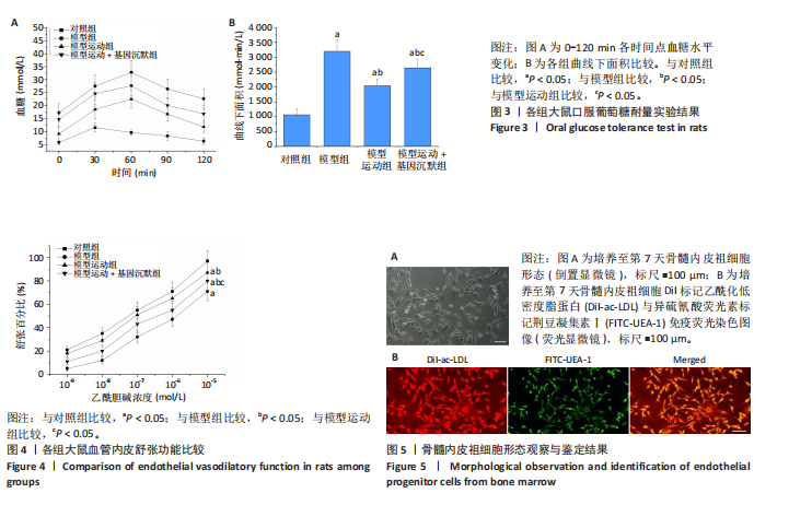
2.1 实验动物数量分析 实验过程中,由于造模失败、死亡、感染等原因共剔除11只动物,最终样本量为n=49,其中对照组n=15、模型组n=11、模型运动组n=13、模型运动+基因沉默组n=10。 2.2 基因沉默效率验证结果 实时荧光定量PCR和Western blot检测结果显示,与空载体组比较,沉默组骨髓内皮祖细胞内胰岛素样生长因子1受体mRNA和蛋白表达分别下调87.1%和92.4%(P < 0.05),见图1。 2.3 各组大鼠运动表现 自主跑轮系统传感器记录显示,大鼠运动较为活跃的时间为晚8点至次日晨8点。8周运动期间,模型运动组和模型运动+基因沉默组动物跑动距离、运动时间和运动速度逐渐增加,并且两组间比较差异无显著性意义(P > 0.05),见图2。 2.4 各组大鼠体质量和血清生化指标比较结果 与对照组比较,模型组大鼠空腹血糖水平、口服葡萄糖耐量实验曲线下面积、血清胰岛素水平和胰岛素抵抗指数升高(P < 0.05),体质量、血清胰岛素样生长因子1水平下降(P < 0.05);与模型组比较,模型运动组空腹血糖水平、口服葡萄糖耐量实验曲线下面积、血清胰岛素水平和胰岛素抵抗指数降低(P < 0.05),体质量、血清胰岛素样生长因子1水平升高(P < 0.05);与模型运动组比较,模型运动+基因沉默组空腹血糖水平、口服葡萄糖耐量实验曲线下面积、血清胰岛素水平和胰岛素抵抗指数升高(P < 0.05),体质量、胰岛素样生长因子1水平无明显变化(P > 0.05),见表1,图3。"

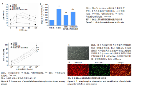
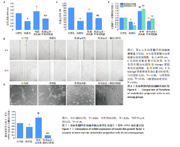
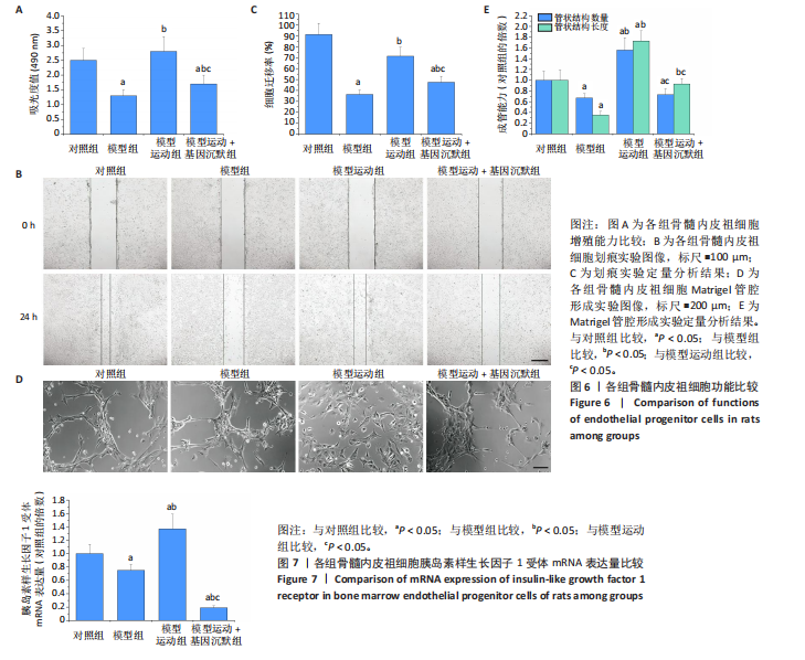
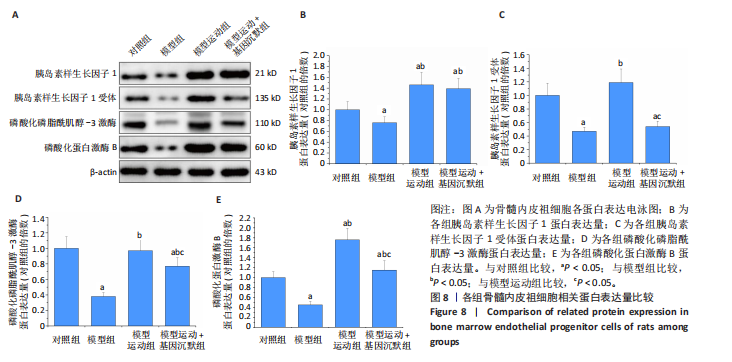
2.5 各组大鼠血管内皮舒张功能比较 内皮依赖性血管舒张剂乙酰胆碱介导的内皮依赖性血管舒张反应是评价内皮功能的重要指标。结果显示,各组大鼠胸主动脉对乙酰胆碱均有浓度依赖性舒张作用,与对照组比较,其他3组血管舒张百分比降低(P < 0.05),说明血管对乙酰胆碱的反应性降低;与模型组比较,模型运动组血管舒张百分比升高(P < 0.05);与模型运动组比较,模型运动+基因沉默组大鼠血管舒张百分比降低(P < 0.05),见图4。 2.6 骨髓内皮祖细胞的形态观察与鉴定结果 倒置显微镜下观察发现培养至第7天时骨髓内皮祖细胞呈现梭形或纺锤形,细胞有明显的极性,两端常伸出细长的伪足,见图5A。荧光显微镜观察显示,Dil-ac-LDL阳性细胞呈现红色荧光,FITC-UEA-1呈现绿色荧光,双阳性细胞(呈现黄色荧光)占贴壁细胞数的(88.6±5.8)%,见图5B。 2.7 各组骨髓内皮祖细胞功能比较 与对照组比较,模型组骨髓内皮祖细胞增殖、迁移和成管能力下降(P < 0.05);与模型组比较,模型运动组骨髓内皮祖细胞增殖、迁移和成管能力增强(P < 0.05);与模型运动组比较,模型运动+基因沉默组骨髓内皮祖细胞各功能均降低(P < 0.05),见图6。 2.8 各组内皮祖细胞胰岛素样生长因子1受体mRNA表达量比较 实时荧光定量PCR检测结果显示,与对照组比较,模型组骨髓内皮祖细胞内胰岛素样生长因子1受体mRNA表达降低(P < 0.05);与模型组比较,模型运动组骨髓内皮祖细胞内胰岛素样生长因子1受体mRNA表达升高(P < 0.05);与模型运动组比较,模型运动+基因沉默组骨髓内皮祖细胞内胰岛素样生长因子1受体mRNA表达降低(P < 0.05),见图7。 2.9 各组骨髓内皮祖细胞相关蛋白表达量比较 Western blot检测结果显示,与对照组比较,模型组骨髓内皮祖细胞内胰岛素样生长因子1、胰岛素样生长因子1受体、磷酸化磷脂酰肌醇-3激酶和磷酸化蛋白激酶B蛋白表达均降低(P < 0.05);与模型组比较,模型运动组骨髓内皮祖细胞内胰岛素样生长因子1、胰岛素样生长因子1受体、磷酸化磷脂酰肌醇-3激酶和磷酸化蛋白激酶B蛋白表达升高(P < 0.05);与模型运动组比较,模型运动+基因沉默组骨髓内皮祖细胞内胰岛素样生长因子1受体、磷酸化磷脂酰肌醇-3激酶和磷酸化蛋白激酶B蛋白表达降低(P < 0.05),胰岛素样生长因子1蛋白表达无明显变化(P > 0.05),见图8。"
"
"

"
| [1] DENG W, ZHAO L, CHEN C, et al. National burden and risk factors of diabetes mellitus in China from 1990 to 2021: Results from the Global Burden of Disease study 2021. J Diabetes. 2024;16(10):e70012. [2] COLE JB, FLOREZ JC. Genetics of diabetes mellitus and diabetes complications. Nat Rev Nephrol. 2020;16(7):377-390. [3] JOSEPH JJ, DEEDWANIA P, ACHARYA T, et al. Comprehensive management of cardiovascular risk factors for adults with type 2 diabetes: A Scientific Statement from the American Heart Association. Circulation. 2022;145(9):e722-e759. [4] MAK A, CHAN J. Endothelial function and endothelial progenitor cells in systemic lupus erythematosus. Nat Rev Rheumatol. 2022;18(5):286-300. [5] HUANG G, CHENG Z, HILDEBRAND A, et al. Diabetes impairs cardioprotective function of endothelial progenitor cell-derived extracellular vesicles via H3K9Ac inhibition. Theranostics. 2022;12(9):4415-4430. [6] KANALEY JA, COLBERG SR, CORCORAN MH, et al. Exercise/physical activity in individuals with type 2 diabetes: A Consensus Statement from the American College of Sports Medicine. Med Sci Sports Exerc. 2022;54(2):353-368. [7] MANNUCCI E, BONIFAZI A, MONAMI M. Comparison between different types of exercise training in patients with type 2 diabetes mellitus: A systematic review and network metanalysis of randomized controlled trials. Nutr Metab Cardiovasc Dis. 2021;31(7):1985-1992. [8] CAVALCANTE SL, LOPES S, BOHN L, et al. Effects of exercise on endothelial progenitor cells in patients with cardiovascular disease: A systematic review and meta-analysis of randomized controlled trials. Rev Port Cardiol (Engl Ed). 2019;38(11):817-827. [9] FERENTINOS P, TSAKIRIDES C, SWAINSON M, et al. The impact of different forms of exercise on endothelial progenitor cells in healthy populations. Eur J Appl Physiol. 2022;122(7):1589-1625. [10] SILVA C. Endothelial progenitor cells and exercise: Working together to target endothelial dysfunction in metabolic syndrome. Arq Bras Cardiol. 2021;117(1):118-119. [11] KOUREK C, KARATZANOS E, PSARRA K, et al. Endothelial progenitor cells mobilization after maximal exercise according to heart failure severity. World J Cardiol. 2020;12(11):526-539. [12] FERENTINOS P, TSAKIRIDES C, SWAINSON M, et al. The impact of different forms of exercise on circulating endothelial progenitor cells in cardiovascular and metabolic disease. Eur J Appl Physiol. 2022;122(4):815-860. [13] WANG M, ZHANG J, GONG N. Role of the PI3K/Akt signaling pathway in liver ischemia reperfusion injury: A narrative review. Ann Palliat Med. 2022;11(2):806-817. [14] LI L, HE D, JIANG K, et al. Effects of forced swimming stress on expression and phosphorylation of PI3K/Akt signal pathway in pancreas of type 2 diabetic rats. Ann Transl Med. 2020;8(16):e1006. [15] DAI X, ZHAI L, SU Q, et al. Effect of aerobic and resistance training on endothelial progenitor cells in mice with type 2 diabetes. Cell Reprogram. 2020;22(4):189-197. [16] HIGASHI Y, GAUTAM S, DELAFONTAINE P, et al. IGF-1 and cardiovascular disease. Growth Horm IGF Res. 2019;45:6-16. [17] OBRADOVIC M, ZAFIROVIC S, SOSKIC S, et al. Effects of IGF-1 on the Cardiovascular System. Curr Pharm Des. 2019;25(35):3715-3725. [18] DE ALCANTARA BORBA D, DA SILVA ALVES E, ROSA J, et al. Can IGF-1 serum levels really be changed by acute physical exercise? A systematic review and meta-analysis. J Phys Act Health. 2020;17(5):575-584. [19] KRAEMER WJ, RATAMESS NA, NINDL BC. Recovery responses of testosterone, growth hormone, and IGF-1 after resistance exercise. J Appl Physiol (1985). 2017;122(3):549-558. [20] 邵俊伟,蔡逊.高脂饮食联合链脲佐菌素建立2型糖尿病大鼠模型的研究进展[J].中国实验动物学报,2014,22(4):90-93. [21] QIAN J, DONG A, KONG M, et al. Suppression of type 1 Insulin-like growth factor receptor expression by small interfering RNA inhibits A549 human lung cancer cell invasion in vitro and metastasis in xenograft nude mice. Acta Biochim Biophys Sin (Shanghai). 2007;39(2):137-147. [22] BUTENAS A, COPP SW, HAGEMAN KS, et al. Effects of comorbid type II diabetes mellitus and heart failure on rat hindlimb and respiratory muscle blood flow during treadmill exercise. J Appl Physiol (1985). 2023; 134(4):846-857. [23] ALIZADE S, FARAMARZI M, BANITALEBI E, et al. Effect of resistance and endurance training with ursolic acid on oxidative stress and cognitive impairment in hippocampal tissue in HFD/STZ-induced aged diabetic rats. Iran J Basic Med Sci. 2023;26(12):1449-1459. [24] MANZANARES G, BRITO-DA-SILVA G, GANDRA PG. Voluntary wheel running: patterns and physiological effects in mice. Braz J Med Biol Res. 2018;52(1):e7830. [25] SAMPATH KUMAR A, MAIYA AG, SHASTRY BA, et al. Exercise and insulin resistance in type 2 diabetes mellitus: A systematic review and meta-analysis. Ann Phys Rehabil Med. 2019;62(2):98-103. [26] YANG Z, SCOTT C A, MAO C, et al. Resistance exercise versus aerobic exercise for type 2 diabetes: A systematic review and meta-analysis. Sports Med. 2014;44(4):487-499. [27] 陈娟娟,郑荣发,莫伟彬,等.运动联合益生菌干预2型糖尿病大鼠糖脂代谢、氧化应激及骨骼肌卫星细胞成肌分化水平的变化[J].中国实验动物学报,2024,32(9):1171-1181. [28] 宋燕娟,马春莲,丁海超,等.中等强度游泳运动调控PPARγ/NF-κB/ADPN通路对2型糖尿病大鼠肝脏糖脂代谢紊乱的影响[J].中国康复医学杂志,2024,39(5):618-627. [29] GHOSH A, GAO L, THAKUR A, et al. Role of free fatty acids in endothelial dysfunction. J Biomed Sci. 2017;24(1):e50. [30] BACH LA. Endothelial cells and the IGF system. J Mol Endocrinol. 2015; 54(1):R1-13. [31] TIEN TY, WU YJ, SU CH, et al. Pannexin 1 modulates angiogenic activities of human endothelial colony-forming cells through IGF-1 mechanism and is a marker of senescence. Arterioscler Thromb Vasc Biol. 2023; 43(10):1935-1951. [32] WEN HJ, LIU GF, XIAO LZ, et al. Involvement of endothelial nitric oxide synthase pathway in IGF‑1 protects endothelial progenitor cells against injury from oxidized LDLs. Mol Med Rep. 2019;19(1):660-666. [33] LIN S, ZHANG Q, SHAO X, et al. IGF-1 promotes angiogenesis in endothelial cells/adipose-derived stem cells co-culture system with activation of PI3K/Akt signal pathway. Cell Prolif. 2017;50(6):e12390. [34] CUI F, HE X. IGF-1 ameliorates streptozotocin-induced pancreatic β cell dysfunction and apoptosis via activating IRS1/PI3K/Akt/FOXO1 pathway. Inflamm Res. 2022;71(5-6):669-680. [35] DAI C, LI N, SONG G, et al. Insulin-like growth factor 1 regulates growth of endometrial carcinoma through PI3k signaling pathway in insulin-resistant type 2 diabetes. Am J Transl Res. 2016;8(8):3329-3336. [36] SUAIFAN G, ALKHAWAJA B, SHEHADEH MB, et al. Glucosamine substituted sulfonylureas: IRS-PI3K-PKC-AKT-GLUT4 insulin signalling pathway intriguing agent. RSC Med Chem. 2024;15(2):695-703. [37] REN BC, ZHANG YF, LIU SS, et al. Curcumin alleviates oxidative stress and inhibits apoptosis in diabetic cardiomyopathy via Sirt1-Foxo1 and PI3K-Akt signalling pathways. J Cell Mol Med. 2020;24(21):12355-12367. [38] HAN Z, HE X, FENG Y, et al. Hsp20 promotes endothelial progenitor cell angiogenesis via activation of PI3K/Akt signaling pathway under hypoxia. Tissue Eng Regen Med. 2022;19(6):1251-1266. [39] YAO J, SHI Z, MA X, et al. lncRNA GAS5/miR-223/NAMPT axis modulates the cell proliferation and senescence of endothelial progenitor cells through PI3K/AKT signaling. J Cell Biochem. 2019;120(9):14518-14530. [40] XIA L, WANG XX, HU XS, et al. Resveratrol reduces endothelial progenitor cells senescence through augmentation of telomerase activity by Akt-dependent mechanisms. Br J Pharmacol. 2008;155(3):387-394. [41] WANG X, JIANG H, GUO L, et al. SDF-1 secreted by mesenchymal stem cells promotes the migration of endothelial progenitor cells via CXCR4/PI3K/AKT pathway. J Mol Histol. 2021;52(6):1155-1164. [42] WANG H, HUANG H, DING SF. Sphingosine-1-phosphate promotes the proliferation and attenuates apoptosis of Endothelial progenitor cells via S1PR1/S1PR3/PI3K/Akt pathway. Cell Biol Int. 2018;42(11):1492-1502. |
| [1] | Cao Xinyan, Yu Zifu, Leng Xiaoxuan, Gao Shiai, Chen Jinhui, Liu Xihua. Effect of repetitive transcranial magnetic stimulation and transcranial direct current stimulation on motor function and gait in children with cerebral palsy: a network meta-analysis [J]. Chinese Journal of Tissue Engineering Research, 2026, 30(6): 1539-1548. |
| [2] | Guo Ying, Tian Feng, Wang Chunfang. Potential drug targets for the treatment of rheumatoid arthritis: large sample analysis from European databases [J]. Chinese Journal of Tissue Engineering Research, 2026, 30(6): 1549-1557. |
| [3] | Yang Zhijie, Zhao Rui, Yang Haolin, Li Xiaoyun, Li Yangbo, Huang Jiachun, Lin Yanping, Wan Lei, HuangHongxing. Postmenopausal osteoporosis: predictive values of muscle mass, grip strength, and appendicular skeletal muscle index [J]. Chinese Journal of Tissue Engineering Research, 2026, 30(5): 1073-1080. |
| [4] | Wen Xiaolong, Weng Xiquan, Feng Yao, Cao Wenyan, Liu Yuqian, Wang Haitao. Effects of inflammation on serum hepcidin and iron metabolism related parameters in patients with type 2 diabetes mellitus: a meta-analysis [J]. Chinese Journal of Tissue Engineering Research, 2026, 30(5): 1294-1301. |
| [5] | Yan Chengbo, Luo Qiuchi, Fan Jiabing, Gu Yeting, Deng Qian, Zhang Junmei. Effect of type 2 diabetes mellitus on orthodontic tooth movement and bone microstructure parameters on the tension side in rats [J]. Chinese Journal of Tissue Engineering Research, 2026, 30(4): 824-831. |
| [6] | Wang Yuhe, Xie Tianyu, Ma Shijia, Wang Yujiao, Li Mengting, Xie Daojun. Regulatory effects of Yuping Shen’an Granules on neuronal autophagy in a mouse model of insomnia [J]. Chinese Journal of Tissue Engineering Research, 2026, 30(35): 9217-9230. |
| [7] | Jiang Li, Peng Guoqiang, Li Sen. Interleukin-10 alleviates inflammatory responses after acute tendon injury [J]. Chinese Journal of Tissue Engineering Research, 2026, 30(34): 8906-8913. |
| [8] | Wang Zhizhuang, Xu Bo, Ma Guoliang, Zhang Dan, Qin Xiaokuan, Feng Minshan, Chen Xin, Yang Kexin, Yang Bowen, Yin He. Application of tissue clearing technology in a rat model of chronic spinal cord injury [J]. Chinese Journal of Tissue Engineering Research, 2026, 30(34): 8939-8945. |
| [9] | Wang Zhengye, Liu Wanlin, Zhao Zhenqun. Mechanism by which vascular endothelial growth factor A targets regulation of angiogenesis in the treatment of steroid-induced osteonecrosis of the femoral head [J]. Chinese Journal of Tissue Engineering Research, 2026, 30(3): 671-679. |
| [10] | Wang Jingfeng, Feng Shuo, Cao Xuan, Li Xiaolin. Lycium barbarum polysaccharide-mediated intestinal flora remodeling improves glycolipid abnormalities in type 2 diabetic rats [J]. Chinese Journal of Tissue Engineering Research, 2026, 30(29): 7592-7602. |
| [11] |
Zhao Yanan, Cao Liquan, Tan Sijie.
A new perspective on exercise for the prevention and treatment of type 2 diabetes mellitus: pyroptosis#br#
#br#
[J]. Chinese Journal of Tissue Engineering Research, 2026, 30(29): 7673-7679.
|
| [12] | Zhang Xiaoxu, Tian Zhenli, Xie Tingting. Roles of pregnane X receptor in sodium arsenite-induced oxidative stress and inflammatory injury in human normal hepatocytes [J]. Chinese Journal of Tissue Engineering Research, 2026, 30(24): 6259-6266. |
| [13] | Gao Jiabin, Li Tianqi, Xu Kun, Zhu Hanmin, Zhou Xi, Li Wei. Mitophagy regulates osteoclasts: a new perspective for osteoporosis treatment [J]. Chinese Journal of Tissue Engineering Research, 2026, 30(23): 5982-5991. |
| [14] | Wang Hengxin, Li Hongkun, Xu Nuo, Li Anping, Wang Xinjing, Zhang Tong. Quercetin promotes osteogenic differentiation of senescent jaw bone marrow mesenchymal stem cells [J]. Chinese Journal of Tissue Engineering Research, 2026, 30(19): 4843-4852. |
| [15] | Liu Enxu, Sun Yu, Duan Jiahao, Yang Lei, Jiang Haobo, Yang Shaofeng. Bidirectional causal interplay between Epstein-Barr virus and ankylosing spondylitis: data analysis based on the UK Biobank and FinnGen databases [J]. Chinese Journal of Tissue Engineering Research, 2026, 30(17): 4542-4547. |
| Viewed | ||||||
|
Full text |
|
|||||
|
Abstract |
|
|||||