Chinese Journal of Tissue Engineering Research ›› 2026, Vol. 30 ›› Issue (33): 8591-8606.doi: 10.12307/2026.477
Previous Articles Next Articles
Mechanism by which curcumin promotes osteogenic differentiation in the treatment of osteoporosis
Yan Xiaolong1, Zhao Canbin2, Shao Jiang3, Sun Hongzhang4, Guan Donghui3, Qin Ying5, Li Xiaoyang3, Chen Dongfeng3
- 1Orthopedics Department, University Town Hospital, Affiliated Hospital of Shandong University of Traditional Chinese Medicine, Jinan 250300, Shandong Province, China; 2First Clinical Medical College, Guangxi University of Traditional Chinese Medicine, Nanning 530200, Guangxi Zhuang Autonomous Region, China; 3Orthopedics Department, Affiliated Hospital of Shandong University of Traditional Chinese Medicine, Jinan 250014, Shandong Province, China; 4Orthopedics Department, Jinan Zhangqiu District Hospital of TCM, Jinan 250200, Shandong Province, China; 5Orthopedics Department, Changqing District Traditional Chinese Medicine Hospital, Jinan 250300, Shandong Province, China
-
Received:2025-11-15Revised:2026-03-09Online:2026-11-28Published:2026-06-09 -
Contact:Shao Jiang, PhD, Attending physician, Orthopedics Department, Affiliated Hospital of Shandong University of Traditional Chinese Medicine, Jinan 250014, Shandong Province, China Co-corresponding author: Sun Hongzhang, MS, Physician, Orthopedics Department, Jinan Zhangqiu District Hospital of TCM, Jinan 250200, Shandong Province, China -
About author:Yan Xiaolong, MS, Associate chief physician, Orthopedics Department, University Town Hospital, Affiliated Hospital of Shandong University of Traditional Chinese Medicine, Jinan 250300, Shandong Province, China -
Supported by:Natural Science Foundation of Shandong Province, No. ZR2022MH147 (to GDH)

CLC Number:
Cite this article
Yan Xiaolong, Zhao Canbin, Shao Jiang, Sun Hongzhang, Guan Donghui, Qin Ying, Li Xiaoyang, Chen Dongfeng. Mechanism by which curcumin promotes osteogenic differentiation in the treatment of osteoporosis[J]. Chinese Journal of Tissue Engineering Research, 2026, 30(33): 8591-8606.
share this article
Add to citation manager EndNote|Reference Manager|ProCite|BibTeX|RefWorks
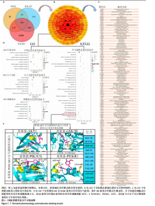
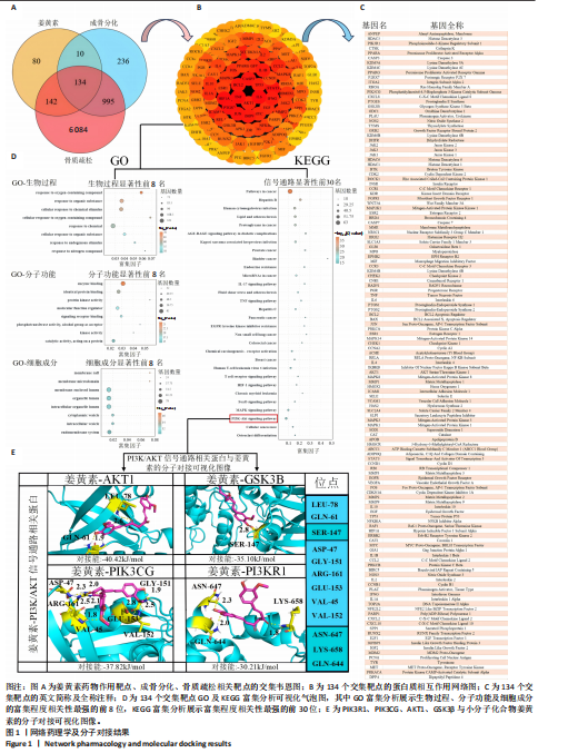
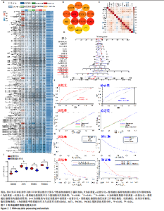
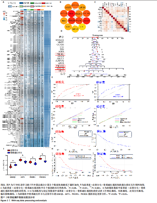
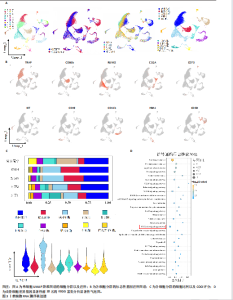
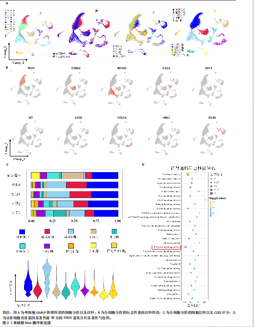
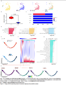
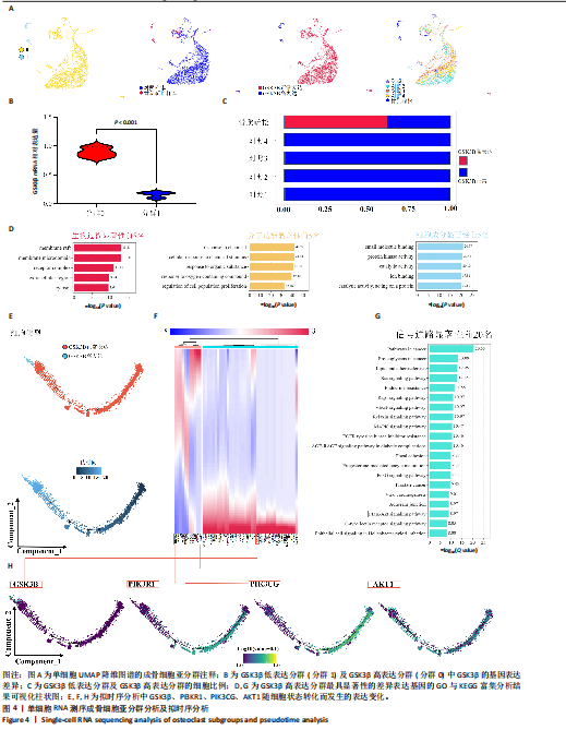
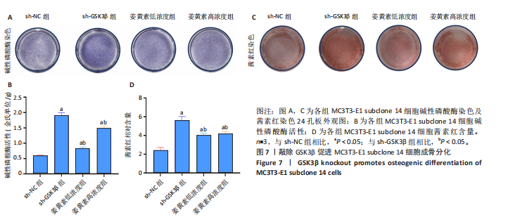
2.1 网络药理学及分子对接结果 2.1.1 姜黄素、成骨分化与骨质疏松靶基因的筛选及交集分析 姜黄素药物作用靶点366个,成骨分化与骨质疏松相关的靶点分别为1 375,7 355个。三者取交集后共得到134个靶点,见图1A。 2.1.2 构建蛋白质相互作用网络图及GO、KEGG富集分析 将134个靶点绘制蛋白质相互作用网络图,见图1B,C。在GO功能富集分析中,涉及细胞对化学刺激的反应、对含氧化合物的反应等生物过程;酶结合、蛋白激酶活性等分子功能;膜阀、膜微区等细胞成分。KEGG通路富集分析前30个信号通路富集情况,见图1D。 2.1.3 分子对接 选取蛋白质相互作用网络图中磷脂酰肌醇3激酶/蛋白激酶B信号通路相关蛋白,结果显示PIK3R1、PIK3CG、AKT1、GSK3β与小分子化合物姜黄素均可良好对接,见图1E。 2.2 生物信息学结果 2.2.1 骨质疏松模型基因集的筛选与构建 广义线性模型提升+随机森林被确定为具有最高平均曲线下面积值(0.692)的最佳模型,见图2A。最佳模型包括14个显著基因(GSK3β、AKT1、PIK3R1、PIK3CG、CCND1、MMP3、HMGCR、APOB、ADIPOQ、STAT3、CAT、ABCC1、SOD1、RB1),定义为姜黄素-成骨分化-骨质疏松基因集。根据14个基因的表达蛋白绘制蛋白质相互作用网络图,其中GSK3β、AKT1、PIK3R1、PIK3CG主要富集于磷脂酰肌醇3激酶/蛋白激酶B信号通路,见图2B。相关性分析显示,GSK3β与AKT1、PIK3R1、PIK3CG之间的负相关较强,见图2C。 2.2.2 骨质疏松临床预测模型的建立 构建诺莫图预测蛋白质相互作用网络基因集与骨质疏松进展的关系,以进一步评估广义线性模型提升+随机森林模型的预测能力。在诺莫图中,每个特征变量的公式对应于一个点值,总值对应于多个骨质疏松风险,见图2D,然后进行受试者工作特征曲线分析,以评估模型的诊断性能。训练集的曲线下面积为0.726,验证集的曲线下面积值为0.734。同时,校准曲线显示该诺莫图模型用于诊断骨质疏松的高准确性。决策分析曲线图、临床影响曲线图均证明了诺莫图模型的预测能力,见图2E-H。最后,进一步评估GSK3β、AKT1、PIK3R1、PIK3CG在训练集中骨质疏松患者样本与非骨质疏松患者样本中的表达差异,发现GSK3β在骨质疏松患者样本中呈现高表达,在非骨质疏松患者样本中呈现低表达,AKT1、PIK3R1、PIK3CG在骨质疏松患者样本中呈现低表达,在非骨质疏松患者样本中呈现高表达,见图2I。 2.3 单细胞转录组学结果 2.3.1 单细胞RNA测序表达谱 单细胞RNA测序数据集中5个样本间具有良好的整合效果,没有显著的批次效应,因此可以进行后续分析。根据标记基因的表达水平,将所有细胞分为20个簇并基于标记基因注释成10种细胞类型:破骨细胞(用TRAP标记)、髓系细胞(用CD66b标记)、成骨细胞(用RUNX2标记)、巨噬细胞(用C1QA标记)、T 细胞(用CD3D标记)、B 细胞(用CD19标记)、单核细胞(用CD11b标记)、肥大细胞(用KIT标记)、红细胞(用HBA1标记)、间充质干细胞(用CD73标记),见图3A,B。此外,评估各个细胞类型在5个样本的细胞分布比例,见图3C。研究结果显示,成骨细胞中COO评分明显高于其他细胞类型。这进一步证实了成骨细胞在骨质疏松发生发展中的重要作用。最后,提取成骨细胞最具显著性的差异表达基因的前30名进行KEGG富集分析,结果显示磷脂酰肌醇3激酶/蛋白激酶B信号通路占据重要位置,排在第18位,见图3D。 2.3.2 细胞状态转换过程中动态基因的鉴定及富集分析 提取通过注释所得的成骨细胞分群进行研究。通过特征性基因筛选发现,可以通过GSK3β的表达差异将成骨细胞分为GSK3β低表达分群及GSK3β高表达分群,GSK3β高表达分群主要集中在骨质疏松样本细胞中,见图4A-C。将GSK3β高表达分群最具显著性的差异表达基因进行了GO与KEGG富集分析,结果显示涉及的基因富集在对含氧化合物的反应、小分子结合、膜阀等方面,且磷脂酰肌醇3激酶/蛋白激酶B信号通路在KEGG富集分析中位列第18位,见图4D,G。选取成骨细胞在由GSK3β低表达分群向GSK3β高表达分群转换过程中最显著的前50个基因绘制基因表达变化图谱,结果发现在细胞状态转换过程中GSK3β低表达分群PIK3R1、PIK3CG、AKT1表达逐渐下降,GSK3β表达逐渐上升,见图4E,F,H。 2.4 细胞实验结果 2.4.1 EdU、CCK-8及细胞克隆形成实验结果 EdU结果显示,与0 μmol/L姜黄素相比,5,10 μmol/L姜黄素未明显抑制MC3T3-E1 subclone 14细胞增殖,而15,20,25 μmol/L姜黄素明显抑制MC3T3-E1 subclone 14细胞增殖(P < 0.05),见图5A,C。CCK-8结果显示,与0 μmol/L姜黄素相比,5,10 μmol/L姜黄素未明显抑制MC3T3-E1 subclone 14细胞活性,而15,20,25 μmol/L姜黄素明显抑制MC3T3-E1 subclone 14细胞活性(P < 0.05),见图5B。细胞克隆形成实验显示,与0 μmol/L姜黄素相比,5,10 μmol/L姜黄素对MC3T3-E1 subclone 14细胞的增殖及细胞形态没有明显影响,而15,20,25 μmol/L姜黄素明显抑制MC3T3-E1 subclone 14细胞增殖,细胞形态出现皱缩及细胞群落数目减少。根据以上结果,选择对MC3T3-E1 subclone 14细胞无明显毒性的5,10 μmol/L姜黄素作为后续研究的药物浓度,见图5D。 2.4.2 慢病毒敲除、免疫荧光及RT-qPCR检测结果 RT-qPCR结果显示,与空白组相比,对照组GSK3β mRNA表达无明显变化;与对照组相比,sh-GSK3β组GSK3β mRNA表达显著下降(P < 0.05),结果提示sh-NC慢病毒不会敲除GSK3β基因,而sh-GSK3β慢病毒可以高效率地敲除GSK3β基因,见图6A,B。免疫荧光显示,与对照组相比,sh-GSK3β组、低浓度姜黄素组及高浓度姜黄素组的β-catenin平均荧光强度显著升高(P < 0.05),见图6C,D。RT-qPCR结果显示,与对照组相比,sh-GSK3β组、低浓度姜黄素组及高浓度姜黄素组的 β-catenin mRNA相对表达量显著升高 (P < 0.05),见图6E,F。 2.4.3 碱性磷酸酶染色、活性测定及茜素红染色、定量分析结果 与对照组相比,sh-GSK3β组、低浓度姜黄素组及高浓度姜黄素组碱性磷酸酶染色着色加深,且碱性磷酸酶活性显著升高(P < 0.05),见图7A,B。与对照组相比,sh-GSK3β组、低浓度姜黄素组及高浓度姜黄素组茜素红染色着色加深,且茜素红相对含量显著升高(P < 0.05),见图7C,D。 2.5 动物实验结果 2.5.1 苏木精-伊红染色及免疫组织化学染色结果 苏木精-伊红染色显示,对照组大鼠股骨远端组织骨小梁结构完整、排列有序且呈淡红色,骨细胞均匀分布于骨陷窝,骨髓腔内造血细胞与脂肪细胞比例正常。模型组大鼠股骨远端组织骨小梁稀疏、纤细且排列紊乱,部分断裂呈游离状,淡红色骨基质减少;骨陷窝内骨细胞数量减少、形态萎缩,核固缩深染;骨髓腔内脂"
"
"
"

"


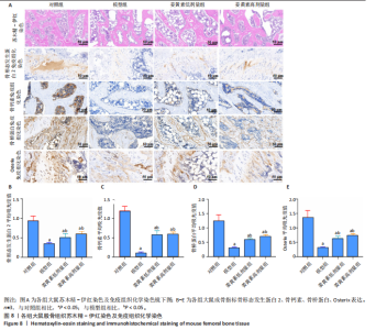
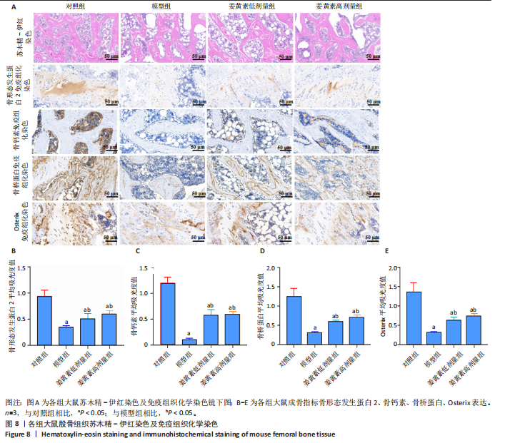
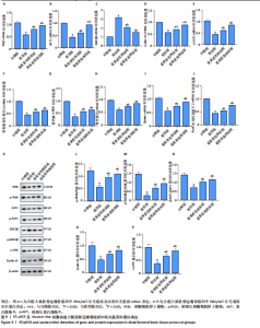
肪细胞增生、体积增大。姜黄素低、高剂量组大鼠股骨远端组织骨小梁数量较模型组增多,排列紊乱程度改善,淡红色骨基质沉积增加;核固缩现象减轻;骨髓腔内脂肪细胞增生受抑、体积缩小,见图8A。 免疫组织化学染色显示,与对照组相比,模型组大鼠成骨相关指标骨形态发生蛋白 2、骨钙素、骨桥蛋白及Osterix的平均吸光度值显著下降(P < 0.05);与模型组相比,姜黄素低、高剂量组大鼠成骨相关指标骨形态发生蛋白 2、骨钙素、骨桥蛋白及Osterix的平均吸光度值显著升高(P < 0.05),见图8B-E。 2.5.2 RT-qPCR及Western blot检测结果 RT-qPCR检测结果显示,与对照组相比,模型组大鼠股骨组织中磷脂酰肌醇3激酶、蛋白激酶B1、Cyclin d1、c-MYC、骨形态发生蛋白 2、骨钙素、骨桥蛋白、Osterix及Runt相关转录因子2 mRNA表达量显著下降,GSK3β mRNA表达量显著升高(P < 0.05)。与模型组相比,姜黄素低、高剂量组大鼠股骨组织中磷脂酰肌醇3激酶、蛋白激酶B1、Cyclin d1、c-MYC、骨形态发生蛋白 2、骨钙素、骨桥蛋白、Osterix及Runt相关转录因子2 mRNA表达量显著升高,GSK3β mRNA表达量显著下降(P < 0.05),见图9A-J。 Western blot检测结果显示,与对照组相比,模型组大鼠股骨组织中磷酸化磷脂酰肌醇3激酶/磷脂酰肌醇3激酶、磷酸化蛋白激酶B/蛋白激酶B1、p-GSK3β/GSK3β、Cyclin d1以及c-MYC的蛋白表达量显著下降(P < 0.05)。与模型组相比,姜黄素低、高剂量组大鼠股骨组织中磷酸化磷脂酰肌醇3激酶/磷脂酰肌醇3激酶、磷酸化蛋白激酶B/蛋白激酶B1、p-GSK3β/GSK3β、Cyclin d1以及c-MYC的蛋白表达量显著上升(P < 0.05),见图9K-P。"


"
"
"

| [1] JOHNSTON CB, DAGAR M. Osteoporosis in Older Adults. Med Clin North Am. 2020;104(5):873-884. [2] 王岩,吕浩,胡芷苜,等.复方补肾活血颗粒干预骨质疏松症模型小鼠:TRIB3/β-catenin轴的作用[J].中国组织工程研究,2026,30(23): 6142-6149. [3] AYERS C, KANSAGARA D, LAZUR B, et al. Effectiveness and Safety of Treatments to Prevent Fractures in People With Low Bone Mass or Primary Osteoporosis: A Living Systematic Review and Network Meta-analysis for the American College of Physicians. Ann Intern Med. 2023;176(2):182-195. [4] 杜健强,殷雪松,秦文秀,等.红景天苷防治原发性骨质疏松症的分子机制研究[J].中国骨质疏松杂志,2025,31(9):1394-1400. [5] ZHOU Y, XU Z, WANG Y, et al. LncRNA MALAT1 mediates osteogenic differentiation in osteoporosis by regulating the miR-485-5p/WNT7B axis. Front Endocrinol (Lausanne). 2023;13:922560. [6] 张世磊,秦川洪,王建旭,等.骨髓间充质干细胞成骨-成脂分化失衡与股骨头坏死:从分子机制到治疗策略[J].中国组织工程研究,2026, 30(13):3350-3358. [7] 龙远,黄元波,夏一锋,等.中药成分调控信号通路促进成骨细胞分化的研究进展[J].现代药物与临床,2025,40(8):2109-2115. [8] 陈心蕾.骨代谢潜在调控基因EGFR在成骨分化过程中的作用研究[D].南京:南京医科大学,2024. [9] 伍伟.Romosozumab通过Wnt/β-catenin信号通路对炎症状态下骨髓间充质干细胞的成骨分化的作用机制研究[D].南昌:南昌大学,2024. [10] 牛黎明,邴雅珺.老年骨质疏松症的中医治疗研究进展[J].中外医药研究,2025,4(19):165-167. [11] LIU H, YI X, TU S, et al. Kaempferol promotes BMSC osteogenic differentiation and improves osteoporosis by downregulating miR-10a-3p and upregulating CXCL12. Mol Cell Endocrinol. 2021;520:111074. [12] ZHAO SJ, KONG FQ, JIE J, et al. Macrophage MSR1 promotes BMSC osteogenic differentiation and M2-like polarization by activating PI3K/AKT/GSK3β/β-catenin pathway. Theranostics. 2020;10(1):17-35. [13] GAO S, CHEN B, ZHU Z, et al. PI3K-Akt signaling regulates BMP2-induced osteogenic differentiation of mesenchymal stem cells (MSCs): A transcriptomic landscape analysis. Stem Cell Res. 2023;66:103010. [14] CHEN J, LIU Z, ZHANG H, et al. YBX1 Promotes MSC Osteogenic Differentiation by Activating the PI3K/AKT Pathway. Curr Stem Cell Res Ther. 2023;18(4):513-521. [15] WANG H, LIANG J, WANG Y, et al. Exploring the effects of naringin on oxidative stress-impaired osteogenic differentiation via the Wnt/β-catenin and PI3K/Akt pathways. Sci Rep. 2024;14(1):14047. [16] ZHANG Z, SHANG W, ZHAO X, et al. Phenytoin regulates osteogenic differentiation of human bone marrow stem cells by PI3K/Akt pathway. Regen Ther. 2023;24:201-210. [17] KOTHA RR, LUTHRIA DL. Curcumin: Biological, Pharmaceutical, Nutraceutical, and Analytical Aspects. Molecules. 2019;24(16):2930. [18] SADEGHI M, DEHNAVI S, ASADIRAD A, et al. Curcumin and chemokines: mechanism of action and therapeutic potential in inflammatory diseases. Inflammopharmacology. 2023;31(3):1069-1093. [19] TOMEH MA, HADIANAMREI R, ZHAO X. A Review of Curcumin and Its Derivatives as Anticancer Agents. Int J Mol Sci. 2019;20(5):1033. [20] ZIA A, FARKHONDEH T, POURBAGHER-SHAHRI AM, et al. The role of curcumin in aging and senescence: Molecular mechanisms. Biomed Pharmacother. 2021;134:111119. [21] YANG S, SUN Y, KAPILEVICH L, et al. Protective effects of curcumin against osteoporosis and its molecular mechanisms: a recent review in preclinical trials. Front Pharmacol. 2023;14:1249418. [22] WANG K. The potential therapeutic role of curcumin in osteoporosis treatment: based on multiple signaling pathways. Front Pharmacol. 2024; 15:1446536. [23] 赵灿斌,秦英,孙宏章,等.姜黄素通过Wnt/β-catenin信号通路调控成骨分化治疗骨质疏松的潜在机制[J].世界中西医结合杂志,2025, 20(3):514-523. [24] BROWN JP. Long-Term Treatment of Postmenopausal Osteoporosis. Endocrinol Metab (Seoul). 2021;36(3):544-552. [25] YAO Y, CAI X, CHEN Y, et al. Estrogen deficiency-mediated osteoimmunity in postmenopausal osteoporosis. Med Res Rev. 2025; 45(2):561-575. [26] GOSSET A, POUILLÈS JM, TRÉMOLLIERES F. Menopausal hormone therapy for the management of osteoporosis. Best Pract Res Clin Endocrinol Metab. 2021;35(6):101551. [27] 李斌,脱伟旺.中医药治疗骨质疏松症研究进展[J].云南中医中药杂志,2025,46(7):95-100. [28] 王铭洋,于冬冬.中医药多维干预绝经后骨质疏松症[J].中西医结合慢性病杂志,2026, 3(1):49-54. [29] 许丰,谢忠建.抗骨质疏松治疗中维生素D的合理使用[J].中国实用内科杂志,2025,45(8):652-655. [30] 李俊伟,秦凡,罗宇茜.老年脊柱退行性改变伴骨质疏松症患者血尿酸、脂代谢及维生素D的变化与预测价值[J].川北医学院学报,2025, 40(7):905-908. [31] 张欣,许云腾,陈西,等.柚皮苷激活FXR/FGF19通路改善绝经后骨质疏松大鼠脂骨代谢紊乱的机制研究[J].中国中药杂志,2025,50(24): 6939-6946. [32] 谭东,谭建,何鑫.血清铁蛋白、雌激素水平与绝经后骨质疏松症患者骨代谢相关指标的关系[J].实用预防医学,2025,32(9):1130-1132. [33] 辛莉.四妙丸通过PI3K/AKT通路改善骨质疏松症的作用及其机制[D].广州:南方医科大学,2025. [34] 邵丹玥,王倍翔,杨旭,等.藿骨补钙颗粒调控Wnt2/β-catenin/PPARγ信号通路改善PMOP大鼠骨密度的机制研究[J].长春中医药大学学报, 2025,41(9):990-995. [35] 王贺贺,李姣.骨髓间充质干细胞衰老与骨髓微环境失衡在老年性骨质疏松中的作用机制[J].遵义医科大学学报,2025,48(8):860-872. [36] 邹远超,尹润宇,杨浩宇,等.中药基于外泌体防治膝骨关节炎与骨质疏松症“共病”机制研究进展[J].中国中医药信息杂志,2025,32(9): 186-191. [37] 毛钰涵,王甲甲.非编码RNA在糖尿病性骨质疏松症中作用的研究进展[J].中国现代医药杂志,2024,26(6):90-94. [38] REID IR, BILLINGTON EO. Drug therapy for osteoporosis in older adults. Lancet. 2022;399(10329):1080-1092. [39] LEBOFF MS, GREENSPAN SL, INSOGNA KL, et al. Correction to: The clinician’s guide to prevention and treatment of osteoporosis. Osteoporos Int. 2022; 33(10):2243. [40] ABD EL-HACK ME, EL-SAADONY MT, SWELUM AA, et al. Curcumin, the active substance of turmeric: its effects on health and ways to improve its bioavailability. J Sci Food Agric. 2021;101(14):5747-5762. [41] TANG K, DENG W, HUANG Z, et al. Neoandrographolide inhibits mature osteoclast differentiation to alleviate bone loss and treat osteoporosis. Front Pharmacol. 2025;16:1466057. [42] AHAMAD S, SAQUIB M, HUSSAIN MK, et al. Targeting Wnt signaling pathway with small-molecule therapeutics for treating osteoporosis. Bioorg Chem. 2025;156:108195. [43] TIPBUNJONG C, KHIMMAKTONG W, HENGPRATOM T, et al. Glabridin Alleviates Oxidative Stress-Induced Osteoporosis by Targeting the Akt/NF-ĸB and Akt/GSK-3β Pathways. Int J Mol Sci. 2025;26(7):2949. [44] WONG SK. Glycogen Synthase Kinase-3 Beta (GSK3β) as a Potential Drug Target in Regulating Osteoclastogenesis: An Updated Review on Current Evidence. Biomolecules. 2024;14(4):502. [45] MA S, LIN J, YANG M, et al. Zhuangyao Jianshen Wan ameliorates senile osteoporosis in SAMP6 mice through Modulation of the GCN5L1-mediated PI3K/Akt/wnt signaling pathway. J Orthop Translat. 2024;49:308-324. [46] LU J, SHI X, FU Q, et al. New mechanistic understanding of osteoclast differentiation and bone resorption mediated by P2X7 receptors and PI3K-Akt-GSK3β signaling. Cell Mol Biol Lett. 2024;29(1):100. [47] ZHOU C, HU G, LI Y, et al. Polydatin accelerates osteoporotic bone repair by inducing the osteogenesis-angiogenesis coupling of bone marrow mesenchymal stem cells via the PI3K/AKT/GSK-3β/β-catenin pathway. Int J Surg. 2025;111(1):411-425. [48] HU J, MAO Z, HE S, et al. Icariin protects against glucocorticoid induced osteoporosis, increases the expression of the bone enhancer DEC1 and modulates the PI3K/Akt/GSK3β/β-catenin integrated signaling pathway. Biochem Pharmacol. 2017;136:109-121. [49] REN M, LIU H, JIANG W, et al. Melatonin Repairs Osteoporotic Bone Defects in Iron-Overloaded Rats through PI3K/AKT/GSK-3β/P70S6k Signaling Pathway. Oxid Med Cell Longev. 2023;2023:7718155. [50] ZHENG F, ZHANG F, WANG F. Inhibition of miR985p promotes high glucoseinduced suppression of preosteoblast proliferation and differentiation via the activation of the PI3K/AKT/GSK3β signaling pathway by targeting BMP2. Mol Med Rep. 2022;26(3):292. [51] LI C, YANG P, LIU B, et al. Prednisolone induces osteocytes apoptosis by promoting Notum expression and inhibiting PI3K/AKT/GSK3β/β-catenin pathway. J Mol Histol. 2021;52(5):1081-1095. [52] RONG X, KOU Y, ZHANG Y, et al. ED-71 Prevents Glucocorticoid-Induced Osteoporosis by Regulating Osteoblast Differentiation via Notch and Wnt/β-Catenin Pathways. Drug Des Devel Ther. 2022;16:3929-3946. [53] YU F, CHANG J, LI J, et al. Protective effects of oridonin against osteoporosis by regulating immunity and activating the Wnt3a/β-catenin/VEGF pathway in ovariectomized mice. Int Immunopharmacol. 2023;118:110011. [54] PARK OJ, KWON Y, KIM J, et al. Muramyl dipeptide alleviates estrogen deficiency-induced osteoporosis through canonical Wnt signaling. J Pathol. 2023;260(2):137-147. [55] SANJAYA SS, PARK J, CHOI YH, et al. Polyphenol extract from Tagetes erecta L. flowers stimulates osteogenesis via β-catenin activation. Phytomedicine. 2025;136:156313. [56] CHEN Y, CHEN L, HUANG R, et al. Investigation for GSK3β expression in diabetic osteoporosis and negative osteogenic effects of GSK3β on bone marrow mesenchymal stem cells under a high glucose microenvironment. Biochem Biophys Res Commun. 2021;534:727-733. |
| [1] | Liu Wenlong, Dong Lei, Xiao Zhengzheng, Nie Yu. Finite element analysis of tibial prosthesis loosening after fixed-bearing unicompartmental knee arthroplasty for osteoporosis [J]. Chinese Journal of Tissue Engineering Research, 2026, 30(9): 2191-2198. |
| [2] | Chen Long, Wang Xiaozhen, Xi Jintao, Lu Qilin. Biomechanical performance of short-segment screw fixation combined with expandable polyetheretherketone vertebral body replacement in osteoporotic vertebrae [J]. Chinese Journal of Tissue Engineering Research, 2026, 30(9): 2226-2235. |
| [3] | Chen Huiting, Zeng Weiquan, Zhou Jianhong, Wang Jie, Zhuang Congying, Chen Peiyou, Liang Zeqian, Deng Weiming. Tail anchoring technique of vertebroplasty in treatment of osteoporotic vertebral compression fractures with intravertebral cleft: a finite element analysis [J]. Chinese Journal of Tissue Engineering Research, 2026, 30(9): 2145-2152. |
| [4] | Zeng Xuan, Weng Rui, Ye Shicheng, Tang Jiadong, Mo Ling, Li Wenchao. Two lumbar rotary manipulation techniques in treating lumbar disc herniation: a finite element analysis of biomechanical differences [J]. Chinese Journal of Tissue Engineering Research, 2026, 30(9): 2153-2161. |
| [5] | Cheng Qisheng, Julaiti·Maitirouzi, Xiao Yang, Zhang Chenwei, Paerhati·Rexiti. Finite element analysis of novel variable-diameter screws in modified cortical bone trajectory of lumbar vertebrae [J]. Chinese Journal of Tissue Engineering Research, 2026, 30(9): 2162-2171. |
| [6] | Wu Yanting, Li Yu, Liao Jinfeng. Magnesium oxide nanoparticles regulate osteogenesis- and angiogenesis-related gene expressions to promote bone defect healing [J]. Chinese Journal of Tissue Engineering Research, 2026, 30(8): 1885-1895. |
| [7] | Hu Xiongke, Liu Shaohua, Tan Qian, Liu Kun, Zhu Guanghui. Shikonin intervention with bone marrow mesenchymal stem cells improves microstructure of femur in aged mice [J]. Chinese Journal of Tissue Engineering Research, 2026, 30(7): 1609-1615. |
| [8] | Li Zhenyu, Zhang Siming, Bai Jiaxiang, Zhu Chen. Osthole improves osteogenic differentiation function of bone marrow mesenchymal stem cells under high-glucose conditions [J]. Chinese Journal of Tissue Engineering Research, 2026, 30(7): 1641-1648. |
| [9] | Jin Dongsheng, Zhao Zhanghong, Zhu Ziyin, Zhang Sen, Sun Zuyan, Deng Jiang. Effects of icariin-loaded microsphere-three-dimensional scaffold on osteogenic differentiation of rabbit bone marrow mesenchymal stem cells [J]. Chinese Journal of Tissue Engineering Research, 2026, 30(7): 1658-1668. |
| [10] | Zou Yulian, Chen Chaopei, Huang Haixia, Lan Yuyan, Liu Min, Huang Ting. Resveratrol promotes osteogenic differentiation of bone marrow mesenchymal stem cells in an inflammatory microenvironment [J]. Chinese Journal of Tissue Engineering Research, 2026, 30(7): 1669-1678. |
| [11] | Lyu Guoqing, Aizimaitijiang·Rouzi, Xiong Daohai. Irisin inhibits ferroptosis in human articular chondrocytes: roles and mechanisms [J]. Chinese Journal of Tissue Engineering Research, 2026, 30(6): 1359-1367. |
| [12] | Wu Zhilin, , He Qin, Wang Pingxi, Shi Xian, Yuan Song, Zhang Jun, Wang Hao . DYRK2: a novel therapeutic target for rheumatoid arthritis combined with osteoporosis based on East Asian and European populations [J]. Chinese Journal of Tissue Engineering Research, 2026, 30(6): 1569-1579. |
| [13] | Zhang Haiwen, Zhang Xian, Xu Taichuan, Li Chao. Bibliometric and visual analysis of the research status and trends of senescence in osteoporosis [J]. Chinese Journal of Tissue Engineering Research, 2026, 30(6): 1580-1591. |
| [14] | Wen Guangwei, Zhen Yinghao, Zheng Taikeng, Zhou Shuyi, Mo Guoye, Zhou Tengpeng, Li Haishan, Lai Yiyi. Effects and mechanisms of isoginkgetin on osteoclastogenesis [J]. Chinese Journal of Tissue Engineering Research, 2026, 30(6): 1348-1358. |
| [15] | Huang Jie, Zeng Hao, Wang Wenchi, Lyu Zhucheng, Cui Wei. Visualization analysis of literature on the effect of lipid metabolism on osteoporosis [J]. Chinese Journal of Tissue Engineering Research, 2026, 30(6): 1558-1568. |
| Viewed | ||||||
|
Full text |
|
|||||
|
Abstract |
|
|||||