Chinese Journal of Tissue Engineering Research ›› 2026, Vol. 30 ›› Issue (11): 2764-2773.doi: 10.12307/2026.317
Previous Articles Next Articles
Moxibustion improves endothelial function in atherosclerotic mice by regulating fatty acid oxidation through mediating mitochondrial autophagy
Pan Li1, Zhu Zhou1, Yan Zhaobo2, Zhang Ning1, Yang Zhihong1, Xiong Jiaojiao1, Yang Xiaofang1
- 1Guizhou University of Traditional Chinese Medicine, Guiyang 550025, Guizhou Province, China; 2Handan Hospital of Integrated Traditional Chinese and Western Medicine, Handan 056005, Hebei Province, China
-
Received:2025-05-06Accepted:2025-06-18Online:2026-04-18Published:2025-09-05 -
Contact:Yang Xiaofang, PhD, Professor, Doctoral supervisor, Guizhou University of Traditional Chinese Medicine, Guiyang 550025, Guizhou Province, China -
About author:Pan Li, PhD candidate, Guizhou University of Traditional Chinese Medicine, Guiyang 550025, Guizhou Province, China -
Supported by:National Natural Science Foundation of China, Nos. 82360978 (to YXF) and 82160937 (to ZN); Guizhou Provincial Science and Technology Plan Project, No. QKH-ZK[2022] General 499 (to YXF)
CLC Number:
Cite this article
Pan Li, Zhu Zhou, Yan Zhaobo, Zhang Ning, Yang Zhihong, Xiong Jiaojiao, Yang Xiaofang. Moxibustion improves endothelial function in atherosclerotic mice by regulating fatty acid oxidation through mediating mitochondrial autophagy[J]. Chinese Journal of Tissue Engineering Research, 2026, 30(11): 2764-2773.
share this article
Add to citation manager EndNote|Reference Manager|ProCite|BibTeX|RefWorks
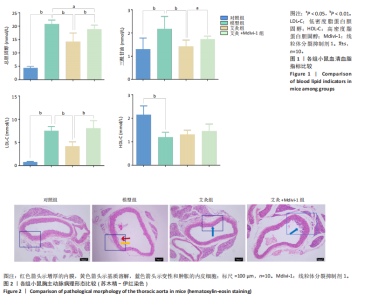
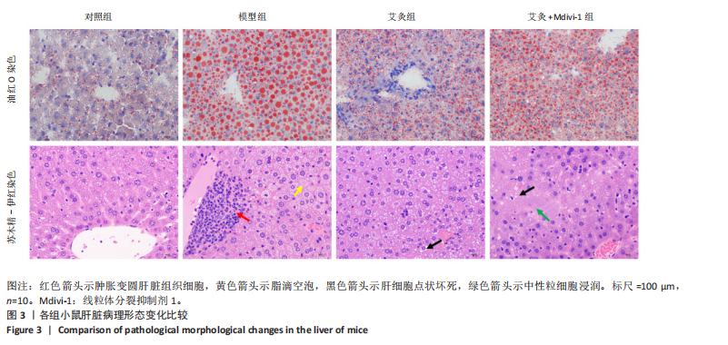
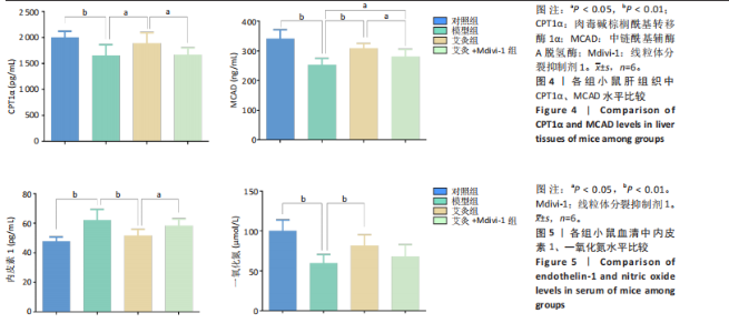
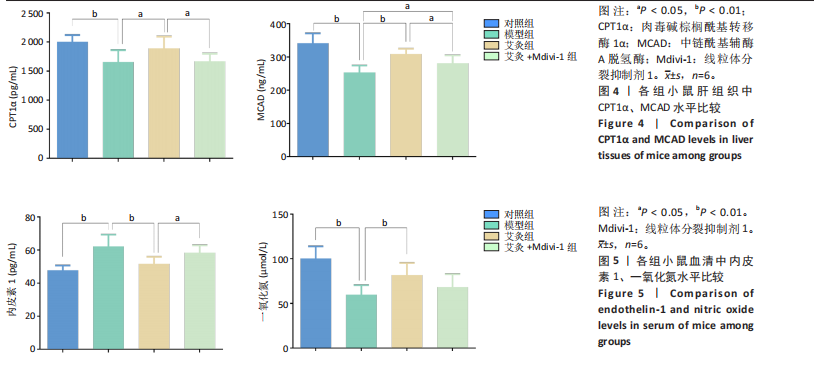
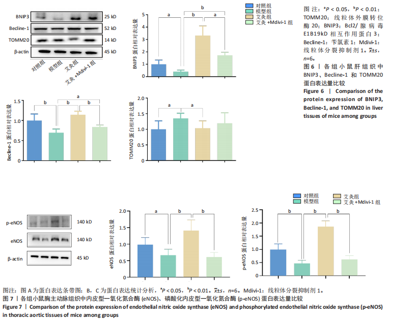
2.1 实验动物数量分析 实验选取10只C57BL/6J小鼠为对照组,30只ApoE-/-小鼠随机分为模型组、艾灸组、艾灸+Mdivi-1组,每组10只,实验过程中小鼠均无死亡。 2.2 各组小鼠血清血脂水平比较 与对照组比较,模型组小鼠血清总胆固醇、三酰甘油、低密度脂蛋白胆固醇浓度均显著升高(P < 0.01),高密度脂蛋白胆固醇浓度明显降低(P < 0.05)。与模型组比较,艾灸组小鼠血清总胆固醇、三酰甘油、低密度脂蛋白胆固醇浓度显著降低(P < 0.01),高密度脂蛋白胆固醇浓度差异无显著性意义(P > 0.05);艾灸+Mdivi-1组小鼠总胆固醇浓度降低(P < 0.05),三酰甘油、低密度脂蛋白胆固醇、高密度脂蛋白胆固醇浓度差异无显著性意义(P > 0.05)。与艾灸组比较,艾灸+ Mdivi-1组小鼠血清总胆固醇、三酰甘油、低密度脂蛋白胆固醇浓度明显升高(P < 0.05,P < 0.01),高密度脂蛋白胆固醇浓度差异无显著性意义(P > 0.05),见图1。 2.3 各组小鼠主动脉弓病理形态比较 对照组小鼠胸主动脉组织结构完整,血管结构较为清晰,血管内皮细胞较为完整,未见动脉粥样硬化病变特征。模型组小鼠胸主动脉血管内壁不平整,内膜增厚,中膜结构紊乱,弹性膜较薄,基质溶解,弹力纤维断裂。干预后,艾灸组小鼠胸主动脉管腔较规则,中膜内弹性膜排列较为整齐,血管内膜、中膜、外膜结构较为清晰,内膜变性和肿胀的内皮细胞较少。艾灸+Mdivi-1组小鼠胸主动脉管腔较规则,血管内壁稍不平整,局部内膜增厚,有少量泡沫细胞和肿胀的内皮细胞,见图2。 2.4 各组小鼠肝脏组织病理形态变化及脂肪样变结果 对照组肝脏组织条理清晰,胞浆内少量脂滴空泡,油红O染色亦显示少量脂滴蓄积;与对照组比,模型组肝脏组织细胞肿胀变圆,出现肝细胞点状坏死,中性粒细胞浸润,可见较多的脂肪空泡及脂肪变性,油红O染色亦显示大量脂滴蓄积;与模型组相比,艾灸组和艾灸+Mdivi-1组肝脏组织肿胀显著减轻,纹理清晰,脂滴沉积减少,脂滴蓄积显著减少;与艾灸组相比,艾灸+ Mdivi-1组肿胀加重,脂滴蓄积增加,见图3。 2.5 各组小鼠肝脏组织CPT-1α、MCAD水平比较 与对照组比较,模型组小鼠肝脏组织中CPT1α、MCAD质量浓度显著降低(P < 0.01);与模型组比较,艾灸组肝脏组织中CPT1α、MCAD质量浓度明显增加(P < 0.05,P < 0.01),艾灸+Mdivi-1组肝脏组织中MCAD质量浓度明显增加(P < 0.05),CPT1α质量浓度差异无显著性意义(P > 0.05);与艾灸组相比,艾灸+ Mdivi-1组肝脏组织中CPT1α、MCAD质量浓度明显降低(P < 0.05),见图4。 2.6 各组小鼠血清一氧化氮、内皮素1水平比较 与对照组相比,模型组小鼠的血清一氧化氮浓度显著降低(P < 0.01),内皮素1质量浓度显著升高(P < 0.01);与模型组比,艾灸组小鼠血清一氧化氮浓度显著升高(P < 0.01),而内皮素1质量浓度显著降低(P < 0.01);与艾灸组相比,艾灸+Mdivi-1组血清内皮素1质量浓度增加(P < 0.05),一氧化氮浓度下降但差异无显著性意义(P > 0.05),见图5。 2.7 各组小鼠肝脏组织BNIP3、Becline-1、TOMM20蛋白表达比较 与对照组比较,模型组小鼠肝脏组织中BNIP3、Becline-1表达量明显降低(P < 0.05,P < 0.01),TOMM20表达量明显升高(P < 0.05);与模型组比较,艾灸组肝脏组织BNIP3、Becline-1表达量显著增加(P < 0.01),TOMM20表达量明显降低(P < 0.05),艾灸+Mdivi-1组肝脏组织BNIP3、Becline-1表达量显著增加(P < 0.05,P < 0.01),TOMM20表达量差异无显著性意义(P > 0.05);与艾灸组相比,艾灸+Mdivi-1组肝脏组织中BNIP3、Becline-1表达量明显下降(P < 0.05,P < 0.01),TOMM20表达量差异无显著性意义(P > 0.05),见图6。 2.8 各组小鼠胸主动脉组织中内皮型一氧化氮合酶、磷酸化内皮型一氧化氮合酶蛋白表达比较 与对照组比较,模型组小鼠胸主动脉组织中内皮型一氧化氮合酶、磷酸化内皮型一氧化氮合酶表达量显著降低(P < 0.05、P < 0.01);与模型组比较,艾灸组小鼠胸主动脉组织中内皮型一氧化氮合酶、磷酸化内皮型一氧化氮合酶表达量明显增加(P < 0.01);与艾灸组相比,艾灸+Mdivi-1组小鼠胸主动脉组织中内皮型一氧化氮合酶、磷酸化内皮型一氧化氮合酶表达量明显降低(P < 0.01),见图7。"


"
"

"
| [1] ZHU Y, XIAN X, WANG Z, et al. Research Progress on the Relationship between Atherosclerosis and Inflammation. Biomolecules. 2018; 8(3):80. [2] DING Y, SUN Y, WANG H, et al.Atherosis-associated lnc_000048 activates PKR to enhance STAT1-mediated polarization of THP-1 macrophages to M1 phenotype. Neural Regen Res. 2024;19(11):2488-2498. [3] YANG M, JIAO H, LI Y, et al. Guanmaitong Granule Attenuates Atherosclerosis by Inhibiting Inflammatory Immune Response in ApoE (-/-) Mice Fed High-Fat Diet. Drug Des Devel Ther. 2022;16:3145-3168. [4] 徐倩, 姜志胜. 动脉粥样硬化机制研究的新认识[J]. 中国动脉硬化杂志, 2024,32(11):921-931. [5] 刘姚钰菁, 沈月红, 瞿旻晔, 等. 从“脉生痰核”理论及脂质炎症学说辨治动脉粥样硬化[J]. 中药药理与临床, 网络首发时间:2024-06-13. [6] 赵然, 赵文廷, 卢秉久. 基于“脾气散精”理论探讨线粒体功能紊乱对代谢相关脂肪性肝病的影响及机制[J]. 河北中医,2024, 46(6):1028-1032. [7] 王伟霖, 王诗瑶, 王钰, 等. 基于NF-κB/p62通路探讨益脉颗粒调控动脉粥样硬化小鼠肝脏线粒体自噬与炎症的分子机制[J]. 辽宁中医药大学学报,2024,26(7):42-47. [8] BARBIER-TORRES L, FORTNER KA, IRUZUBIETA P, et al. Silencing hepatic MCJ attenuates non-alcoholic fatty liver disease (NAFLD) by increasing mitochondrial fatty acid oxidation. Nat Commun. 2020;11(1):3360. [9] 石情, 冉苏叶, 宋铃榆, 等. NLRP6过表达通过AMPK/CPT1A/PGC1A通路促进肝细胞脂肪氧化分解改善非酒精性脂肪肝[J]. 南方医科大学学报,2025,45(1):118-125. [10] 肖怡琴. Zfp217通过脂肪酸氧化影响脂质代谢的研究[D]. 武汉:华中农业大学,2024. [11] ZHANG L, YANG B, YU B. Paeoniflorin Protects against Nonalcoholic Fatty Liver Disease Induced by a High-Fat Diet in Mice. Biol Pharm Bull. 2015;38(7):1005-1011. [12] YANG Y, OWUSU FB, WU H, et al. Mitochondria as therapeutic targets for Natural Products in the treatment of Cardiovascular Diseases. J Ethnopharmacol. 2025;345:119588. [13] TIAN HY, YU DJ, XIE T, et al. Cordycepin alleviates metabolic dysfunction-associated liver disease by restoring mitochondrial homeostasis and reducing oxidative stress via Parkin-mediated mitophagy. Biochem Pharmacol. 2025;232:116750. [14] 文晓宾. 羟基酪醇调控PI3K/Akt-Nrf2信号通路和线粒体自噬缓解仔猪肠道氧化损伤[D]. 北京:中国农业科学院, 2024. [15] 余欣慧, 韩欣雨, 韩萌. 补肾安胎冲剂通过激活细胞自噬促进ox-LDL诱导PMVEC损伤模型血管生成的作用机制研究[J]. 中国中药杂志,网络首发时间:2025-04-10. doi: 10.19540/j.cnki.cjcmm.20250409.705 [16] 李金科, 杨凯凯, 杨善儒, 等. 血管老化机制的研究进展[J]. 中华老年心脑血管病杂志,2025,27(4):527-529. [17] LU D, LI Y, NIU X, et al. STAT2/SLC27A3/PINK1-Mediated Mitophagy Remodeling Lipid Metabolism Contributes to Pazopanib Resistance in Clear Cell Renal Cell Carcinoma. Research (Wash D C). 2024;7:539. [18] 王舒舒, 周桂庭, 林莉雯, 等. 脑心清介导AMPK/SIRT1通路促进脂肪酸氧化改善非酒精性脂肪肝[J]. 中药新药与临床药理,2024, 35(10):1531-1541. [19] 郭向宇, 王浩, 杨戈. 延胡索乙素改善肝细胞脂质积累及氧化应激的作用机制[J]. 中国老年学杂志,2025,45(7):1712-1717. [20] 闫朝勃, 伍先明, 张宁, 等. 艾灸对ApoE~(-/-)动脉粥样硬化小鼠SIRT1/FOXO3a信号通路的影响[J]. 针刺研究,2024,49(4):376-383. [21] 吴冰心, 杨孝芳, 闫朝勃, 等. 温阳通脉灸法通过线粒体途径对ApoE-/-动脉粥样硬化小鼠内皮相关凋亡因子的影响[J]. 中华中医药学刊,网络首发时间:2025-04-21. [22] 伍先明, 张宁, 朱洲, 等. 艾灸对ApoE-/-动脉粥样硬化小鼠沉默信息调节因子1/内皮型一氧化氮合酶信号通路的影响[J]. 针刺研究,网络首发时间:2025-02-12. doi: 10.13702/j.1000-0607.20240618 [23] 中国针灸学会.实验动物常用穴位名称与定位第3部分:小鼠[J]. 针刺研究,2021,46(5):445-446. [24] 夏冉. 基于线粒体质量控制探究艾灸治疗气虚血瘀型慢性心力衰竭的机制及临床疗效观察[D]. 合肥:安徽中医药大学,2022. [25] 王阶,李军,董艳,等.泛血管疾病的中医内涵及防治策略[J]. 中国实验方剂学杂志,2025,31(7):1-14. [26] 安贺. PPP2CB通过LOX-1/MAPK/ERK轴促进动脉粥样硬化中的肝脂累积[D]. 十堰:湖北医药学院,2024. [27] CUI Y, LIU J, HUANG C, et al. Moxibustion at CV4 alleviates atherosclerotic lesions through activation of the LXRalpha/ABCA1 pathway in apolipoprotein-E-deficient mice. Acupunct Med. 2019;37(4):237-243. [28] KIAMEHR M, VIIRI LE, VIHERVAARA T, et al.Lipidomic profiling of patient-specific iPSC-derived hepatocyte-like cells. Dis Model Mech. 2017;10(9):1141-1153. [29] 李超伟. 解偶联剂BAM15促进线粒体自噬改善动物肝脏脂肪代谢紊乱的机制研究[D]. 咸阳:西北农林科技大学, 2023. [30] 杜敏, 陶丽宇, 冯骁腾, 等. 基于线粒体能量代谢障碍探讨从脾论治“双心疾病”的机制[J]. 北京中医药大学学报,2021,44(9):860-864. [31] 张伟, 卢秉久, 张艳. 基于“肝心同病”探析代谢相关脂肪性肝病合并动脉粥样硬化的病机与治疗思路[J]. 北京中医药大学学报, 2023,46(5):697-704. [32] 潘萌, 史晓燕. 线粒体自噬在肝脏相关疾病发生发展中的作用[J]. 临床肝胆病杂志,2024,40(2):413-418. [33] 任甜甜, 刘如秀, 李翠芝, 等. 基于“虚气留滞”与线粒体自噬探讨慢性心力衰竭的病机与治疗[J]. 中医学报,2025,40(4):728-734. [34] 郑标, 王琼, 肖玉波, 等. 线粒体自噬在动脉粥样硬化中作用的研究进展[J]. 中国病理生理杂志,2025,41(3):585-592. [35] 何炜, 李玉明, 周欣, 等. 短时高强度间歇运动训练心肌梗死模型大鼠心室重构及线粒体变化[J]. 中国组织工程研究,2016,20(40): 5986-5993. [36] 吴昀紫, 郗博洲, 马亮亮, 等. 线粒体质量控制与肿瘤研究进展[J]. 中国医学前沿杂志(电子版),2024,16(2):78-87. [37] 张嘉祺, 王茜婷, 陈夏欢, 等. 线粒体融合分裂与自噬在动脉粥样硬化中的作用及研究进展[J]. 中华老年心脑血管病杂志,2024, 26(8):974-977. [38] 宋明箫, 陈君顺子, 王宁伟, 等. 耐力训练大鼠抗动脉粥样硬化线粒体自噬通路中抗增殖蛋白2的作用[J].中国组织工程研究, 2025,29(11):2294-2300. [39] 范靖琪. 调神针法调控线粒体自噬改善APP/PS1小鼠认知功能的研究[D]. 广州:广州中医药大学,2023. [40] BERTOLIN G, FERRANDO-MIGUEL R, JACOUPY M, et al. The TOMM machinery is a molecular switch in PINK1 and PARK2/PARKIN-dependent mitochondrial clearance. Autophagy. 2013;9(11):1801-1817. [41] 丁烨, 魏佳乐, 熊阳. 线粒体自噬在乳腺癌发生发展中的作用及其调控机制[J]. 中国药理学与毒理学杂志,2024,38(7):533-541. [42] AHMADPOUR ST, DESQUIRET-DUMAS V, YIKILMAZ U, et al.Doxorubicin-Induced Autophagolysosome Formation Is Partly Prevented by Mitochondrial ROS Elimination in DOX-Resistant Breast Cancer Cells. Int J Mol Sci. 2021;22(17):9283. [43] 王天源, 王晓慧. 有氧运动对糖尿病大鼠PPARα信号通路的影响及其与PPARγ关系[J]. 中国应用生理学杂志,2020,36(4):312-317. [44] WANG Y, VISCARRA J, KIM SJ, et al. Transcriptional regulation of hepatic lipogenesis. Nat Rev Mol Cell Biol. 2015;16(11):678-689. [45] HOUTEN SM, VIOLANTE S, VENTURA FV, et al. The Biochemistry and Physiology of Mitochondrial Fatty Acid beta-Oxidation and Its Genetic Disorders. Annu Rev Physiol. 2016;78:23-44. [46] 王爽. DGAT2/PLIN5介导脂肪肝奶牛肝细胞脂滴-线粒体互作调控脂肪酸代谢机制研究[D]. 大庆:黑龙江八一农垦大学,2024. [47] 文佩华. 肝豆汤调控JAK2-STAT5通路及脂肪酸β氧化改善Wilson病模型肝脏脂肪变性的机制研究[D]. 安徽中医药大学,2024. [48] BOUGHALEB H, LOBYSHEVA I, DEI ZF, et al. Biological Assessment of the NO-Dependent Endothelial Function. Molecules. 2022;27(22):7921. [49] WU XM, ZHANG N, LI JS, et al. Purinergic receptors mediate endothelial dysfunction and participate in atherosclerosis. Purinergic Signal. 2023; 19(1):265-272. [50] MOUNT PF, KEMP BE, POWER DA.Regulation of endothelial and myocardial NO synthesis by multi-site eNOS phosphorylation. J Mol Cell Cardiol. 2007;42(2):271-279. [51] 刘冬, 王琨. 线粒体分裂抑制剂Mdivi-1通过抑制线粒体自噬促进炎症微环境下髓核细胞衰老退变的研究[J]. 现代医学,2022, 50(6):735-741. |
| [1] | Chen Yulin, He Yingying, Hu Kai, Chen Zhifan, Nie Sha Meng Yanhui, Li Runzhen, Zhang Xiaoduo , Li Yuxi, Tang Yaoping. Effect and mechanism of exosome-like vesicles derived from Trichosanthes kirilowii Maxim. in preventing and treating atherosclerosis [J]. Chinese Journal of Tissue Engineering Research, 2026, 30(7): 1768-1781. |
| [2] | Wang Zhenze, Liu Fende, Zhang Rui, Li Wujun. Mesenchymal stem cells in treatment of arteriosclerosis obliterans of lower extremities: systematic review and meta-analysis [J]. Chinese Journal of Tissue Engineering Research, 2026, 30(7): 1869-1876. |
| [3] | Qi Xiang, Cao Shan, Chen Jian, Zhang Yijia, Liu Keke, Xu Zifu, Liu Wang, Fu Xiaoxiao, Yin Xiaolei. Screening of genes related to mitochondrial dysfunction and ferroptosis in atherosclerosis and target prediction of regulatory traditional Chinese medicine [J]. Chinese Journal of Tissue Engineering Research, 2026, 30(10): 2641-2652. |
| [4] | Zhang Yibo, Lu Jianqi, Mao Meiling, Pang Yan, Dong Li, Yang Shangbing, Xiao Xiang. Exploring the causal relationship between rheumatoid arthritis and coronary atherosclerosis: a Mendel randomized study involving serum metabolites and inflammatory factors [J]. Chinese Journal of Tissue Engineering Research, 2025, 29(在线): 1-9. |
| [5] | Li Huayuan, Li Chun, Liu Junwei, Wang Ting, Li Long, Wu Yongli. Effect of warm acupuncture on PINK1/Parkin pathway in the skeletal muscle of rats with chronic fatigue syndrome [J]. Chinese Journal of Tissue Engineering Research, 2025, 29(8): 1618-1625. |
| [6] | Zhu Hanmin, Wang Song, Xiao Wenlin, Zhang Wenjing, Zhou Xi, He Ye, Li Wei, . Mitophagy regulates bone metabolism [J]. Chinese Journal of Tissue Engineering Research, 2025, 29(8): 1676-1683. |
| [7] | Gao Yang, Qin Hewei, Liu Dandan. ACSL4 mediates ferroptosis and its potential role in atherosclerotic cardiovascular disease [J]. Chinese Journal of Tissue Engineering Research, 2025, 29(6): 1239-1247. |
| [8] | Hu Shujuan, Liu Dang, Ding Yiting, Liu Xuan, Xia Ruohan, Wang Xianwang. Ameliorative effect of walnut oil and peanut oil on atherosclerosis [J]. Chinese Journal of Tissue Engineering Research, 2025, 29(30): 6482-6488. |
| [9] | Zhang Guoan, Shi Jian, Song Baoguo, Huang Xiaoyan. Histone deacetylase 1 gene inhibits pyroptosis of human umbilical vein endothelial cells and alleviates atherosclerosis and inflammatory response [J]. Chinese Journal of Tissue Engineering Research, 2025, 29(25): 5351-5361. |
| [10] | Zhu Liuhui, Zhang Xinyue, Zhu Zhouhai, Yang Xinglong, Guan Ying, Liu Bin. Coiled-coil-helix-coiled-coil-helix domain-containing 2 inhibits apoptosis of Parkinson’ s disease SH-SY5Y cells by promoting mitochondrial autophagy [J]. Chinese Journal of Tissue Engineering Research, 2025, 29(25): 5403-5413. |
| [11] | Yang Jun, Yin Peng, Zheng Zhonghua. Mechanism of stachydrine-induced autophagy in improving atherosclerosis in high-fat-fed mice [J]. Chinese Journal of Tissue Engineering Research, 2025, 29(24): 5140-5147. |
| [12] | Zhang Yibo, Lu Jianqi, Mao Meiling, Pang Yan, Dong Li, Yang Shangbing, Xiao Xiang. Rheumatoid arthritis and coronary atherosclerosis: data analysis of serum metabolite and inflammatory factor in the European population [J]. Chinese Journal of Tissue Engineering Research, 2025, 29(24): 5263-5271. |
| [13] | Li Qingyin, Li Linhua, Zhang Chunle, Fu Ping. Endothelial progenitor cell and mesenchymal stem cell therapy for vascular stent-associated diseases [J]. Chinese Journal of Tissue Engineering Research, 2025, 29(19): 4091-4101. |
| [14] | Cao Panxia, Peng Zining, Liu Shanshan, Fei Tiantian, Liang Tengyun, Zhang Mengwen, Wu Hong. AMP-activated protein kinase mediates macrophage fatty acid oxidation: an approach to prevent and treat atherosclerosis with traditional Chinese medicine [J]. Chinese Journal of Tissue Engineering Research, 2025, 29(18): 3906-3914. |
| [15] | Wang Jingying, , Ren Binbin, Ma Suna, Yang Yueyue, , Wu Song, , Guan Mengya, . Mechanism of alpha-synuclein in mitochondrial damage induced by Parkinson’s disease [J]. Chinese Journal of Tissue Engineering Research, 2025, 29(17): 3668-3674. |
| Viewed | ||||||
|
Full text |
|
|||||
|
Abstract |
|
|||||