Chinese Journal of Tissue Engineering Research ›› 2025, Vol. 29 ›› Issue (34): 7293-7300.doi: 10.12307/2025.888
Previous Articles Next Articles
Effect of crocin hydrogel on chondrocytes and MC3T3-E1 cells
Yin Hang1, Song Kui2
- 1School of Physical Education, Wuhan Business University, Wuhan 430056, Hubei Province, China; 2First Affiliated Hospital of Jishou University, Jishou 416000, Hunan Province, China
-
Received:2024-06-27Accepted:2024-09-05Online:2025-12-08Published:2025-01-17 -
Contact:Yin Hang, MS, Associate professor, School of Physical Education, Wuhan Business University, Wuhan 430056, Hubei Province, China -
About author:Yin Hang, MS, Associate professor, School of Physical Education, Wuhan Business University, Wuhan 430056, Hubei Province, China -
Supported by:Hunan Natural Science Foundation Project, No. 2023JJ30609 (to SK)
CLC Number:
Cite this article
Yin Hang, Song Kui. Effect of crocin hydrogel on chondrocytes and MC3T3-E1 cells[J]. Chinese Journal of Tissue Engineering Research, 2025, 29(34): 7293-7300.
share this article
Add to citation manager EndNote|Reference Manager|ProCite|BibTeX|RefWorks
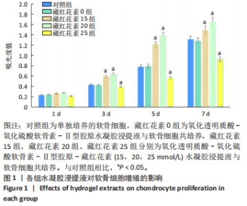
2.1 水凝胶浸提液对软骨细胞增殖的影响 各组软骨细胞CCK-8检测结果见图1。培养1 d后,5组软骨细胞增殖没有明显差异;培养3-7 d后,与对照组相比,藏红花素0组软骨细胞增殖吸光度值无明显差异(P > 0.05),藏红花素15组、藏红花素20组软骨细胞增殖吸光度值升高(P均< 0.05),藏红花素25组细胞增殖吸光度值降低(P < 0.05)。"


2.2 水凝胶浸提液对软骨细胞迁移的影响 与对照组相比,藏红花素0组对软骨细胞迁移无明显影响,藏红花素15组、藏红花素20组、藏红花素25组可促进软骨细胞的迁移,其中以藏红花素25组效果最明显,见图2A。定量分析结果显示,对照组与藏红花素0组迁移细胞数量比较无统计学差异(P均> 0.05),藏红花素15组、藏红花素20组、藏红花素25组迁移细胞数量均多于对照组、藏红花素0组(P均< 0.05),藏红花素25组迁移细胞数量多于藏红花素15组、藏红花素20组(P均< 0.05),藏红花素20组迁移细胞数量多于藏红花素15组(P < 0.05),见图2B。"

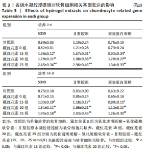
2.3 水凝胶浸提液对软骨细胞相关基因表达的影响 RT-qPCR检测结果显示,培养7 d后,与对照组相比,藏红花素0组SOX9、Ⅱ型胶原、聚集蛋白聚糖mRNA表达无显著变化(P均> 0.05),藏红花素15组、藏红花素20组、藏红花素25组SOX9、Ⅱ型胶原、聚集蛋白聚糖mRNA表达均显著升高(P均< 0.05);藏红花素20组、藏红花素25组SOX9、Ⅱ型胶原、聚集蛋白聚糖mRNA表达均高于藏红花素15组(P均< 0.05),藏红花素25组SOX9、Ⅱ型胶原mRNA表达均高于藏红花素20组(P均< 0.05),见表3。培养14 d后,与对照组相比,藏红花素0组SOX9、Ⅱ型胶原、聚集蛋白聚糖mRNA表达无明显变化(P均> 0.05),藏红花素15组、藏红花素20组、藏红花素25组SOX9、Ⅱ型胶原、聚集蛋白聚糖mRNA表达均升高(P均< 0.05);藏红花素20组、藏红花素25组SOX9、Ⅱ型胶原、聚集蛋白聚糖mRNA表达均高于藏红花素15组(P均< 0.05),藏红花素25组SOX9、Ⅱ型胶原、聚集蛋白聚糖mRNA表达与藏红花素20组相比无显著变化(P均> 0.05),见表3。"

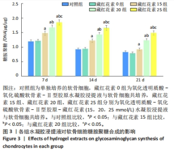
2.4 水凝胶浸提液对软骨细胞糖胺聚糖合成的影响 培养7,14,21 d后,与对照组比较,藏红花素0组软骨细胞糖胺聚糖合成量均无明显变化(P均> 0.05),藏红花素15组、藏红花素20组、藏红花素25组软骨细胞糖胺聚糖合成量均增加(P均< 0.05);藏红花素25组软骨细胞糖胺聚糖合成量多于藏红花素15组、藏红花素20组(P均< 0.05),藏红花素20组软骨细胞糖胺聚糖合成量多于藏红花素15组(P < 0.05),见图3。"

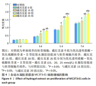
2.5 水凝胶浸提液对MC3T3-E1细胞增殖的影响 各组MC3T3-E1细胞增殖CCK-8检测结果见图4。培养1 d后,5组MC3T3-E1细胞增殖吸光度值无明显差异(P > 0.05)。培养3 d后,与对照组比较,藏红花素0组MC3T3-E1细胞增殖吸光度值无明显变化(P > 0.05),藏红花素15组、藏红花素20组、藏红花素25组细胞增殖吸光度值升高(P均< 0.05),并且藏红花素20组、藏红花素25组细胞增殖吸光度值高于藏红花素15组(P均< 0.05),前两组比较无明显差异(P > 0.05)。培养5,7 d后,与对照组比较,藏红花素0组MC3T3-E1细胞增殖无明显变化(P > 0.05),藏红花素15组、藏红花素20组、藏红花素25组细胞增殖吸光度值升高(P均< 0.05),藏红花素20组、藏红花素25组细胞增殖高于藏红花素15组(P均< 0.05),藏红花素25组细胞增殖吸光度值高于藏红花素20组(P < 0.05)。"

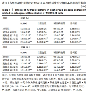
2.6 水凝胶浸提液对MC3T3-E1细胞成骨分化相关基因表达的影响 RT-qPCR检测结果显示,成骨诱导培养7,14 d后,与对照组比较,藏红花素0组RUNX2、Ⅰ型胶原、碱性磷酸酶、骨钙素mRNA无明显变化(P均> 0.05),藏红花素15组、藏红花素20组、藏红花素25组RUNX2、Ⅰ型胶原、碱性磷酸酶、骨钙素mRNA表达均升高(P均< 0.05);藏红花素20组RUNX2、Ⅰ型胶原、碱性磷酸酶、骨钙素mRNA表达高于藏红花素15组(P均< 0.05),藏红花素25组RUNX2、Ⅰ型胶原、碱性磷酸酶、骨钙素mRNA表达高于藏红花素20组(P均< 0.05),见表4。"

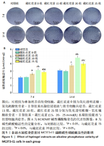
2.7 水凝胶浸提液对MC3T3-E1细胞碱性磷酸酶活性的影响 BCIP/NBT碱性磷酸酶染色显示,藏红花素15组、藏红花素20组、藏红花素25组染色较深,对照组与藏红花素0组染色较浅,其中藏红花素20组染色最深,见图5A。碱性磷酸酶活性检测结果显示,相同时间下,对照组与藏红花素0组碱性磷酸酶活性比较无明显差异(P > 0.05),藏红花素15组、藏红花素20组、藏红花素25组碱性磷酸酶活性高于对照组、藏红花素0组(P均< 0.05),藏红花素20组碱性磷酸酶活性高于藏红花素15组、藏红花素25组(P均< 0.05),藏红花素25组碱性磷酸酶活性高于藏红花素15组(P < 0.05),见图5B,与BCIP/NBT碱性磷酸酶染色结果基本一致。"

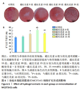
2.8 水凝胶浸提液对MC3T3-E1细胞矿化形成的影响 茜素红染色显示5组均可见矿化结节形成,对照组与藏红花素0组矿化结节形成面积最小,藏红花素20组矿化结节形成面积最大,见图6。"

| [1] ZHANG M, HU W, CAI C, et al. Advanced application of stimuli-responsive drug delivery system for inflammatory arthritis treatment. Mater Today Bio. 2022;14:100223. [2] KRYCH AJ, SARIS DBF, STUART MJ, et al. Cartilage Injury in the Knee: Assessment and Treatment Options. J Am Acad Orthop Surg. 2020;28(22): 914-922. [3] CHEN Y, WANG X, TAO S, et al. Research advances in smart responsive-hydrogel dressings with potential clinical diabetic wound healing properties. Mil Med Res. 2023;10(1):37. [4] LEI Y, WANG Y, SHEN J, et al. Injectable hydrogel microspheres with self-renewable hydration layers alleviate osteoarthritis. Sci Adv. 2022;8(5): eabl6449. [5] ZHOU C, WANG C, XU K, et al. Hydrogel platform with tunable stiffness based on magnetic nanoparticles cross-linked GelMA for cartilage regeneration and its intrinsic biomechanism. Bioact Mater. 2022;25:615-628. [6] CARDONEANU A, MACOVEI LA, BURLUI AM, et al. Temporomandibular Joint Osteoarthritis: Pathogenic Mechanisms Involving the Cartilage and Subchondral Bone, and Potential Therapeutic Strategies for Joint Regeneration. Int J Mol Sci. 2022;24(1):171. [7] DING SL, ZHAO XY, XIONG W, et al. Cartilage Lacuna-Inspired Microcarriers Drive Hyaline Neocartilage Regeneration. Adv Mater. 2023;35(30):e2212114. [8] UZIELIENE I, BIRONAITE D, PACHALEVA J, et al. Chondroitin Sulfate-Tyramine-Based Hydrogels for Cartilage Tissue Repair. Int J Mol Sci. 2023;24(4):3451. [9] LEI J, YAN S, ZHOU Y, et al. Abnormal expression of chondroitin sulfate sulfotransferases in the articular cartilage of pediatric patients with Kashin-Beck disease. Histochem Cell Biol. 2020;153(3):153-164. [10] KILMER CE, WALIMBE T, PANITCH A, et al. Incorporation of a Collagen-Binding Chondroitin Sulfate Molecule to a Collagen Type I and II Blend Hydrogel for Cartilage Tissue Engineering. ACS Biomater Sci Eng. 2022;8(3): 1247-1257. [11] AO Y, TANG W, TAN H, et al. Hydrogel composed of type II collagen, chondroitin sulfate and hyaluronic acid for cartilage tissue engineering. Biomed Mater Eng. 2022;33(6):515-523. [12] HEYDARI M, ZARE M, BADIE MR, et al. Crocin as a vision supplement. Clin Exp Optom. 2023;106(3):249-256. [13] BAKSHI HA, QUINN GA, NASEF MM, et al. Crocin Inhibits Angiogenesis and Metastasis in Colon Cancer via TNF-alpha/NF-kB/VEGF Pathways. Cells. 2022;11(9):1502. [14] TAO W, RUAN J, WU R, et al. A natural carotenoid crocin exerts antidepressant action by promoting adult hippocampal neurogenesis through Wnt/beta-catenin signaling. J Adv Res. 2023;43:219-231. [15] HOSSEINI SS, NAZIFI P, AMINI M, et al. Crocin Suppresses Colorectal Cancer Cell Proliferation by Regulating miR-143/145 and KRAS/RREB1 Pathways. Anticancer Agents Med Chem. 2023;23(17):1916-1923. [16] 邓新超,钱亮,邹曼.藏红花素调节Hippo-YAP信号通路抑制膝骨关节炎大鼠软骨细胞凋亡[J].中国骨质疏松杂志,2023,29(4):538-543,598. [17] POURSAMIMI J, SHARIATI-SARABI Z, TAVAKKOL-AFSHARI J, et al. A Significant Increase in the Gene Expression of GATA-3 Following the Treatment of Osteoarthritis Patients with Crocin. Iran J Allergy Asthma Immunol. 2022;21(1):35-43. [18] VAFAEI S, WU X, TU J, et al. The Effects of Crocin on Bone and Cartilage Diseases.Front Pharmacol. 2022;12:830331. [19] REKABI A, RAM A, NAZARI A, et al. Does crocin create new hope for the treatment of oral problems? A focus on periodontitis. Mol Biol Rep. 2024; 51(1):224. [20] AMIRBEKYAN KY, SHAHINYAN GA, GHAZOYAN HH, et al. Fluorescence anisotropy studies on the Hoechst 33258-DNA interaction: the solvent effect. J Biomol Struct Dyn. 2021;39(13):4902-4906. [21] BOOS MA, GRINSTAFF MW, LAMANDÉ SR, et al. Contrast-Enhanced Micro-Computed Tomography for 3D Visualization and Quantification of Glycosaminoglycans in Different Cartilage Types .Cartilage. 2021;13(2_suppl): 486S-494S. [22] YAMASHITA A, TSUMAKI N. Recent progress of animal transplantation studies for treating articular cartilage damage using pluripotent stem cells. Dev Growth Differ. 2021;63(1):72-81. [23] ZHANG FX, LIU P, DING W, et al. Injectable Mussel-Inspired highly adhesive hydrogel with exosomes for endogenous cell recruitment and cartilage defect regeneration. Biomaterials. 2021;278:121169. [24] LI Q, YU H, ZHAO F, et al. 3D Printing of Microenvironment-Specific Bioinspired and Exosome-Reinforced Hydrogel Scaffolds for Efficient Cartilage and Subchondral Bone Regeneration. Adv Sci (Weinh). 2023; 10(26):e2303650. [25] COMAS B, RIZZA LS, RUSECKAITE RA, et al. Schiff base crosslinked gelatin-Spirulina platensis protein concentrate films with enhanced antioxidant activity. J Food Sci. 2023;88(3):1075-1088. [26] QIN Z, HUANG Y, XIAO S, et al. Preparation and Characterization of High Mechanical Strength Chitosan/Oxidized Tannic Acid Composite Film with Schiff Base and Hydrogen Bond Crosslinking.Int J Mol Sci. 2022;23(16):9284. [27] LEI Y, PENG J, DAI Z, et al. Articular Cartilage Fragmentation Improves Chondrocyte Migration by Upregulating Membrane Type 1 Matrix Metalloprotease. Cartilage. 2021;13(2_suppl):1054S-1063S. [28] SONG H, PARK KH. Regulation and function of SOX9 during cartilage development and regeneration. Semin Cancer Biol. 2020;67(Pt 1):12-23. [29] HASEEB A, KC R, ANGELOZZI M, et al. SOX9 keeps growth plates and articular cartilage healthy by inhibiting chondrocyte dedifferentiation/osteoblastic redifferentiation. Proc Natl Acad Sci U S A. 2021;118(8):e2019152118. [30] BAY-JENSEN AC, MOBASHERI A, THUDIUM CS, et al. Blood and urine biomarkers in osteoarthritis - an update on cartilage associated type II collagen and aggrecan markers. Curr Opin Rheumatol. 2022;34(1):54-60. [31] HAN G, BOZ U, ERITEN M, et al. Glycosaminoglycan depletion increases energy dissipation in articular cartilage under high-frequency loading. J Mech Behav Biomed Mater. 2020;110:103876. [32] HU G, YU Y, SHARMA D, et al. Glutathione limits RUNX2 oxidation and degradation to regulate bone formation. JCI Insight. 2023;8(16):e166888. [33] SAHIN E, ORHAN C, BALCI TA, et al. Magnesium Picolinate Improves Bone Formation by Regulation of RANK/RANKL/OPG and BMP-2/Runx2 Signaling Pathways in High-Fat Fed Rats. Nutrients. 2021;13(10):3353. [34] NALESSO G, SHERWOOD J, BERTRAND J, et al. WNT-3A modulates articular chondrocyte phenotype by activating both canonical and noncanonical pathways. J Cell Biol. 2011;193(3):551-564. [35] YAO Q, WU X, TAO C, et al. Osteoarthritis: pathogenic signaling pathways and therapeutic targets. Signal Transduct Target Ther. 2023;8(1):56. [36] HOUSMANS BAC, VAN DEN AKKER GGH, NEEFJES M, et al. Direct comparison of non-osteoarthritic and osteoarthritic synovial fluid-induced intracellular chondrocyte signaling and phenotype changes. Osteoarthritis Cartilage. 2023;31(1):60-71. |
| [1] | Liu Yang, Liu Donghui , Xu Lei, Zhan Xu, Sun Haobo, Kang Kai. Role and trend of stimuli-responsive injectable hydrogels in precise myocardial infarction therapy [J]. Chinese Journal of Tissue Engineering Research, 2026, 30(8): 2072-2080. |
| [2] | Wang Zheng, Cheng Ji, Yu Jinlong, Liu Wenhong, Wang Zhaohong, Zhou Luxing. Progress and future perspectives on the application of hydrogel materials in stroke therapy [J]. Chinese Journal of Tissue Engineering Research, 2026, 30(8): 2081-2090. |
| [3] | Guo Yuchao, Ni Qianwei, Yin Chen, Jigeer·Saiyilihan, Gao Zhan . Quaternized chitosan hemostatic materials: synthesis, mechanism, and application [J]. Chinese Journal of Tissue Engineering Research, 2026, 30(8): 2091-2100. |
| [4] | Sun Lei, Zhang Qi, Zhang Yu. Pro-osteoblastic effect of chlorogenic acid protein microsphere/polycaprolactone electrospinning membrane [J]. Chinese Journal of Tissue Engineering Research, 2026, 30(8): 1877-1884. |
| [5] | Wang Qisa, Lu Yuzheng, Han Xiufeng, Zhao Wenling, Shi Haitao, Xu Zhe. Cytocompatibility of 3D printed methyl acrylated hyaluronic acid/decellularized skin hydrogel scaffolds [J]. Chinese Journal of Tissue Engineering Research, 2026, 30(8): 1912-1920. |
| [6] | Liu Hongjie, Mu Qiuju, Shen Yuxue, Liang Fei, Zhu Lili. Metal organic framework/carboxymethyl chitosan-oxidized sodium alginate/platelet-rich plasma hydrogel promotes healing of diabetic infected wounds [J]. Chinese Journal of Tissue Engineering Research, 2026, 30(8): 1929-1939. |
| [7] | Li Hao, Tao Hongcheng, Zeng Ping, Liu Jinfu, Ding Qiang, Niu Chicheng, Huang Kai, Kang Hongyu. Mitogen-activated protein kinase signaling pathway regulates the development of osteoarthritis: guiding targeted therapy with traditional Chinese medicine [J]. Chinese Journal of Tissue Engineering Research, 2026, 30(6): 1476-1485. |
| [8] | Liu Xinyue, Li Chunnian, Li Yizhuo, Xu Shifang. Regeneration and repair of oral alveolar bone defects [J]. Chinese Journal of Tissue Engineering Research, 2026, 30(5): 1247-1259. |
| [9] | Huang Liuyan, Zhang Wenxi, Chen Shuwen, Yu Shimei, Dai Zhong, Zuo Changqing. Forskolin promotes C2C12 myoblast differentiation via regulating the ERK and Akt signaling pathways [J]. Chinese Journal of Tissue Engineering Research, 2026, 30(5): 1114-1121. |
| [10] | Yang Xiao, Bai Yuehui, Zhao Tiantian, Wang Donghao, Zhao Chen, Yuan Shuo. Cartilage degeneration in temporomandibular joint osteoarthritis: mechanisms and regenerative challenges [J]. Chinese Journal of Tissue Engineering Research, 2026, 30(4): 926-935. |
| [11] | Huang Xinxu, Zhang Xin, Wang Jian. Living microecological hydrogels promote skin wound healing [J]. Chinese Journal of Tissue Engineering Research, 2026, 30(2): 489-498. |
| [12] | Liu Xiaohong, Zhao Tian, Mu Yunping, Feng Wenjin, Lyu Cunsheng, Zhang Zhiyong, Zhao Zijian, Li Fanghong. Acellular dermal matrix hydrogel promotes skin wound healing in rats [J]. Chinese Journal of Tissue Engineering Research, 2026, 30(2): 395-403. |
| [13] | Li Congcong, Wufanbieke·Baheti, Zhao Li, Chen Xiaotao, Kong Chuifan, Yu Min. Physicochemical properties and biocompatibility of hydroxyapatite/graphene oxide/interleukin-4 composite coating materials [J]. Chinese Journal of Tissue Engineering Research, 2026, 30(2): 404-413. |
| [14] | Wang Yu, Fan Minjie, Zheng Pengfei. Application of multistimuli-responsive hydrogels in bone damage repair: special responsiveness and diverse functions [J]. Chinese Journal of Tissue Engineering Research, 2026, 30(2): 469-479. |
| [15] | Ma Wenjing, Zhang Jinyu, Jiang Mingxia, Xiu Bingshui, Bai Rui, Liu Yuhan, Chen Xuyi, Yuan Zengqiang, Liu Zhiqiang. Scaffold-free three-dimensional human umbilical cord mesenchymal stem cell secretome repairs mouse skin injury [J]. Chinese Journal of Tissue Engineering Research, 2026, 30(1): 68-77. |
| Viewed | ||||||
|
Full text |
|
|||||
|
Abstract |
|
|||||