Chinese Journal of Tissue Engineering Research ›› 2025, Vol. 29 ›› Issue (25): 5382-5389.doi: 10.12307/2025.529
Previous Articles Next Articles
Protective effect of paeoniflorin on angiotensin II-induced fibrosis in cardiac fibroblasts
Ji Yaqiong1, 2, Ning Zhongping1, 2
- 1Shanghai University of Traditional Chinese Medicine, Shanghai 201203, China; 2Department of Cardiology, Zhoupu Hospital Affiliated to Shanghai University of Medicine & Health Science, Shanghai 201318, China
-
Received:2024-04-20Accepted:2024-07-08Online:2025-09-08Published:2024-12-25 -
Contact:Ning Zhongping, Master, Chief physician, Shanghai University of Traditional Chinese Medicine, Shanghai 201203, China; Department of Cardiology, Zhoupu Hospital Affiliated to Shanghai University of Medicine & Health Science, Shanghai 201318, China -
About author:Ji Yaqiong, Master candidate, Shanghai University of Traditional Chinese Medicine, Shanghai 201203, China; Department of Cardiology, Zhoupu Hospital Affiliated to Shanghai University of Medicine & Health Science, Shanghai 201318, China -
Supported by:Key Discipline Group Construction Project of Pudong New Area Health and Health Commission, No. PWZxq2022-11 (to NZP); Epidemiological Investigation of Atrial Fibrillation in Pudong New Area and Prospective Cohort Study on the Whole Process Management of Atrial Fibrillation under the Mode of Graded Diagnosis and Treatment, No. PKJ2021-Y33 (to NZP); Pudong New Area Health Committee Peak Discipline Construction, No. PWYgf2021-04 (to NZP)
CLC Number:
Cite this article
Ji Yaqiong, Ning Zhongping. Protective effect of paeoniflorin on angiotensin II-induced fibrosis in cardiac fibroblasts[J]. Chinese Journal of Tissue Engineering Research, 2025, 29(25): 5382-5389.
share this article
Add to citation manager EndNote|Reference Manager|ProCite|BibTeX|RefWorks
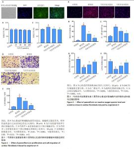
2.1 芍药苷对血管紧张素Ⅱ诱导的心肌成纤维细胞增殖、迁移的影响 2.1.1 SD大鼠心肌成纤维细胞的鉴定 采用细胞免疫荧光染色法观察SD大鼠心肌成纤维细胞的特异性波形蛋白的表达,在倒置显微镜下可观察到心肌成纤维细胞的细胞核被DAPI染成蓝色,而心肌成纤维细胞的特异性波形蛋白则被染成绿色(图1A)。 2.1.2 芍药苷对血管紧张素Ⅱ诱导的心肌成纤维细胞增殖的影响 为了评价不同剂量的芍药苷对心肌成纤维细胞增殖的影响,选用 0,10,30,50,100 μmol/L芍药苷作用于心肌成纤维细胞48 h,采用CCK-8检测细胞活力,结果显示,与对照组相比,不同剂量芍药苷对心肌成纤维细胞的活性无显著影响(图1B)。此外,分别用50,100 μmol/L芍药苷处理心肌成纤维细胞2 h,再用1 μmol/L 血管紧张素Ⅱ刺激48 h,采用CCK-8检测细胞活力,结果显示,与对照组相比,模型组细胞活力增强;与模型组相比,芍药苷呈剂量依赖性降低心肌成纤维细胞活性(图1C)。 2.1.3 芍药苷对血管紧张素Ⅱ诱导的心肌成纤维细胞迁移的影响 将心肌成纤维细胞用芍药苷(50,100 μmol/L)预处理2 h,再用1 μmol/L血管紧张素Ⅱ处理48 h,采用Transwell小室检测心肌成纤维细胞的迁移能力,结果显示,与模型组相比,芍药苷(50,100 μmol/L)呈剂量依赖性抑制血管紧张素Ⅱ诱导的心肌成纤维细胞迁移能力的增加(图1D,E)。 2.2 芍药苷对血管紧张素Ⅱ诱导的心肌成纤维细胞活性氧生成和氧化应激的影响 2.2.1 芍药苷对血管紧张素Ⅱ诱导的心肌成纤维细胞活性氧生成的影响 DHE染色检测各组心肌成纤维细胞内活性氧水平。与对照组相比,模型组活性氧的生成明显增强(P < 0.001);而与模型组相比,芍药苷(50,100 μmol/L)呈剂量依赖性抑制血管紧张素Ⅱ诱导的心肌成纤维细胞活性氧生成的增加(P < 0.001)(图2A,B)。 2.2.2 芍药苷对血管紧张素Ⅱ诱导的心肌成纤维细胞氧化应激的影响 丙二醛、超氧化物歧化酶和过氧化氢酶等属于心肌组织氧化应激标志物,代表心肌组织氧化应激能力[32]。与对照组相比,模型组心肌成纤维细胞裂解液中丙二醛水平显著升高(P < 0.001),超氧化物歧化酶和过氧化氢酶活性显著降低(P < 0.001)。而与模型组相比,芍药苷呈剂量依赖性抑制丙二醛水平的增加(P < 0.001),提高超氧化物歧化酶和过氧化氢酶的活性(P < 0.01或P < 0.001)(图2C-E)。"

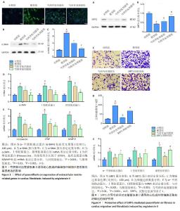
2.3 芍药苷对血管紧张素Ⅱ诱导的心肌成纤维细胞中细胞外基质相关基因表达的影响 2.3.1 纤维化相关基因α-平滑肌肌动蛋白的表达 免疫荧光、Western blot以及RT-qPCR检测α-平滑肌肌动蛋白的表达。与对照组相比,模型组α-平滑肌肌动蛋白荧光强度显著增强、mRNA以及蛋白表达水平均升高(P < 0.001),而与模型组相比,芍药苷组(50,100 μmol/L) α-平滑肌肌动蛋白荧光强度显著减弱、mRNA和蛋白表达显著降低(P < 0.001),具有剂量依赖性(图3A-D)。 2.3.2 其他细胞外基质相关基因的表达 与对照组相比,模型组Ⅰ型胶原蛋白、Ⅲ型胶原蛋白、纤维连接蛋白、结缔组织生长因子、基质金属蛋白酶9 mRNA表达显著升高(P < 0.001);与模型组相比,芍药苷组(50,100 μmol/L)上述基因表达显著降低(P < 0.01或P < 0.001),具有剂量依赖性(图3D,E)。 2.4 SIRT1介导芍药苷对血管紧张素Ⅱ诱导的心肌成纤维细胞迁移和纤维化的保护作用 2.4.1 芍药苷对心肌成纤维细胞SIRT1表达的影响 Western blot检测结果显示,与对照组相比,血管紧张素Ⅱ降低了SIRT1的表达,而芍药苷处理显著增加心肌成纤维细胞SIRT1的表达(图4A,B)。 2.4.2 Transwell小室检测心肌成纤维细胞的迁移情况 与对照组相比,血管紧张素Ⅱ显著提高了心肌成纤维细胞的迁移能力,而芍药苷抑制了心肌成纤维细胞的迁移能力。与芍药苷高剂量组相比,SIRT1抑制剂预处理增强了心肌成纤维细胞的迁移能力(图4C,D)。 2.4.3 细胞外基质相关基因α-平滑肌肌动蛋白、Ⅰ型胶原蛋白和Ⅲ型胶原蛋白的表达 与对照组相比,模型组α-平滑肌肌动蛋白、Ⅰ型胶原蛋白和Ⅲ型胶原蛋白的mRNA 表达升高;与模型组相比,芍药苷显著抑制α-平滑肌肌动蛋白、Ⅰ型胶原蛋白和Ⅲ型胶原蛋白的 mRNA表达,而EX527(SIRT1抑制剂)组上述基因表达显著上调(图4E)。"

| [1] 谢君,符德玉,芦波,等.基于circRNA探讨中医药在心肌纤维化中的研究进展[J].中华中医药学刊,2022,40(3):134-138. [2] ZHAO X, KWAN JYY, YIP K, et al. Targeting metabolic dysregulation for fibrosis therapy. Nat Rev Drug Discov. 2020;19(1):57-75. [3] GAIDAI O, CAO Y, LOGINOV S. Global Cardiovascular Diseases Death Rate Prediction. Curr Probl Cardiol. 2023;48(5):101622. [4] 姜伊雯,王耀晟.成纤维细胞骨架蛋白变化与心肌纤维化关联机制研究进展[J].上海医学,2023,46(12):854-858. [5] LÓPEZ B, RAVASSA S, MORENO MU, et al. Diffuse myocardial fibrosis: mechanisms, diagnosis and therapeutic approaches. Nat Rev Cardiol. 2021;18(7):479-498. [6] LIU M, LÓPEZ DE JUAN ABAD B, CHENG K. Cardiac fibrosis: Myofibroblast-mediated pathological regulation and drug delivery strategies. Adv Drug Deliv Rev. 2021;173:504-519. [7] NAKANO T, KISHIMOTO H, TOKUMOTO M. Direct and indirect effects of fibroblast growth factor 23 on the heart. Front Endocrinol (Lausanne). 2023;14:1059179. [8] BHULLAR SK, DHALLA NS. Angiotensin II-Induced Signal Transduction Mechanisms for Cardiac Hypertrophy. Cells. 2022;11(21):3336. [9] LI CY, ZHANG JR, HU WN, et al. Atrial fibrosis underlying atrial fibrillation (Review). Int J Mol Med. 2021;47(3):9. [10] XIAO Z, REDDY DPK, XUE C, et al. Profiling of miR-205/P4HA3 Following Angiotensin II-Induced Atrial Fibrosis: Implications for Atrial Fibrillation. Front Cardiovasc Med. 2021;8:609300. [11] FRANGOGIANNIS NG. Cardiac fibrosis. Cardiovasc Res. 2021;117(6): 1450-1488. [12] 张素华,栗志英,蔡安盛,等.双氢青蒿素对血管紧张素Ⅱ诱导的心肌成纤维细胞分泌胶原及激活PI3K/AKT通路的影响[J].中国实验诊断学,2024,28(4):483-485. [13] 闫保娥,朱敏杰,方敏,等.姜黄素在心肌纤维化中的作用及机制研究进展[J].心脏杂志,2024,36(4):446-451. [14] 尹茂山,牟艳玲. Sirt1与心肌保护[J].生命科学,2015,27(5):601-608. [15] 闫景顺,朱林平,张红霞,等.中医药调控心肌纤维化相关信号通路研究进展[J].中国实验方剂学杂志,2024,30(13):230-239. [16] 俞瀛.心房颤动住院患者的临床特征与中医证候要素规律分析[D].北京:北京中医药大学,2022. [17] 黄文娟,熊丽辉.张锡纯治疗心悸辨证用药特色探析[J].吉林中医药,2012,32(1):83-84. [18] 崔一然,唐仕欢,刘欣,等.基于数据挖掘的心悸伴失眠方证对应中成药用药规律分析[J].中华中医药杂志,2015,30(5):1792-1799. [19] 侯明桥,张春媛,邱玲.参松养心胶囊联合美托洛尔治疗老年冠心病心律失常患者的效果观察[J].疑难病杂志,2015,14(5):508-510. [20] 张璨,林鹏,杨玉,等.芍药苷抗肿瘤作用机制的研究进展[J].癌变·畸变·突变,2024,36(2):164-167. [21] REN S, WANG Y, ZHANG Y, et al. Paeoniflorin alleviates AngII-induced cardiac hypertrophy in H9c2 cells by regulating oxidative stress and Nrf2 signaling pathway. Biomed Pharmacother. 2023;165:115253. [22] LIU Y, HE CY, YANG XM, et al. Paeoniflorin Coordinates Macrophage Polarization and Mitigates Liver Inflammation and Fibrogenesis by Targeting the NF-[Formula: see text]B/HIF-1α Pathway in CCl4-Induced Liver Fibrosis. Am J Chin Med. 2023;51(5):1249-1267. [23] 王东轶.芍药苷脂质体调节巨噬细胞极化对类风湿关节炎滑膜炎症的作用和机制研究[D].南京:南京中医药大学,2022. [24] MA Y, LANG X, YANG Q, et al. Paeoniflorin promotes intestinal stem cell-mediated epithelial regeneration and repair via PI3K-AKT-mTOR signalling in ulcerative colitis. Int Immunopharmacol. 2023;119:110247. [25] WU J, ZHANG D, HU L, et al. Paeoniflorin alleviates NG-nitro-L-arginine methyl ester (L-NAME)-induced gestational hypertension and upregulates silent information regulator 2 related enzyme 1 (SIRT1) to reduce H2O2-induced endothelial cell damage. Bioengineered. 2022;13(2):2248-2258. [26] ZENG K, XI W, QIAO Y, et al. Paeoniflorin inhibits epithelial mesenchymal transformation and oxidative damage of lens epithelial cells in diabetic cataract via sirtuin 1 upregulation. Bioengineered. 2022;13(3): 5903-5914. [27] 何婷,陈军,庞牧,等.TGF-β1/Smads信号通路在SIRT1抗心肌纤维化中的作用及机制研究[J].华中科技大学学报(医学版),2024, 53(1):45-51. [28] WANG AJ, TANG Y, ZHANG J, et al. Cardiac SIRT1 ameliorates doxorubicin-induced cardiotoxicity by targeting sestrin 2. Redox Biol. 2022;52:102310. [29] 孙韬,宋爽,陈磊,等.芍药苷对TGF-β1诱导的HK-2细胞纤维化及PI3K/AKT通路的影响[J].河南医学高等专科学校学报,2023, 35(6):611-616. [30] 王甜甜,王魏,杨翠华,等.大黄酸对高糖诱导的H9c2心肌细胞损伤的保护作用及机制[J].中西医结合心脑血管病杂志,2024, 22(3):459-463. [31] PFAFFL MW. A new mathematical model for relative quantification in real-time RT-PCR. Nucleic Acids Res. 2001;29(9):e45. [32] 尚茹茹,童曼琳,刘晓红.高良姜素减轻异丙肾上腺素诱导心脏纤维化的作用机制[J].中西医结合心脑血管病杂志,2023,21(24): 4524-4529. [33] REN C, ZHAO X, LIU K, et al. Research progress of natural medicine Astragalus mongholicus Bunge in treatment of myocardial fibrosis. J Ethnopharmacol. 2023;305:116128. [34] 赵世杰,王文荣,王婧一,等.芍药苷对单侧输尿管梗阻大鼠肾间质纤维化的作用[J].中国中西医结合肾病杂志,2018,19(11):985-987. [35] 江舜祥,涂彬,宋凯,等.过表达sFRP3对小鼠原代心肌成纤维细胞活化增殖的影响[J].安徽医科大学学报,2024,59(5):809-814. [36] 帅壮,唐锴,刘茂,等.加入芍药苷的乳鼠心脏成纤维细胞增殖、凋亡、纤维化及TGF-β1 mRNA和Smad3 mRNA表达观察[J].山东医药, 2020,60(18):40-43. [37] VALIENTE-ALANDI I, SCHAFER AE, BLAXALL BC. Extracellular matrix-mediated cellular communication in the heart. J Mol Cell Cardiol. 2016;91:228-237. [38] ZHANG Y, YANG X, HAN C, et al. Paeoniflorin-6’O-benzene sulfonate suppresses fibroblast-like synoviocytes proliferation and migration in rheumatoid arthritis through regulating GRK2-Gβγ interaction. Exp Ther Med. 2022;24(2):523. [39] KHOSRAVI M, POURSALEH A, GHASEMPOUR G, et al. The effects of oxidative stress on the development of atherosclerosis. Biol Chem. 2019;400(6):711-732. [40] NEUMAN RB, BLOOM HL, SHUKRULLAH I, et al. Oxidative stress markers are associated with persistent atrial fibrillation. Clin Chem. 2007;53(9): 1652-1657. [41] AN D, ZENG Q, ZHANG P, et al. Alpha-ketoglutarate ameliorates pressure overload-induced chronic cardiac dysfunction in mice. Redox Biol. 2021;46:102088. [42] 王健康,王彬,郭家娟.基于氧化应激探究中医药防治心肌纤维化的研究进展[J].中西医结合心脑血管病杂志,2024,22(7): 1256-1261. [43] JANUARY CT, WANN LS, ALPERT JS, et al. 2014 AHA/ACC/HRS guideline for the management of patients with atrial fibrillation: a report of the American College of Cardiology/American Heart Association Task Force on practice guidelines and the Heart Rhythm Society. Circulation. 2014;130(23):e199-267. [44] HAN F, ZHOU D, YIN X, et al. Paeoniflorin protects diabetic mice against myocardial ischemic injury via the transient receptor potential vanilloid 1/calcitonin gene-related peptide pathway. Cell Biosci. 2016;6:37. [45] 李雪,赵雅静,张芹,等.心肌纤维化调控机制的研究进展[J].中国医药,2024,19(2):285-288. [46] 王海强,黄耀添,赵黎,等. 张应力对深筋膜Ⅰ、Ⅲ型胶原影响的实验研究[J].中国临床康复,2002,6(2):208-209. [47] 赵倩茹,曹梦菲,孙侠,等.Yes相关蛋白在血管紧张素Ⅱ诱导的心肌纤维化中的作用及机制研究[J].中华老年心脑血管病杂志, 2022,24(3):306-310. [48] 刘旺,周琴怡,莫志勇,等.SIRT1调控氧化应激的研究进展[J].中南医学科学杂志,2021,49(2):239-243. |
| [1] | Lai Pengyu, Liang Ran, Shen Shan. Tissue engineering technology for repairing temporomandibular joint: problems and challenges [J]. Chinese Journal of Tissue Engineering Research, 2025, 29(在线): 1-9. |
| [2] | Zhao Jiyu, Wang Shaowei. Forkhead box transcription factor O1 signaling pathway in bone metabolism [J]. Chinese Journal of Tissue Engineering Research, 2025, 29(9): 1923-1930. |
| [3] | Li Kaiying, Wei Xiaoge, Song Fei, Yang Nan, Zhao Zhenning, Wang Yan, Mu Jing, Ma Huisheng. Mechanism of Lijin manipulation regulating scar formation in skeletal muscle injury repair in rabbits [J]. Chinese Journal of Tissue Engineering Research, 2025, 29(8): 1600-1608. |
| [4] | Yu Jingbang, Wu Yayun. Regulatory effect of non-coding RNA in pulmonary fibrosis [J]. Chinese Journal of Tissue Engineering Research, 2025, 29(8): 1659-1666. |
| [5] | He Guanghui, Yuan Jie, Ke Yanqin, Qiu Xiaoting, Zhang Xiaoling. Hemin regulates mitochondrial pathway of oxidative stress in mouse chondrocytes [J]. Chinese Journal of Tissue Engineering Research, 2025, 29(6): 1183-1191. |
| [6] | He Bo, Chen Wen, Ma Suilu, He Zhijun, Song Yuan, Li Jinpeng, Liu Tao, Wei Xiaotao, Wang Weiwei, Xie Jing . Pathogenesis and treatment progress of flap ischemia-reperfusion injury [J]. Chinese Journal of Tissue Engineering Research, 2025, 29(6): 1230-1238. |
| [7] | Lu Ranran, Zhou Xu, Zhang Lijie, Yang Xinling. Dimethyl fumarate alleviates nerve damage in a mouse model of Parkinson’s disease [J]. Chinese Journal of Tissue Engineering Research, 2025, 29(5): 989-994. |
| [8] | Shui Jing, He Yu, Jiang Nan, Xu Kun, Song Lijuan, Ding Zhibin, Ma Cungen, Li Xinyi. Astrocytes regulate remyelination in central nervous system [J]. Chinese Journal of Tissue Engineering Research, 2025, 29(36): 7889-7897. |
| [9] | Sima Xinli, Liu Danping, Qi Hui. Effect and mechanism of metformin-modified bone marrow mesenchymal stem cell exosomes on regulating chondrocytes [J]. Chinese Journal of Tissue Engineering Research, 2025, 29(36): 7728-7734. |
| [10] | Zhang Xiaoyu, Wei Shanwen, Fang Jiawei, Ni Li. Prussian blue nanoparticles restore mitochondrial function in nucleus pulposus cells through antioxidation [J]. Chinese Journal of Tissue Engineering Research, 2025, 29(34): 7318-7325. |
| [11] | Pan Chun, Fan Zhencheng, Hong Runyang, Shi Yujie, Chen Hao. Effect and mechanism of polystyrene microplastics on prostate in male mice [J]. Chinese Journal of Tissue Engineering Research, 2025, 29(34): 7353-7361. |
| [12] | Su Yongkun, Sun Hong, Liu Miao, Yang Hua, Li Qingsong. Development of novel antioxidants and antioxidant combination carried by nano-hydrogel systems in treatment of intervertebral disc degeneration [J]. Chinese Journal of Tissue Engineering Research, 2025, 29(34): 7376-7384. |
| [13] | Wu Qingyun, Su Qiang. Antioxidant nanomedicine-mediated targeted therapy for myocardial ischemia-reperfusion injury [J]. Chinese Journal of Tissue Engineering Research, 2025, 29(34): 7431-7438. |
| [14] | Zhong Min, Wang Cheng, Fan Zhenhai, Li Linyan, Yu Limei. Effect and mechanism of perinatal mesenchymal stem cells and their combination with hydrogels in treatment of intrauterine adhesions [J]. Chinese Journal of Tissue Engineering Research, 2025, 29(31): 6792-6799. |
| [15] | Tian Yushi, Fu Qiang, Li Ji . Bioinformatics identification and validation of mitochondrial genes related to acute myocardial infarction [J]. Chinese Journal of Tissue Engineering Research, 2025, 29(31): 6697-6707. |
| Viewed | ||||||
|
Full text |
|
|||||
|
Abstract |
|
|||||