Chinese Journal of Tissue Engineering Research ›› 2024, Vol. 28 ›› Issue (27): 4265-4272.doi: 10.12307/2024.560
Safety risk assessment of in vitro heart in antitumor drug development
Zheng Shuangjia, Zhao Ting, Ren Cuixia, Wang Baoqiang, Chen Lanlan, Lin Moxu, Li Yingji, Zhang Xu
- Neurocardiology Center, Ice-Biosci, Beijing 100000, China
-
Received:2023-09-12Accepted:2023-11-08Online:2024-09-28Published:2024-01-26 -
Contact:Zhang Xu, MD, Neurocardiology Center, Ice-Biosci, Beijing 100000, China -
About author:Zheng Shuangjia, Master, Neurocardiology Center, Ice-Biosci, Beijing 100000, China -
Supported by:Beijing Municipal Science and Technology Commission Science and Technology Program, No. Z211100002521003 (to LYJ)
CLC Number:
Cite this article
Zheng Shuangjia, Zhao Ting, Ren Cuixia, Wang Baoqiang, Chen Lanlan, Lin Moxu, Li Yingji, Zhang Xu. Safety risk assessment of in vitro heart in antitumor drug development[J]. Chinese Journal of Tissue Engineering Research, 2024, 28(27): 4265-4272.
share this article
Add to citation manager EndNote|Reference Manager|ProCite|BibTeX|RefWorks
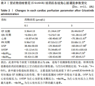
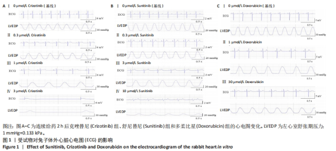
2.1 实验动物数量分析 31只兔均进入结果分析,无脱落。 2.2 不同抗肿瘤药物对兔体外心脏灌流的影响 表2和图1A结果显示,在同一心脏中与给药前相比,1 μmol/L和3 μmol/L的Crizotinib有明显降低心率(P < 0.05和P < 0.01),延长PR间期(P < 0.05),延长QT间期的作用(P < 0.05和P < 0.01);3 μmol/L的Crizotinib有明显缩短QTc间期(P < 0.01),降低左心室收缩压(P < 0.01),降低左心室内压最大上升速率和最大下降速率的作用(P < 0.01)。"
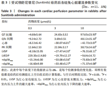
表3和图1B结果显示,在同一心脏中与给药前相比,3 μmol/L和10 μmol/L的Sunitinib明显降低心率(P < 0.01),延长QT间期(P < 0.01),降低左心室收缩压(P < 0.05和P < 0.01),降低左心室内压最大上升速率(P < 0.05和P < 0.01)和最大下降速率的作用(P < 0.01);10 μmol/L的Sunitinib有明显延长PR间期(P < 0.01)和延长QTc间期的作用(P < 0.01)。"
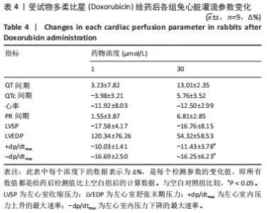
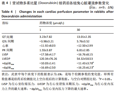
表4和图1C结果显示,在同一心脏中与给药前相比,30 μmol/L Doxorubicin的药效有QT间期延长和心率降低的趋势,但是没有显著性差异(P > 0.05),有明显降低左心室内压最大上升速率和最大下降速率的作用(P < 0.05)。"
"

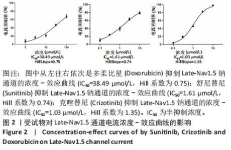
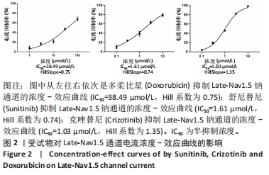
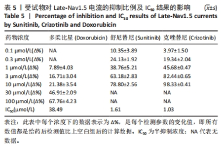
2.3 不同抗肿瘤药物对离子通道的影响 2.3.1 受试物对各通道的抑制比例及IC50结果 图2为受试物对Late-Nav1.5通道电流的浓度-效应曲线;表5结果显示Doxorubicin在100 μmol/L下抑制率为(67.76±4.23)%,IC50值为38.49 μmol/L。而Sunitinib和Crizotinib在3 μmol/L时抑制率超过50%,分别为(63.18±2.83)%、(82.44±0.65)%,两个化合物的IC50值分别为1.61 μmol/L和1.03 μmol/L。"
"
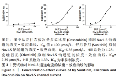
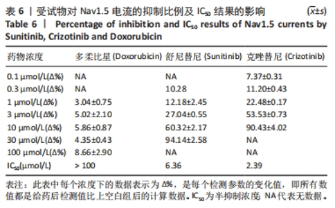
图3为受试物对Nav1.5通道电流的浓度-效应曲线;表6结果显示Doxorubicin在100 μmol/L下抑制率为(8.66±2.90)%,在测试浓度范围内IC50值为> 100 μmol/L;Sunitinib在10 μmol/L时抑制率超过50%,为(60.32±2.17)%,IC50值为6.36 μmol/L;Crizotinib在3 μmol/L时抑制率超过50%,为(53.53±0.73)%,IC50值为2.39 μmol/L。"

"
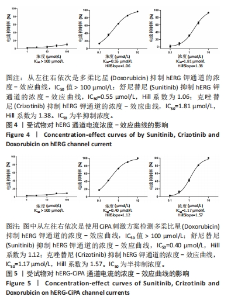
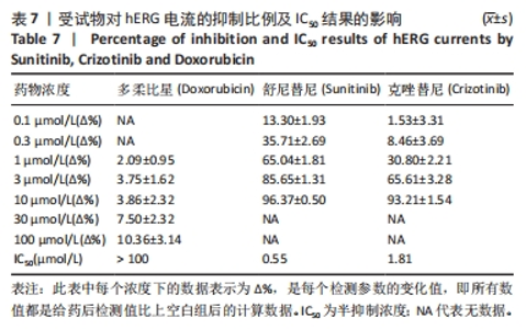
图4,5为受试物对hERG通道和hERG-CiPA通道电流的浓度-效应曲线。表7结果显示Doxorubicin在100 μmol/L下抑制率为(10.36± 3.14)%,在测试浓度范围内IC50值为> 100 μmol/L;Sunitinib在1 μmol/L时抑制率超过50%,为(65.04%±1.81)%,IC50值为0.55 μmol/L;Crizotinib在3 μmol/L时抑制率超过50%,为(65.61±3.28)%,IC50值为1.81 μmol/L。"
"
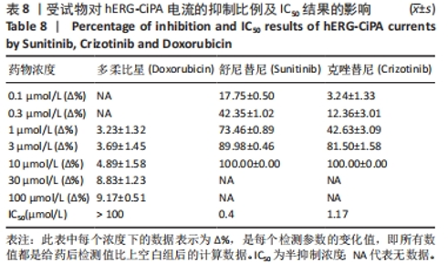
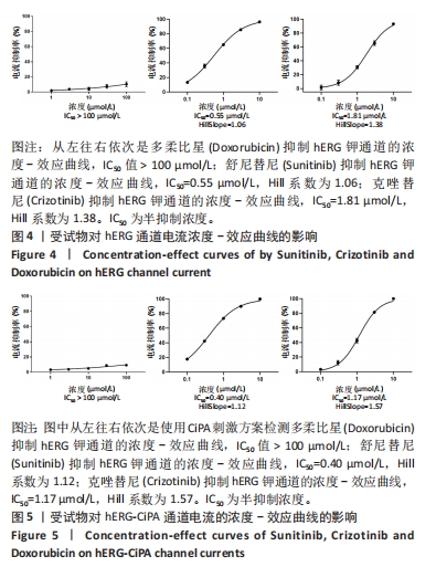
表8和图5的结果为hERG-CiPA结果,与hERG结果较一致。 结果显示:Doxorubicin对hERG通道具有弱抑制或无抑制作用;Sunitinib对hERG通道具有强抑制作用;Crizotinib对hERG通道具有中度抑制作用。实验采用文献报道的标准判断受试物对hERG的抑制作用[12] :极强抑制:IC50 < 0.1 μmol/L;强抑制:0.1 μmol/L ≤ IC50 ≤ 1 μmol/L;中度抑制:1 μmol/L < IC50 ≤ 10 μmol/L;弱抑制或无抑制:IC50 > 10 μmol/L。"
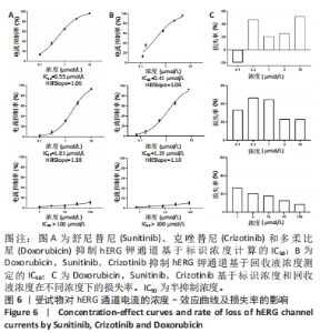
图6结果显示Sunitinib损失率范围为-20.00%-51.70%,Crizotinib损失率范围为23.00%-46.67%,Doxorubicin损失率范围为9.06%-26.67%。结合供试品结果对每种受试物经灌流系统的回收液和标识浓度进行对比,每种受试物的5个浓度点存在不同程度的损失。"
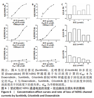

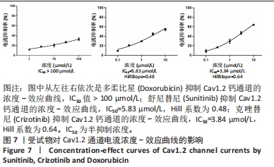
图7为受试物对Cav1.2通道电流的浓度-效应曲线。表9结果显示,Doxorubicin在100 μmol/L下抑制率为(32.74±3.07)%,在测试浓度范围内IC50值为> 100 μmol/L;Sunitinib在10 μmol/L时抑制率超过50%,IC50值为5.83 μmol/L;Crizotinib在10 μmol/L时抑制率超过50%,IC50值为3.84 μmol/L。 2.3.2 受试物对hiPSC-CMs动作电位的影响 表10结果显示,Doxorubicin在测试浓度范围内对hiPSC-CMs的动作电位时程APD90延长(10.85±1.70)%,对APD30和APD50无明显变化,同时,对hiPSC-CMs动作电位的幅度(APA)和静息电位(RMP)也无明显变化。"
"
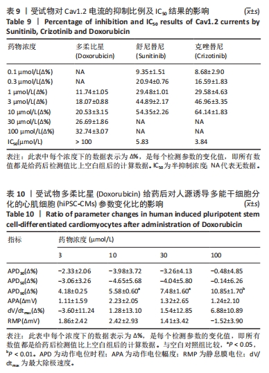
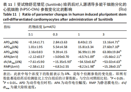
表11结果显示,Sunitinib一定程度延长了hiPSC-CMs动作电位时程,在1 μmol/L浓度下,对APD90延长(36.99±11.99)%,在3 μmol/L浓度下,对APD90延长(63.00±19.84)%,同时减小了hiPSC-CMs动作电位的幅度APA(-15.66±2.11)%和心肌细胞的静息电位RMP(-15.28±0.15)%,并减缓了细胞动作电位最大除极速度dV/dtmax (-53.02±4.63)%,差异均有显著性意义(P < 0.05),结合离子通道检测结果1 μmol/L和3 μmol/L浓度超过了APD90风险阈值(10%-15%)[13-16],有致心律失常的风险。"
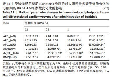
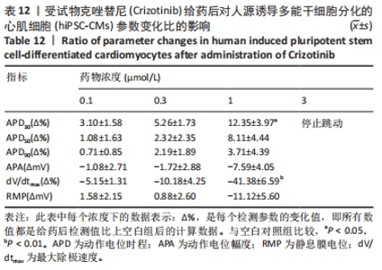
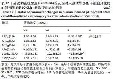
表12结果显示,Crizotinib在1 μmol/L浓度及以下,对hiPSC-CMs动作电位时程,幅度、静息电位没有太大变化,同时受试物在3 μmol/L时会导致细胞节律不稳定,导致细胞停止自发跳动。"
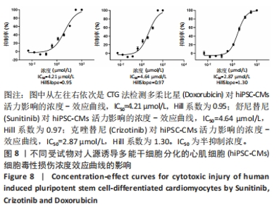
2.4 细胞毒性实验结果 图8结果显示,3种受试物在各测试浓度对hiPSC-CMs中ATP含量有较明显损失,具有剂量依赖性的心脏毒性[17],IC50在低微摩尔范围内。根据ATP产生的剂量反应数据,在iPSC-CMs细胞中Doxorubicin的IC50=4.21 μmol/L,Sunitinib的IC50=4.64 μmol/L,Crizotinib的IC50=2.87 μmol/L。"

| [1] 司丹妮,李彩娥,陈馨嵘,等.酪氨酸激酶抑制剂诱导的心脏毒性发病情况、评估和监测研究进展[J]. 山东医药,2022,62(33):97-102. [2] SELTZER JH, GINTANT G, AMIRI-KORDESTANI L, et al. Assessing cardiac safety in oncology drug development. Am Heart J. 2019;214:125. [3] SHYAM SUNDER S, SHARMA UC, POKHAREL S. Adverse effects of tyrosine kinase inhibitors in cancer therapy: pathophysiology, mechanisms and clinical management. Signal Transduct Target Ther. 2023;8(1):262 [4] WANG H, WANG Y, LI J, et al. Three tyrosine kinase inhibitors cause cardiotoxicity by inducing endoplasmic reticulum stress and inflammation in cardiomyocytes. BMC Med. 2023;21(1):147 [5] KADDOURA R, DABDOOB WA, AHMED K, et al. A practical guide to managing cardiopulmonary toxicities of tyrosine kinase inhibitors in chronic myeloid leukemia. Front Med (Lausanne). 2023;10:1163137. [6] YAMAMICHI Y, KUSUOKA H, MORISHITA K, et al. Metabolism of iodine-123-BMIPP in perfused rat hearts. J Nucl Med. 1995;36(6):1043-1050. [7] RUMSEY WL, PATEL B, LINDER KE. Effect of graded hypoxia on retention of technetium-99m-nitroheterocycle in perfused rat heart. J Nucl Med. 1995;36(4): 632-636. [8] KUBURAS R, GHARANEI M, HAUSSMANN I, et al. Metformin protects against sunitinib-induced cardiotoxicity: investigating the role of AMPK. J Cardiovasc Pharmacol. 2022;79(6):799-807. [9] OBEJERO-PAZ CA, BRUENING-WRIGHT A, KRAMER J, et al. Quantitative profiling of the effects of vanoxerine on human cardiac ion channels and its application to cardiac risk. Sci Rep. 2015;5:17623. [10] JONSSON MK, DUKER G, TROPP C, et al. Quantified proarrhythmic potential of selected human embryonic stem cell-derived cardiomyocytes. Stem Cell Res. 2010;4(3):189-200. [11] GILLIE DJ, NOVICK SJ, DONOVAN BT, et al. Development of a high-throughput electrophysiological assay for the human ether-à-go-go related potassium channel hERG. J Pharmacol Toxicol Methods. 2013;67(1):33-44. [12] COHEN JD, BABIARZ JE, ABRAMS RM, et al. Use of human stem cell derived cardiomyocytes to examine sunitinib mediated cardiotoxicity and electrophysiological alterations. Toxicol Appl Pharmacol. 2011;257(1):74-83. [13] WHITMIRE ML, BRYAN P, HENRY TR, et al. Nonclinical dose formulation analysis method validation and sample analysis. AAPS J. 2010;12:628-634. [14] ROCHE O, TRUBE G, ZUEGGE J, et al. A virtual screening method for prediction of the hERG potassium channel liability of compound libraries. Chembiochem. 2002;3(5):455-459. [15] GINTANT GA, LIMBERIS JT, MCDERMOTT JS, et al. The canine Purkinje fiber: an in vitro model system for acquired long QT syndrome and drug-induced arrhythmogenesis. J Cardiovasc Pharmacol. 2001;37(5):607-618. [16] TAKASUNA K, KATSUYOSHI C, MANABE S. Pre-clinical QT risk assessment in pharmaceutical companies-issues of current QT risk assessment. Biomol Ther. 2009;17(1):1-11. [17] SHARMA A, BURRIDGE PW, MCKEITHAN WL, et al. High-throughput screening of tyrosine kinase inhibitor cardiotoxicity with human induced pluripotent stem cells. Sci Transl Med. 2017;9(377):eaaf2584. [18] NAZEYROLLAS P, PREVOST A, BACCARD N, et al. Effects of amifostine on perfused isolated rat heart and on acute doxorubicin-induced cardiotoxicity. Cancer Chemother Pharmacol. 1999;43:227-232. [19] TOKARSKA-SCHLATTNER M, ZAUGG M, DA SILVA R, et al. Acute toxicity of doxorubicin on isolated perfused heart:response of kinases regulating energy supply. Am J Physiol Heart Circ Physiol. 2005;289:H37-H47. [20] WANG YX, KORTH M. Effects of doxorubicin on excitation-concentration coupling in guinea pig ventricular myocardium. Circ Res. 1995;76:645-653. [21] STEINBERG JS, COHEN AJ, WASSERMAN AG, et al. Acute arrhythmogenicity of doxorubicin administration. Cancer. 1987;60:1213-1218. [22] DUCROQ J, MOHA OU MAATI H, GUILBOT S, et al. Dexrazoxane protects the heart from acute doxorubicin-induced QT prolongation: a key role for IKs. Br J Pharmacol. 2010;159:93-101. [23] FAIVRE S, DELBALDO C, VERA K, et al. Safety, pharmacokinetic, and antitumor activity of SU11248, a novel oral multitarget tyrosine kinase inhibitor, in patients with cancer. J Clin Oncol. 2006;24(1):25-35. [24] KHOSRAVAN R, TOH M, LAFARGUE J, et al. Sunitinib pharmokinetic (PK) and safety data in subejcts with renal impairment and hemodialysis. 2008. [25] CHU TF, RUPNICK MA, KERKELA R, et al. Cardiotoxicity associated with tyrosine kinase inhibitor sunitinib. Lancet. 2007;370:2011-2019. [26] DI LORENZO G, AUTORINO R, BRUNI G, et al. Cardiovascular toxicity following sunitinib therapy in metastatic renal cell carcinoma: a multicenter analysis. Ann Oncol. 2009;20:1535-1542. [27] COHEN JD, BABIARZ JE, ABRAMS RM, et al. Use of human stem cell derived cardiomyocytes to examine sunitinib mediated cardiotoxicity and electrophysiological alterations. Toxicol Appl Pharmacol. 2011;257(1):74-83. [28] OBRIEN Z, F MOGHADDAM M. A systematic analysis of physicochemical and ADME properties of all small molecule kinase inhibitors approved by US FDA from January 2001 to October 2015. Curr Med Chem. 2017;24(29):3159-3184. [29] KURATA Y, MIYAUCHI N, SUNO M, et al. Correlation of plasma crizotinib trough concentration with adverse events in patients with anaplastic lymphoma kinase positive non-small-cell lung cancer. J Pharm Health Care Sci. 2015;1(1):1-5. [30] LESTER RM. Update on ICH E14/S7B cardiac safety regulations: the expanded role of preclinical assays and the “double-negative” scenario. Clin Pharmacol Drug Dev. 2021;10(9):964-973. [31] CURIGLIANO G, SPITALERI G, DE BRAUD F, et al. QTc prolongation assessment in anticancer drug development: clinical and methodological issues. Ecancermedicalscience. 2009;3:130. [32] PANG L, SAGER P, YANG X, et al. Workshop report: FDA workshop on improving cardiotoxicity assessment with human-relevant platforms. Circ Res. 2019;125(9):855-867. [33] SHARMA A, BURRIDGE PW, MCKEITHAN WL, et al. High-throughput screening of tyrosine kinase inhibitor cardiotoxicity with human induced pluripotent stem cells. Sci Transl Med. 2017;9(377):eaaf2584. [34] SELTZER JH, GINTANT G, AMIRI-KORDESTANI L, et al. Assessing cardiac safety in oncology drug development. Am Heart J. 2019;214:125-133. [35] XIAO L, SALEM JE, CLAUSS S, et al. Ibrutinib-mediated atrial fibrillation attributable to inhibition of C-Terminal Src kinase. Circulation. 2020;142(25):2443-2455. |
| [1] | Zhang Boya, Duan Hongmei, Bai Tianyu, Hao Fei, Hao Peng, Zhao Wen, Gao Yudan, Li Xiaoguang, Yang Zhaoyang. Neurotrophic factor 3-chitosan carrier induces neural stem cells to differentiate into neuronal subtypes and their electrophysiological properties [J]. Chinese Journal of Tissue Engineering Research, 2023, 27(25): 4020-4027. |
| [2] | Zhang Xin, Lu Ying, Yao Qingqiang, Zhu Yishen. Preparation and in vitro evaluation of self-assembling peptide hydrogel loaded with doxorubicin [J]. Chinese Journal of Tissue Engineering Research, 2021, 25(16): 2488-2493. |
| [3] | Su Liping, Liu Li, Lu Ziyang, Su Tianyuan, Hu Xiayun, Pu Hongwei. Diethylmorphine effect on action potential and calcium ion current of neonatal rat cardiomyocytes in vitro [J]. Chinese Journal of Tissue Engineering Research, 2020, 24(26): 4201-4207. |
| [4] | Zhang Wenjing, Wang Jia, Tian Mengting, Han Xiangzhen, Zhou Qiqi, He Huiyu. Effects of bone morphogenetic protein 2 and basic fibroblast growth factor 2 on proliferation and osteogenic differentiation of rat bone marrow mesenchymal stem cell sheet [J]. Chinese Journal of Tissue Engineering Research, 2020, 24(1): 65-71. |
| [5] | Li Yongheng, Cui Yan, Zhang Zhiyu. Inhibitory effects of doxorubicin-loaded chitosan nanoparticles on osteosarcoma in mice [J]. Chinese Journal of Tissue Engineering Research, 2019, 23(26): 4194-4199. |
| [6] |
Xing Li-na, Zhang Xue-jun, Wang Ying, Niu Zhi-yun, Wang Fu-xu, Wen Shu-peng.
Graphene oxide loaded with doxorubicin: a killer for multiple myeloma cells
[J]. Chinese Journal of Tissue Engineering Research, 2016, 20(38): 5636-5641.
|
| [7] | Yang Huan-dan, Zhang Rui-feng, Feng Dong-jin, Zhu Bing-bing, Lv Juan . Effects of bone marrow mesenchymal stem cell transplantation on CD4+CD25+ regulatory T cells in rats with primary nephrotic syndrome [J]. Chinese Journal of Tissue Engineering Research, 2014, 18(1): 33-38. |
| [8] | Wang Dan, Jiang Hang-hang. Characteristics and experimental application of polylactic acid copolymer composite sustained-release drug materials [J]. Chinese Journal of Tissue Engineering Research, 2013, 17(8): 1473-1480. |
| [9] | Wang Qiang, He Jin-shan, Xiong Chuan-zhi, Feng Xin-min, Wang Jing-cheng, Yan Lian-qi, Chen Peng-tao, Cai Jun. Effects of the L-type calcium channels on chondrocytes in response to basic fibroblast growth factor [J]. Chinese Journal of Tissue Engineering Research, 2013, 17(50): 8654-8659. |
| [10] | Qu Shao-zheng, Li Shu-zhong, Gao Jia-ke, Zhang Jin-feng. Isolation and identification of osteosarcoma stem cells [J]. Chinese Journal of Tissue Engineering Research, 2013, 17(14): 2641-2648. |
| Viewed | ||||||
|
Full text |
|
|||||
|
Abstract |
|
|||||