Chinese Journal of Tissue Engineering Research ›› 2022, Vol. 26 ›› Issue (22): 3480-3486.doi: 10.12307/2022.274
Previous Articles Next Articles
Theaflavin-3-gallate modified nano-hydroxyapatite/polycaprolactone composite porous scaffold in bone defect repair
Liu Ming, Wang Kai
- First Department of Orthopedics, Qinghai Provincial People’s Hospital, Xining 810000, Qinghai Province, China
-
Received:2020-12-29Revised:2021-01-26Accepted:2021-06-06Online:2022-08-08Published:2022-01-11 -
Contact:Wang Kai, Chief physician, First Department of Orthopedics, Qinghai Provincial People’s Hospital, Xining 810000, Qinghai Province, China -
About author:Liu Ming, Master, Associate chief physician, First Department of Orthopedics, Qinghai Provincial People’s Hospital, Xining 810000, Qinghai Province, China
CLC Number:
Cite this article
Liu Ming, Wang Kai. Theaflavin-3-gallate modified nano-hydroxyapatite/polycaprolactone composite porous scaffold in bone defect repair[J]. Chinese Journal of Tissue Engineering Research, 2022, 26(22): 3480-3486.
share this article
Add to citation manager EndNote|Reference Manager|ProCite|BibTeX|RefWorks

2.1 TF-3G的细胞毒性 培养72 h后CCK-8检测结果显示,0.86,4.30,8.60,17.20,34.4,68.8,86,106,126,146 mg/L TF-3G溶液组的相对细胞增殖率分别为95.35%,107.35%,118.09%,125.34%,110.72%,93.27%,88.79%,74.13%,68.13%,59.23%,显示0.86-86 mg/L TF-3G溶液的毒性分级为0级,106 mg/L TF-3G溶液的毒性为1级,而126,146 mg/L TF-3G溶液的细胞毒性为2级;阴性对照组的相对细胞增殖率为98.62%,阳性对照组为11.38%,说明此次实验结果符合细胞毒性反应国家分级标准。 2.2 TF-3G对细胞增殖活性的影响 培养第1天时,各组之间细胞增殖活性比较差异无显著性意义(P > 0.05);培养第3天时,34.4,68.8,86,106 mg/L组的细胞增殖活性低于0 mg/L组(P < 0.05),其余各浓度组细胞增殖活性与0 mg/L组比较差异无显著性意义(P > 0.05);培养第5天时,4.30,8.60 mg/L组的细胞增殖活性高于0 mg/L浓度组(P < 0.05),17.20,34.4,68.8,86,106 mg/L组的细胞增殖活性低于0 mg/L组(P < 0.05),0.86 mg/L组的增殖活性与0 mg/L组比较差异无显著性意义(P > 0.05),并且8.60 mg/L组细胞增殖活性高于0.86,4.30 mg/L组(P < 0.05);培养第7天时,0.86,4.30,8.60 mg/L组的细胞增殖活性高于0 mg/L组(P < 0.05),17.20,34.4,68.8,86,106 mg/L组的细胞增殖活性低于0 mg/L组(P < 0.05),并且8.60 mg/L组的细胞增殖活性高于0.86,4.30 mg/L组(P < 0.05),见表3。各组细胞增殖情况见图1。"


"


2.3 TF-3G的促成骨作用 根据细胞增殖活性实验结果,选择0,0.86,4.30,8.60 mg/L的TF-3G溶液进行成骨诱导实验。碱性磷酸酶染色显示骨髓间充质干细胞被染成黑色,并且随着培养时间的延长染色逐渐加深,同一时间下随着TF-3G溶液质量浓度的增加而颜色加深,见图2。定量分析结果显示,培养第7,10天时,0.86,4.30,8.60 mg/L组的碱性磷酸酶活性均高于0 mg/L组(P < 0.05),同时8.60 mg/L组高于0.86,4.30 mg/L组(P < 0.05)。 "


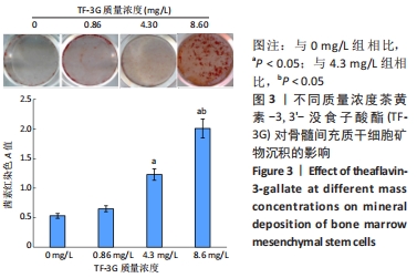
培养21 d后的茜素红染色显示骨髓间充质干细胞被染成橙红色,随着TF-3G溶液质量浓度的升高颜色加深,见图3。定量分析结果显示,4组间矿物沉积比较差异有显著性意义(P < 0.05),其中以8.60 mg/L组的矿物沉积最多。"
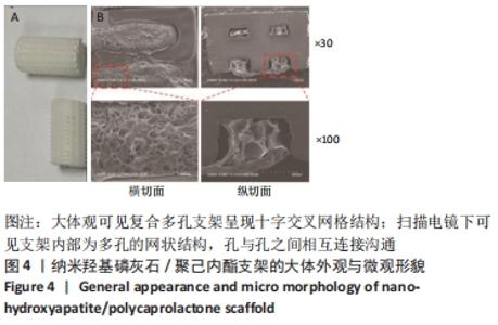
2.4 nHA/PCL复合多孔支架微观形貌 大支架观可见nHA/PCL复合多孔支架呈现十字交叉网格结构,见图4A。扫描电镜下观察支架的横切面和纵切面可见,支架内部为多孔的网状结构,孔与孔之间相互连接沟通,见图4B。"
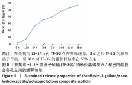
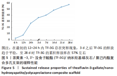
2.5 TF-3G修饰支架的缓释性能 由图5可见,在最初的12-24 h内TF-3G存在突释现象,3 d之后TF-3G的释放趋于平稳,至28 d时TF-3G的累积释放率在57%左右,说明TF-3G修饰的支架具有一定的缓释性能。"

2.6 复合多孔支架体内修复骨缺损实验结果 2.6.1 影像学观察 X射线片检查显示,空白对照组术后4周时可见骨缺损部位低密度影,缺损断端较清晰;术后12周时骨缺损部位仍呈较低密度影,与术后4周时相比无明显变化,断端骨髓腔已闭合。nHA/PCL组术后4周时可见骨缺损部位较低密度影,与周围骨质界限清晰可见;术后12周时骨缺损部位密度较术后4周时升高,与周围骨质界限仍清晰。TF-3G/nHA/PCL组术后4周时骨缺损部位密度高于nHA/PCL组,术后12周时骨缺损部位密度进一步增高,与周围骨质界限模糊。3组术后的X射线片检查图像,见图6。"
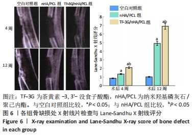

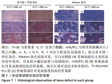
术后4,12周的Lane-Sandhu X射线评分显示,3组间评分比较差异有显著性意义,两实验组高于空白对照组(P < 0.05),并且TF-3G/nHA/PCL组的评分高于nHA/PCL组(P < 0.05),见图6。 2.6.2 组织学观察 结合苏木精-伊红染色、Masson染色结果可见,空白对照组骨缺损部位未见明显的骨样组织,可见极少量的血管与结缔组织;nHA/PCL组骨缺损部位可见成熟度较低的骨基质,部分区域可见较多软骨组织;TF-3G/nHA/PCL组骨缺损部位可见成熟度较高的骨基质,见图7。"

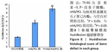
Huddleston组织学评分结果显示,3组间评分比较差异有显著性意义,两实验组高于空白对照组(P < 0.05),并且TF-3G/nHA/PCL组高于nHA/PCL组(P < 0.05),见图8。"

2.7 复合多孔支架的生物相容性 由动物体内骨缺损修复组织学结果可知,TF-3G/nHA/PCL复合多孔支架具有良好的生物相容性。"

| [1] GRAHAM SM, LEONIDOU A, ASLAM-PERVEZ N, et al. Biological therapy of bone defects: the immunology of bone allo-transplantation. Expert Opin Biol Ther. 2010;10(6):885-901. [2] AHMED GA, ISHAQUE B, RICKERT M, et al. Allogeneic bone transplantation in hip revision surgery : Indications and potential for reconstruction. Orthopade. 2018;47(1):52-66. [3] CHU W. Treatment of nonunion with autologous bone transplantation combined with platelet-rich plasma and extracorporeal shock wave. Zhongguo Gu Shang. 2019;32(5):434-439. [4] 余幸鸽,林开利.基于天然水凝胶的生物材料在骨组织工程中的应用[J].中国生物工程杂志,2020,40(5):69-77. [5] HOLZAPFEL BM, RUDERT M, HUTMACHER DW. Scaffold-based Bone Tissue Engineering. Orthopade. 2017;46(8):701-710. [6] ZHOU X, SHI G, FAN B, et al. Polycaprolactone electrospun fiber scaffold loaded with iPSCs-NSCs and ASCs as a novel tissue engineering scaffold for the treatment of spinal cord injury. Int J Nanomedicine. 2018;13:6265-6277. [7] 武小童,何儿,刘来俊,等.基于聚己内酯纤维的组织工程支架研究进展[J].中国生物医学工程学报,2020,39(5):611-620. [8] TEOH SH, GOH BT, LIM J. Three-Dimensional Printed Polycaprolactone Scaffolds for Bone Regeneration Success and Future Perspective. Tissue Eng Part A. 2019;25(13-14):931-935. [9] ALEMÁN-DOMÍNGUEZ ME, GIUSTO E, ORTEGA Z, et al. Three-dimensional printed polycaprolactone-microcrystalline cellulose scaffolds. J Biomed Mater Res B Appl Biomater. 2019;107(3):521-528. [10] DAAS I, BADR S, OSMAN E. Comparison between Fluoride and Nano-hydroxyapatite in Remineralizing Initial Enamel Lesion: An in vitro Study. J Contemp Dent Pract. 2018;19(3):306-312. [11] GIZER M, KÖSE S, KARAOSMANOGLU B, et al. The Effect of Boron-Containing Nano-Hydroxyapatite on Bone Cells.Biol Trace Elem Res. 2020;193(2):364-376. [12] 胡超然,邱冰,周祝兴,等.3D打印聚己内酯/纳米羟基磷灰石复合支架与骨髓间充质干细胞的体外生物相容性[J].中国组织工程研究,2020,24(4):589-595. [13] NAUDOT M, GARCIA GARCIA A, JANKOVSKY N, et al. The combination of a poly-caprolactone/nano-hydroxyapatite honeycomb scaffold and mesenchymal stem cells promotes bone regeneration in rat calvarial defects. J Tissue Eng Regen Med. 2020;14(11):1570-1580. [14] ROBERTS EAH, CARTWRIGHT RA, OLDSCHOOL M. The phenolic substances of manufactured tea. I. —Fractionation and paper chromatography of water‐soluble substances. J Sci Food Agric. 1957; 8(2):72-80. [15] 尚希福,路玉峰,张文志,等.茶黄素对人骨髓基质干细胞的成骨诱导作用[J].山东医药,2010,50(19):7-9. [16] 赵传勇,丁艳芳,张文志,等.骨髓间充质干细胞联合茶黄素修复激素性股骨头坏死[J].中国组织工程研究,2015,19(32):5210-5214. [17] Oka Y, Iwai S, Amano H, et al. Tea Polyphenols Inhibit Rat Osteoclast Formation and Differentiation. J Pharmacol Sci. 2012;118(1):55-64. [18] 辛红美,许洁,汪长东.淫羊藿苷促进MC3T3-E1成骨分化通过Hedgehog信号通路[J].中国药理学通报,2020,36(5):616-620. [19] 向声燚,焦志伟,马昊鹏,等.聚己内酯/纳米羟基磷灰石复合材料的3D打印及性能[J].工程塑料应用,2018,46(8):122-125,130. [20] JIN Y, ZHANG W, LIU Y, et al. rhPDGF-BB via ERK pathway osteogenesis and adipogenesis balancing in ADSCs for critical-sized calvarial defect repair. Tissue Eng Part A. 2014;20(23-24):3303-3313. [21] INODA H, YAMAMOTO G, HATTORI T. Histological investigation of osteoinductive properties of rh-BMP2 in a rat calvarial bone defect model. J Craniomaxillofac Surg. 2004;32(6):365-369. [22] 李树源,周琦石,周宏亮,等.强骨胶囊辅助诱导膜技术治疗感染性骨髓炎并骨缺损临床观察[J].山东医药,2019,59(19):73-76. [23] HUDDLESTON PM, STECKELBERG JM, HANSSEN AD, et al. Ciprofloxacin inhibition of experimental fracture healing. J Bone Joint Surg Am. 2000; 82(2):161-173. [24] FENNEMA EM, TCHANG LAH, YUAN H, et al. Ectopic bone formation by aggregated mesenchymal stem cells from bone marrow and adipose tissue: A comparative study. J Tissue Eng Regen Med. 2018;12(1): e150-e158. [25] 董晨露.骨髓间充质干细胞成骨分化的研究与进展[J].中国骨伤杂志,2019,32(3):288-292. [26] NISHIKAWA K, IWAMOTO Y, KOBAYASHI Y, et al. DNA methyltransferase 3a regulates osteoclast differentiation by coupling to an S-adenosylmethionine-producing metabolic pathway. Nat Med. 2015; 21(3):281-287. [27] CHAI S, WAN L, WANG JL, et al. Gushukang inhibits osteocyte apoptosis and enhances BMP-2/Smads signaling pathway in ovariectomized rats. Phytomedicine. 2019;64:153063. [28] FENG C, XIAO L, YU JC, et al. Simvastatin promotes osteogenic differentiation of mesenchymal stem cells in rat model of osteoporosis through BMP-2/Smads signaling pathway. Eur Rev Med Pharmacol Sci. 2020;24(1):434-443. [29] MOON SH, KIM I, KIM SH. Mollugin enhances the osteogenic action of BMP-2 via the p38-Smad signaling pathway. Arch Pharm Res. 2017; 40(11):1328-1335. [30] CHUNG YH, HUANG YH, CHU TH, et al. BMP-2 restoration aids in recovery from liver fibrosis by attenuating TGF-beta1 signaling. Lab Invest. 2018;98(8):999-1013. [31] REN C, GONG W, LI F, et al. Pilose antler aqueous extract promotes the proliferation and osteogenic differentiation of bone marrow mesenchymal stem cells by stimulating the BMP-2/Smad1, 5/Runx2 signaling pathway. Chin J Nat Med. 2019;17(10):756-767. [32] YANG G, YUAN G, LI X, et al. BMP-2 induction of Dlx3 expression is mediated by p38/Smad5 signaling pathway in osteoblastic MC3T3-E1 cells. J Cell Physiol. 2014;229(7):943-954. [33] YODTHONG T, KEDJARUNE-LEGGAT U, SMYTHE C, etal.l-Quebrachitol Promotes the Proliferation, Differentiation, and Mineralization of MC3T3-E1 Cells: Involvement of the BMP-2/Runx2/MAPK/Wnt/beta-Catenin Signaling Pathway. Molecules. 2018;23(12):3086. [34] MOON SH, KIM I, KIM SH. Mollugin enhances the osteogenic action of BMP-2 via the p38-Smad signaling pathway. Arch Pharm Res. 2017; 40(11):1328-1335. [35] 金光辉,孙晓飞,夏琰,等.选择性激光烧结法构建纳米羟基磷灰石与聚己内酯复合材料人工骨支架[J].第二军医大学学报,2015, 36(12): 1289-1294. [36] 金光辉,张馨雯,孙晓飞,等.组织工程化纳米羟基磷灰石/聚己内酯人工骨支架修复兔桡骨大段骨缺损的实验研究[J].中华损伤与修复杂志(电子版),2015(1):43-49. [37] QIN T, LI X, LONG H, et al. Bioactive Tetracalcium Phosphate Scaffolds Fabricated by Selective Laser Sintering for Bone Regeneration Applications. Materials (Basel). 2020;13(10):2268. |
| [1] | Xue Yadong, Zhou Xinshe, Pei Lijia, Meng Fanyu, Li Jian, Wang Jinzi . Reconstruction of Paprosky III type acetabular defect by autogenous iliac bone block combined with titanium plate: providing a strong initial fixation for the prosthesis [J]. Chinese Journal of Tissue Engineering Research, 2022, 26(9): 1424-1428. |
| [2] | Gao Yujin, Peng Shuanglin, Ma Zhichao, Lu Shi, Cao Huayue, Wang Lang, Xiao Jingang. Osteogenic ability of adipose stem cells in diabetic osteoporosis mice [J]. Chinese Journal of Tissue Engineering Research, 2022, 26(7): 999-1004. |
| [3] | Wu Weiyue, Guo Xiaodong, Bao Chongyun. Application of engineered exosomes in bone repair and regeneration [J]. Chinese Journal of Tissue Engineering Research, 2022, 26(7): 1102-1106. |
| [4] | Zhou Hongqin, Wu Dandan, Yang Kun, Liu Qi. Exosomes that deliver specific miRNAs can regulate osteogenesis and promote angiogenesis [J]. Chinese Journal of Tissue Engineering Research, 2022, 26(7): 1107-1112. |
| [5] | Chen Xiaoxu, Luo Yaxin, Bi Haoran, Yang Kun. Preparation and application of acellular scaffold in tissue engineering and regenerative medicine [J]. Chinese Journal of Tissue Engineering Research, 2022, 26(4): 591-596. |
| [6] | Kang Kunlong, Wang Xintao. Research hotspot of biological scaffold materials promoting osteogenic differentiation of bone marrow mesenchymal stem cells [J]. Chinese Journal of Tissue Engineering Research, 2022, 26(4): 597-603. |
| [7] | Wang Ruanbin, Cheng Liqian, Chen Kai. Application and value of polymer materials in three-dimensional printing biological bones and scaffolds [J]. Chinese Journal of Tissue Engineering Research, 2022, 26(4): 610-616. |
| [8] | Li Hui, Chen Lianglong. Application and characteristics of bone graft materials in the treatment of spinal tuberculosis [J]. Chinese Journal of Tissue Engineering Research, 2022, 26(4): 626-630. |
| [9] | Tan Guozhong, Tu Xinran, Guo Liyang, Zhong Jialin, Zhang Yang, Jiang Qianzhou. Biosafety evaluation of three-dimensional printed gelatin/sodium alginate/58S bioactive glass scaffolds for bone defect repair [J]. Chinese Journal of Tissue Engineering Research, 2022, 26(4): 521-527. |
| [10] | He Guanyu, Xu Baoshan, Du Lilong, Zhang Tongxing, Huo Zhenxin, Shen Li. Biomimetic orientated microchannel annulus fibrosus scaffold constructed by silk fibroin [J]. Chinese Journal of Tissue Engineering Research, 2022, 26(4): 560-566. |
| [11] | Guan Jian, Jia Yanfei, Zhang Baoxin , Zhao Guozhong. Application of 4D bioprinting in tissue engineering [J]. Chinese Journal of Tissue Engineering Research, 2022, 26(3): 446-455. |
| [12] | Han Zhi, Wang Zhimiao, Gaxi Sijia, Lu Qingling, Guo Tao. Tissue engineered cartilage constructed by polyurethane composite chondrocytes [J]. Chinese Journal of Tissue Engineering Research, 2022, 26(22): 3455-3459. |
| [13] | Tang Zhenzhou, Gu Yong, Chen Liang. Preparation of modified dextran composite hydrogel with osteogenetic effect and in vitro experiment [J]. Chinese Journal of Tissue Engineering Research, 2022, 26(22): 3521-3527. |
| [14] | Meng Lulu, Liu Hao, Liu Han, Zhang Jun, Li Ruixin, Gao Lilan. Mechanical properties of silk fibroin/type I collagen/hydroxyapatite scaffolds based on low-temperature 3D printing [J]. Chinese Journal of Tissue Engineering Research, 2022, 26(22): 3550-3555. |
| [15] | Guo Xiaopeng, Liu Yingsong, Shang Hui. Silk fibroin/nano hydroxyapatite composite combined with icariin can promote the proliferation and differentiation of bone marrow mesenchymal stem cells into nucleus pulposus like cells [J]. Chinese Journal of Tissue Engineering Research, 2022, 26(22): 3528-3534. |
| Viewed | ||||||
|
Full text |
|
|||||
|
Abstract |
|
|||||