Chinese Journal of Tissue Engineering Research ›› 2021, Vol. 25 ›› Issue (26): 4106-4111.doi: 10.12307/2021.118
Previous Articles Next Articles
Regulatory effects of LncRNA MEG3 on the proliferation and apoptosis of nucleus pulposus cells in rat intervertebral disc
Cheng Ning1, Zhang Zhizhong2
- 1Anyang Vocational and Technical College, Anyang 455000, Henan Province, China;
2Anyang Tumor Hospital, the Fourth Affiliated Hospital of Henan University of Science and Technology, Anyang 455000, Henan Province, China
-
Received:2019-12-24Revised:2019-12-26Accepted:2020-09-03Online:2021-09-18Published:2021-04-25 -
Contact:Cheng Ning, Lecturer, Anyang Vocational and Technical College, Anyang 455000, Henan Province, China E-mail:chenwenxix@163.com
CLC Number:
Cite this article
Cheng Ning, Zhang Zhizhong. Regulatory effects of LncRNA MEG3 on the proliferation and apoptosis of nucleus pulposus cells in rat intervertebral disc[J]. Chinese Journal of Tissue Engineering Research, 2021, 25(26): 4106-4111.
share this article
Add to citation manager EndNote|Reference Manager|ProCite|BibTeX|RefWorks

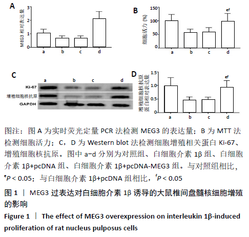
2.1 白细胞介素1β诱导的大鼠椎间盘髓核细胞中MEG3的表达量 采用实时荧光定量PCR检测白细胞介素1β诱导的大鼠椎间盘髓核细胞中MEG3的表达量,结果显示与对照组(1.02±0.20)比较,白细胞介素1β组细胞中MEG3的表达量(0.63±0.21)显著降低(t=4.034,P<0.05)。 2.2 MEG3过表达对白细胞介素1β诱导的大鼠椎间盘髓核细胞增殖的影响 将pcDNA-MEG3转染至大鼠椎间盘髓核细胞后,结果显示与对照组比较,白细胞介素1β组细胞活力显著降低(P<0.05),Ki-67、增殖细胞核抗原蛋白水平显著降低(P<0.05);与白细胞介素1β+pcDNA组比较,白细胞介素1β+pcDNA-MEG3组细胞活力显著升高(P<0.05),Ki-67、增殖细胞核抗原蛋白水平显著升高(P<0.05),见图1。"

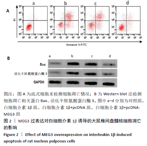
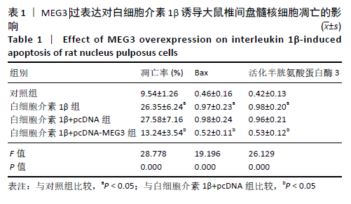
2.3 MEG3过表达对白细胞介素1β诱导的大鼠椎间盘髓核细胞凋亡的影响 将pcDNA-MEG3转染至大鼠椎间盘髓核细胞后,结果显示与对照组比较,白细胞介素1β组细胞凋亡率显著升高(P<0.05),Bax、活化半胱氨酸蛋白酶3蛋白水平显著升高(P<0.05);与白细胞介素1β+pcDNA组比较,白细胞介素1β+pcDNA-MEG3组细胞凋亡率显著降低(P<0.05),Bax、活化半胱氨酸蛋白酶3蛋白水平显著降低(P<0.05),见图2及表1。"

"

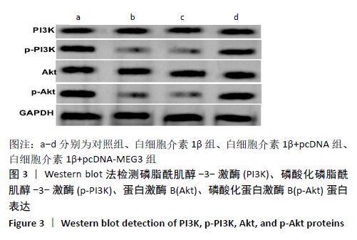
2.4 MEG3过表达对白细胞介素1β诱导的大鼠椎间盘髓核细胞中PI3K/Akt信号通路相关蛋白表达的影响 将pcDNA-MEG3转染至大鼠椎间盘髓核细胞后,采用Western blot法检测PI3K/Akt信号通路相关蛋白表达量,结果显示与对照组比较,白细胞介素1β组p-PI3K、p-Akt蛋白水平显著降低(P<0.05);与白细胞介素1β+pcDNA组比较,白细胞介素1β+pcDNA-MEG3组p-PI3K、p-Akt蛋白水平显著升高(P<0.05),对照组、白细胞介素1β组、白细胞介素1β+pcDNA组、白细胞介素1β+pcDNA-MEG3组间PI3K、Akt蛋白水平比较差异无显著性意义(P>0.05),见图3及表2。"

"


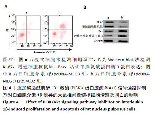
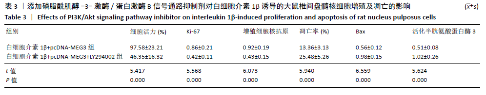
2.5 添加PI3K/Akt信号通路抑制剂对白细胞介素1β诱导的大鼠椎间盘髓核细胞增殖及凋亡的影响 添加PI3K/Akt信号通路抑制剂LY294002后,结果显示与白细胞介素1β+pcDNA-MEG3组比较,白细胞介素1β+pcDNA-MEG3+LY294002组细胞活力显著降低(P<0.05),细胞凋亡率显著升高(P<0.05),Ki-67、增殖细胞核抗原蛋白水平显著降低(P<0.05),Bax、活化半胱氨酸蛋白酶3蛋白水平显著升高(P<0.05),见图4及表3。"

"
| [1]VERGROESEN PP, KINGMA I, EMANUEL KS, et al. Mechanics and biology in intervertebral disc degeneration: a vicious circle. Osteoarthritis Cartilage. 2015;23(7): 1057-1070. [2]KERMANI HR, HOBOUBATI H, ESMAEILI-MAHANI S, et al. Induction of intervertebral disc cell apoptosis and degeneration by chronic unpredictable stress. J Neurosurg Spine. 2014;20(5): 578-584. [3]LI N, GAO Q, ZHOU W, et al. MicroRNA-129-5p affects immune privilege and apoptosis of nucleus pulposus cells via regulating FADD in intervertebral disc degeneration. Cell Cycle. 2020;11(1): 1-16. [4]SHEN J, FANG J, HAO J, et al. SIRT1 Inhibits the Catabolic Effect of IL-1β Through TLR2/SIRT1/NF-κB Pathway in Human Degenerative Nucleus Pulposus Cells. Pain Phys. 2016;19(1): E215-E216. [5]TONG P, PENG QH, GU LM, et al. LncRNA-MEG3 alleviates high glucose induced inflammation and apoptosis of retina epithelial cells via regulating miR-34a/SIRT1 axis. Exp Mol Pathol. 2019;107(1): 102-109. [6] 孙中仪, 晏鹏, 王圣杰,等. 长链非编码RNA在人退变椎间盘组织中的基因表达谱[J]. 中华医学杂志, 2017, 97(33):2582-2586. [7] 王毅峰. LncRNA在椎间盘退变过程中调控髓核细胞凋亡的功能及其机制研究[D].上海:第二军医大学, 2016. [8]Bai X, Guo X, Zhang F, et al. Resveratrol Combined with 17β-Estradiol Prevents IL-1β Induced Apoptosis in Human Nucleus Pulposus Via The PI3K/AKT/Mtor and PI3K/AKT/GSK-3β Pathway. J Invest Surg. 2020; 10(1): 1-8. [9] 陈江, 刘志超, 张帆,等. 身痛逐瘀汤对人髓核细胞模型PI3K/Akt信号通路Bad、Caspase-9、GSK-3β表达的影响[J]. 中国中医药信息杂志, 2019, 26(9):48-53. [10] 鲁花, 于露, 甄欢欢,等. 骨形态发生蛋白9激活PI3K/Akt信号通路抑制退变髓核细胞的炎症反应和凋亡[J]. 第三军医大学学报, 2016, 38(18):2047-2052. [11]吴瑞凯, 齐义营, 茹选良, 等. 血清饥饿对大鼠椎间盘髓核细胞凋亡的影响及其机制研究[J]. 中华全科医学, 2016, 14(6): 889-892. [12]张海丹, 李培武, 张玲. 五味子乙素通过调控p38MAPK信号通路对IL-1β诱导的髓核细胞凋亡的影响[J]. 医学分子生物学杂志, 2019, 16(2): 119-124. [13]郑文凯, 黄智, 达逸峰, 等. 胸腺基质促淋巴细胞生成素介导PI3K/Akt通路抑制髓核细胞凋亡[J]. 中华骨科杂志, 2019, 39(6): 346-353. [14]DING F, SHAO ZW, XIONG LM. Cell death in intervertebral disc degeneration. Apoptosis. 2013; 18(7): 777-785. [15]KITYA D, PUNCHAK M, BAJUNIRWE F. The Role of Conventional Myelography in the Diagnosis and Treatment of Degenerative Spine Diseases in Low-Income Communities: a Prospective Study. World Neurosurg. 2017;104(1): 161-166. [16]WANG J, CHEN H, CAO P, et al. Inflammatory cytokines induce caveolin-1/β-catenin signalling in rat nucleus pulposus cell apoptosis through the p38 MAPK pathway. Cell Prolif. 2016; 49(3): 362-372. [17]CHAI X, SI H, SONG J, et al. miR-486-5p Inhibits Inflammatory Response, Matrix Degradation and Apoptosis of Nucleus Pulposus Cells Through Directly Targeting FOXO1 in Intervertebral Disc Degeneration. Cell Physiol Biochem. 2019; 52(1): 109-118. [18]WANG K, CHEN T, YING X, et al. Ligustilide alleviated IL-1β induced apoptosis and extracellular matrix degradation of nucleus pulposus cells and attenuates intervertebral disc degeneration in vivo. Int Immunopharmacol. 2019; 69(1): 398-407. [19]TAN Y, YAO X, DAI Z, et al. Bone morphogenetic protein 2 alleviated intervertebral disc degeneration through mediating the degradation of ECM and apoptosis of nucleus pulposus cells via the PI3K/Akt pathway. Int J Mol Med. 2019; 43(1): 583-592. [20]TANG P, GU JM, XIE ZA, et al. Honokiol alleviates the degeneration of intervertebral disc via suppressing the activation of TXNIP-NLRP3 inflammasome signal pathway. Free Radic Biol Med. 2018; 120(1): 368-379. [21]WU X, LIU Y, GUO X, et al. Prolactin inhibits the progression of intervertebral disc degeneration through inactivation of the NF-κB pathway in rats. Cell Death Dis. 2018; 9(2): 98-108. [22]LI G, LIU Y, MENG F, et al. LncRNA MEG3 inhibits rheumatoid arthritis through miR-141 and inactivation of AKT/mTOR signalling pathway. J Cell Mol Med. 2019; 23(10): 7116-7120. [23]LI Y, ZHANG S, ZHANG C, et al. LncRNA MEG3 inhibits the inflammatory response of ankylosing spondylitis by targeting miR-146a. Mol Cell Biochem. 2020; 466(1-2): 17-24. [24]PANG X, FENG G, SHANG W, et al. Inhibition of lncRNA MEG3 protects renal tubular from hypoxia-induced kidney injury in acute renal allografts by regulating miR-181b/TNF-α signaling pathway. J Cell Biochem. 2019; 120(8): 12822-12831. [25]楚广民, 张建波, 孙淼淼. RNA干扰沉默SOX9对肾细胞癌786-O细胞体外增殖、凋亡及裸鼠成瘤能力的影响[J]. 分子诊断与治疗杂志, 2018, 10(3): 174-179. [26]蒋汉霞, 刘志杰. 紫草素调控PTEN/AKT通路抑制宫颈癌细胞增殖、侵袭研究[J]. 热带医学杂志, 2019, 19(7): 822-826. [27]韩敦富, 尹荷珊, 管晨彤, 等. IL-1β诱导椎间盘细胞凋亡通路再认识[J]. 福建医科大学学报, 2019, 53(4): 224-228. [28]NAN LP, WANG F, RAN D, et al. Naringin alleviates H2O2-induced apoptosis via the PI3K/Akt pathway in rat nucleus pulposus-derived mesenchymal stem cells. Connect Tissue Res. 2019;11(1): 1-4. [29]TAN Y, YAO X, DAI Z, et al. Bone morphogenetic protein 2 alleviated intervertebral disc degeneration through mediating the degradation of ECM and apoptosis of nucleus pulposus cells via the PI3K/Akt pathway. Int J Mol Med. 2019; 43(1): 583-592. [30]GAO J, ZHANG Q, SONG L. Resveratrol Enhances Matrix Biosynthesis of Nucleus Pulposus Cells Through Activating Autophagy via the PI3K/Akt Pathway Under Oxidative Damage. Biosci Rep. 2018; 38(4): 1-10. [31]GONG C, PAN W, HU W, et al. Bone Morphogenetic protein-7 Retards Cell Subculture-Induced Senescence of Human Nucleus Pulposus Cells Through Activating the PI3K/Akt Pathway. Biosci Rep. 2019; 39(3): 1-12. [32]LI P, GAN Y, XU Y, et al. The Inflammatory Cytokine TNF-α Promotes the Premature Senescence of Rat Nucleus Pulposus Cells via the PI3K/Akt Signaling Pathway. Sci Rep. 2017; 7(1): 42938-42948. [33]ZHANG X, HU Z, HAO J, et al. Low Intensity Pulsed Ultrasound Promotes the Extracellular Matrix Synthesis of Degenerative Human Nucleus Pulposus Cells Through FAK/PI3K/Akt Pathway. Spine (Phila Pa 1976). 2016; 41(5): 248-254. [34]NAN LP, WANG F, RAN D, et al. Naringin Alleviates H 2 O 2-induced Apoptosis via the PI3K/Akt Pathway in Rat Nucleus Pulposus-Derived Mesenchymal Stem Cells. Connect Tissue Res. 2019; 1(10): 1-14. [35]JIANG Y, XIE Z, YU J, et al. Resveratrol Inhibits IL-1β-mediated Nucleus Pulposus Cell Apoptosis Through Regulating the PI3K/Akt Pathway. Biosci Rep. 2019; 39(3): 1-13. [36]MING-YAN Y, JING Z, SHU-QIN G, et al. Liraglutide Inhibits the Apoptosis of Human Nucleus Pulposus Cells Induced by High Glucose Through PI3K/Akt/caspase-3 Signaling Pathway. Biosci Rep. 2019;39(8): 1-10. [37]YANG Y, WANG X, LIU Z, et al. Osteogenic protein-1 Attenuates Nucleus Pulposus Cell Apoptosis Through Activating the PI3K/Akt/mTOR Pathway in a Hyperosmotic Culture. Biosci Rep. 2018; 38(6): 1-10. [38]WU X, LI S, WANG K, et al. TNF-α Regulates ITGβ1 and SYND4 Expression in Nucleus Pulposus Cells: Activation of FAK/PI3K Signaling. Inflammation. 2019; 42(5): 1575-1584. [39]RISBUD MV, FERTALA J, VRESILOVIC EJ, et al. Nucleus Pulposus Cells Upregulate PI3K/Akt and MEK/ERK Signaling Pathways Under Hypoxic Conditions and Resist Apoptosis Induced by Serum Withdrawal. Spine (Phila Pa 1976). 2005; 30(8): 882-889. [40]LIU G, CAO P, CHEN H, et al. MiR-27a Regulates Apoptosis in Nucleus Pulposus Cells by Targeting PI3K. PLoS One. 2013; 8(9): e75251-e75261. [41]TIAN D, LIU J, CHEN L, et al. The Protective Effects of PI3K/Akt Pathway on Human Nucleus Pulposus Mesenchymal Stem Cells Against Hypoxia and Nutrition Deficiency. J Orthop Surg Res. 2020;15(1): 29-39. [42]CHENG CC, UCHIYAMA Y, HIYAMA A, et al. PI3K/AKT Regulates Aggrecan Gene Expression by Modulating Sox9 Expression and Activity in Nucleus Pulposus Cells of the Intervertebral Disc. J Cell Physiol. 2009; 221(3): 668-676. [43]YANG SD, MA L, YANG DL, et al. Combined Effect of 17β-estradiol and Resveratrol Against Apoptosis Induced by interleukin-1β in Rat Nucleus Pulposus Cells via PI3K/Akt/caspase-3 Pathway. Peer J. 2016;4(1): 1640-1650. [44]HUANG D, PENG Y, MA K, et al. Puerarin Relieved Compression-Induced Apoptosis and Mitochondrial Dysfunction in Human Nucleus Pulposus Mesenchymal Stem Cells via the PI3K/Akt Pathway. Stem Cells Int. 2020;2020(1): 1-10. |
| [1] | Chen Jinping, Li Kui, Chen Qian, Guo Haoran, Zhang Yingbo, Wei Peng. Meta-analysis of the efficacy and safety of tranexamic acid in open spinal surgery [J]. Chinese Journal of Tissue Engineering Research, 2021, 25(9): 1458-1464. |
| [2] | Zhang Chong, Liu Zhiang, Yao Shuaihui, Gao Junsheng, Jiang Yan, Zhang Lu. Safety and effectiveness of topical application of tranexamic acid to reduce drainage of elderly femoral neck fractures after total hip arthroplasty [J]. Chinese Journal of Tissue Engineering Research, 2021, 25(9): 1381-1386. |
| [3] | Wang Haiying, Lü Bing, Li Hui, Wang Shunyi. Posterior lumbar interbody fusion for degenerative lumbar spondylolisthesis: prediction of functional prognosis of patients based on spinopelvic parameters [J]. Chinese Journal of Tissue Engineering Research, 2021, 25(9): 1393-1397. |
| [4] | Chai Le, Lü Jianlan, Hu Jintao, Hu Huahui, Xu Qingjun, Yu Jinwei, Quan Renfu. Signal pathway variation after induction of inflammatory response in rats with acute spinal cord injury [J]. Chinese Journal of Tissue Engineering Research, 2021, 25(8): 1218-1223. |
| [5] | Geng Qiudong, Ge Haiya, Wang Heming, Li Nan. Role and mechanism of Guilu Erxianjiao in treatment of osteoarthritis based on network pharmacology [J]. Chinese Journal of Tissue Engineering Research, 2021, 25(8): 1229-1236. |
| [6] | Liu Xiangxiang, Huang Yunmei, Chen Wenlie, Lin Ruhui, Lu Xiaodong, Li Zuanfang, Xu Yaye, Huang Meiya, Li Xihai. Ultrastructural changes of the white zone cells of the meniscus in a rat model of early osteoarthritis [J]. Chinese Journal of Tissue Engineering Research, 2021, 25(8): 1237-1242. |
| [7] | Shu Wenbo, Chen Mengchi, Li Hua, Huang Liqian, Huang Binbin, Zhang Wenhai, Wu Yachen, Wang Zefeng, Li Qiaoli, Liu Peng. Correlation between body fat distribution and characteristics of daily physical activity in college students [J]. Chinese Journal of Tissue Engineering Research, 2021, 25(8): 1277-1283. |
| [8] | Ji Zhixiang, Lan Changgong. Polymorphism of urate transporter in gout and its correlation with gout treatment [J]. Chinese Journal of Tissue Engineering Research, 2021, 25(8): 1290-1298. |
| [9] | Gao Yan, Zhao Licong, Zhao Hongzeng, Zhu Yuanyuan, Li Jie, Sang Deen. Alteration of low frequency fluctuation amplitude at brain-resting state in patients with chronic discogenic low back pain [J]. Chinese Journal of Tissue Engineering Research, 2021, 25(8): 1160-1165. |
| [10] | Wu Xun, Meng Juanhong, Zhang Jianyun, Wang Liang. Concentrated growth factors in the repair of a full-thickness condylar cartilage defect in a rabbit [J]. Chinese Journal of Tissue Engineering Research, 2021, 25(8): 1166-1171. |
| [11] | Liu Zhichao, Zhang Fan, Sun Qi, Kang Xiaole, Yuan Qiaomei, Liu Genzhe, Chen Jiang. Morphology and activity of human nucleus pulposus cells under different hydrostatic pressures [J]. Chinese Journal of Tissue Engineering Research, 2021, 25(8): 1172-1176. |
| [12] | Shen Jinbo, Zhang Lin. Micro-injury of the Achilles tendon caused by acute exhaustive exercise in rats: ultrastructural changes and mechanism [J]. Chinese Journal of Tissue Engineering Research, 2021, 25(8): 1190-1195. |
| [13] | Zeng Zhen, Hu Jingwei, Li Xuan, Tang Linmei, Huang Zhiqiang, Li Mingxing. Quantitative analysis of renal blood flow perfusion using contrast-enhanced ultrasound in rats with hemorrhagic shock during resuscitation [J]. Chinese Journal of Tissue Engineering Research, 2021, 25(8): 1201-1206. |
| [14] | Tang Hui, Yao Zhihao, Luo Daowen, Peng Shuanglin, Yang Shuanglin, Wang Lang, Xiao Jingang. High fat and high sugar diet combined with streptozotocin to establish a rat model of type 2 diabetic osteoporosis [J]. Chinese Journal of Tissue Engineering Research, 2021, 25(8): 1207-1211. |
| [15] | Lun Zhigang, Jin Jing, Wang Tianyan, Li Aimin. Effect of peroxiredoxin 6 on proliferation and differentiation of bone marrow mesenchymal stem cells into neural lineage in vitro [J]. Chinese Journal of Tissue Engineering Research, 2021, 25(7): 1014-1018. |
| Viewed | ||||||
|
Full text |
|
|||||
|
Abstract |
|
|||||