|
[1] 程斌,宋焕瑾.同种异体椎间融合器的研发及其应用[J].中国组织工程研究,2015,19(53):8656.
[2] 朱领军,刘佳,鲍小刚,等.可降解脊柱椎间融合器的研究进展[J].局解手术学杂志,2019,28(1):71-75.
[3] 马向阳,杨进城,邱锋,等.寰枢椎脱位后路植骨材料的选择与融合效果评价[J].中国骨科临床与基础研究杂志,2015,7(1):5-9.
[4] 廖文胜,刘玉峰,鲍恒,等. 经口前路寰枢椎侧块关节间固定融合器的研制及生物力学分析[J].郑州大学学报(医学版), 2013, 48(5):658-661.
[5] 张皓南,尹庆水,刘桂英,等. 经口前路寰枢椎侧块关节融合器的力学稳定性评价[J].中国临床解剖学杂志, 2014,3(3):348-350.
[6] ZHANG BC, LIU HB, CAI XH, et al. Biomechanical comparison of a novel transoral atlantoaxial anchored cage with established fixation technique -a finite element analysis. BMC Musculoskelet Disord. 2015;22(16):261.
[7] LI S, NI B, XIE N, et al. Biomechanical evaluation of an atlantoaxial lateral mass fusion cage with C1-C2 pedicle fixation. Spine (Phila Pa 1976). 2010;35(14):E624-E632.
[8] YIN QS, WANG JH. Current trends in management of atlantoaxial dislocation. Orthop Surg. 2015;7(3):189-199.
[9] HUANG DG, HAO DJ, HE BR, et al. Posterior atlantoaxial fixation: a review of all techniques. Spine J. 2015;15(10): 2271-2281.
[10] SIM HB, LEE JW, PARK JT, et al. Biomechanical evaluations of various c1-c2 posterior fixation techniques. Spine (Phila Pa 1976). 2011; 36(6):E401-E407.
[11] PARK J, SCHEER JK, LIM TJ, et al. Biomechanical analysis of Goel technique for C1-2 fusion. J Neurosurg Spine. 2011; 14(5):639-646.
[12] MELCHER RP, PUTTLITZ CM, KLEINSTUECK FS, et al. Biomechanical testing of posterior atlantoaxial fixation techniques. Spine (Phila Pa 1976). 2002; 27(22):2435-2440.
[13] 马向阳,尹庆水,吴增晖,等.寰枢椎后路四种钉棒固定方法的三维稳定性评价[J].中国脊柱脊髓杂志,2008,18(6):464-468.
[14] CLAYBROOKS R, KAYANJA M, MILKS R, et al. Atlantoaxial fusion: a biomechanical analysis of two C1-C2 fusion techniques. Spine J. 2007;7(6):682-688.
[15] 马向阳,尹庆水,吴增晖,等. 多种寰枢椎后路钉棒固定技术的临床组合应用[J]. 中国骨科临床与基础研究杂志,2010,2(1): 12-16.
[16] BHOWMICK DA, BENZEL EC. Posterior atlantoaxial fixation with screw-rod constructs: safety, advantages, and shortcomings. World Neurosurg. 2014; 81(2):288-289.
[17] HWANG IC, KANG DH, HAN JW, et al. Clinical experiences and usefulness of cervical posterior stabilization with polyaxial screw-rod system. J Korean Neurosurg Soc. 2007; 42(4): 311-316.
[18] 邹小宝,马向阳,杨进城,等. 经口咽前路减压侧块关节融合器植骨融合联合颈椎压力固定器治疗难复性寰枢椎脱位[J].中国脊柱脊髓杂志, 2016,26(11):961-966.
[19] WEI G, WANG Z, AI F, et al. Treatment of basilar invagination with klippel-feil syndrome: atlantoaxial joint distraction and fixation with transoral atlantoaxial reduction plate. Neurosurgery. 2016;78:492-498.
[20] YIN Q, AI F, ZHANG K, et al. Irreducible anterior atlantoaxial dislocation: one-stage treatment with a transoral atlantoaxial reduction plate fixation and fusion. Report of 5 cases and review of the literature. Spine (Phila Pa 1976). 2005;30: E375-E381.
[21] Yin QS, Li XS, Bai ZH, et al. An 11-Year Review of the TARP Procedure in the Treatment of Atlantoaxial Dislocation. Spine (Phila Pa 1976). 2016;41:E1151-E1158.
[22] XIA H, YIN Q, AI F, et al. Treatment of basilar invagination with atlantoaxial dislocation: atlantoaxial joint distraction and fixation with transoral atlantoaxial reduction plate (TARP) without odontoidectomy. Eur Spine J. 2014;23:1648-1655.
[23] GOEL A, KULKARNI AQ, SHARMA P. Reduction of fixed atlantoaxial dislocation in 24 cases: technical note. J Neurosurg Spine. 2005;2(4):505-509.
[24] 李胜华,汪康,夏良政,等.后路寰枢椎侧块关节融合器置入的临床应用解剖[J].中国骨与关节损伤杂志,2012,27(5):385-388.
[25] MATSUMOTO M, CHIBA K, TSUJI T, et al. Use of a titanium mesh cage for posterior atlantoaxial arthrodesis. Technical note. J Neurosurg. 2002;96(1 Suppl):127-130.
[26] CHUN HJ, OH SH, YI HJ, et al. Efficacy and durability of the titanium mesh cage spacer combined with transarticular screw fixation for atlantoaxial instability in rheumatoid arthritis patients. Spine (Phila Pa 1976). 2009;34(22):2384-2388.
[27] RYU JI, BAK KH, YI HJ, et al. Evaluation of the efficacy of titanium mesh cages with posterior C1 lateral mass and C2 pedicle screw fixation in patients with atlantoaxial instability. World Neurosurg. 2016;90:103-108.
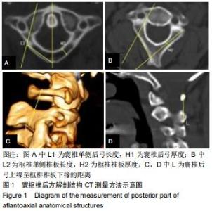
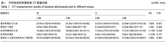
[28] 王宾宾,马向阳,段明阳,等.成人寰椎后弓部分结构测量及意义[J].中国临床解剖学杂志,2017,35(6):610-614.
[29] KELLY BP, GLASER JA, DIANGELO DJ. Biomechanical comparison of a novel C1 posterior locking plate with the harms technique in a C1-C2 fixation model. Spine (Phila Pa 1976). 2008;33(24):920-925.
[30] MA XY, YIN QS, WU ZH, et al.C2 anatomy and dimensions relative to translaminar screw placement in an asian population. Spine (Phila Pa 1976). 2010; 35(6):704-708.
|