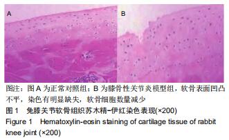
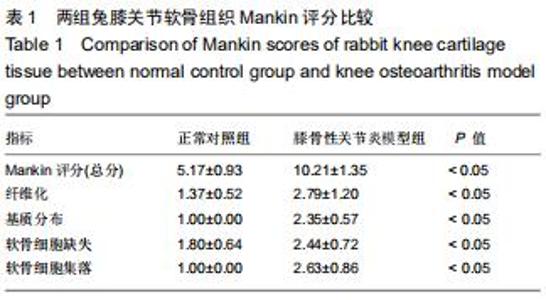
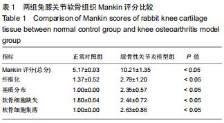
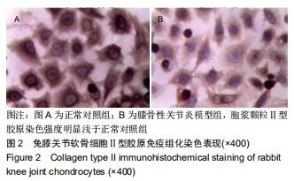
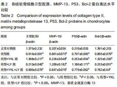
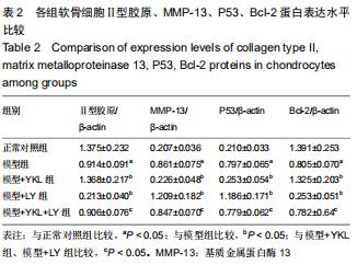
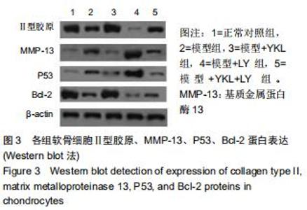
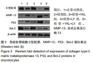
|
[1] HUNTER DJ, BIERMA-ZEINSTRA S.Osteoarthritis. Lancet. 2019;393(10182):1745-1759.
[2] 魏国,梁杰.骨性关节炎发病分子机制的研究进展[J].实用医学杂志,2015,31(24):4148-4150.
[3] 袁芳,何晓瑾,邱亦江,等.骨关节炎的软骨细胞凋亡机制[J].实用医学杂志,2015,31(4):666-668.
[4] 苏晓恩,孔志强,朱娟,等.膝关节骨关节炎软骨中YKL-40、IL-1β的表达及相关性探讨[J].重庆医学,2017,46(4):480-482.
[5] DUNDAR U, ASIK G, ULASLI AM, et al. Assessment of pulsed electromagnetic field therapy with Serum YKL-40 and ultrasonography in patients with knee osteoarthritis.Int J Rheum Dis.2016;19(3):287-293.
[6] GUAN J, LIU Z, LI F, et al. Increased Synovial Fluid YKL-40 Levels are Linked with Symptomatic Severity in Knee Osteoarthritis Patients.Clin Lab.2015;61(8): 991-997.
[7] 贺元,廖明芳,曲乐丰.YKL-40在炎症性疾病中的作用及其信号通路研究进展[J].医学研究生学报,2016,29(8):883-888.
[8] SZYCHLINSKA MA, TROVATO FM, DI RM, et al. Co-expression and co-localization of cartilage glycoproteins CHI3L1 and lubricin in osteoarthritic cartilage: morphological, immunohistochemical and gene expression profiles.Int J Mol Sci.2016;17(3):359-378.
[9] YE E, JIANG Z, LU X, et al. GZD824 suppresses the growth of human B cell precursor acute lymphoblastic leukemia cells by inhibiting the SRC kinase and PI3K/AKT pathways. Oncotarget. 2016;8(50):87002-87015.
[10] JEAN YH, WEN ZH, CHANG YC, et al. Increase in excitatory amino acid concentration and transporters expression in osteoarthritic knees of anterior cruciate ligament transected rabbits.Osteoarthritis Cartilage.2008;16(12):1442-1449.
[11] GURKAN I, RANGANATHAN A,YANG X, et al. Modification of osteoarthritis in the guinea pig with pulsed low-intensity ultrasound treatment.Osteoarthritis Cartilage.2010;18(5): 724-733.
[12] 黄艺林,刘洪柏,张鸣生.低能量体外冲击波对兔膝骨关节炎软骨细胞修复和重塑能力的影响[J].生物医学工程与临床,2016, 20(6):557-561.
[13] 武中庆,邓闵军,郭攀,等.右归饮对兔膝骨关节炎软骨细胞凋亡中Fas/FasL蛋白表达的影响[J].浙江创伤外科,2016,21(2): 202-205.
[14] CHIMENTI MS, TRIGGIANESE P, CONIGLIARO P, et al. The interplay between inflammation and metabolism in rheumatoid arthritis.Cell Death Dis.2015;1887(6):2041-2051.
[15] 钟文婕,柳占彪.YKL-40在骨关节炎中的研究进展[J].医学综述, 2013,19(3):401-403.
[16] 贺斌,陶海鹰,卫爱林,等.PI3K/Akt信号通路在羧甲基壳聚糖保护一氧化氮诱导软骨细胞凋亡中的作用及机制研究[J].中华风湿病学杂志,2015,19(3):170-175.
[17] DENG F, MA YX, LIANG L, et al. The pro-apoptosis effect of sinomenine in renal carcinoma via inducing autophagy through inactivating PI3K/AKT/mTOR pathway. Biomed Pharmacother. 2018;97:1269-1274.
[18] CHEN X, BIAN M, ZHANG C, et al. Dihydroartemisinin inhibits ER stress-mediated mitochondrial pathway to attenuate hepatocyte lipoapoptosis via blocking the activation of the PI3K/Akt pathway. Biomed Pharmacother.2018;97: 975-984.
[19] LIBREROS S, IRAGAVARAPU-CHARYULU V. YKL-40/CHI3L1 drives inflammation on the road of tumor progression.J Leukoc Biol.2015;98(6):931-936.
[20] SHAO R, TAYLOR SL, OH DS, et al. Vascular heterogeneity and targeting: the role of YKL-40 in glioblastoma vascularization. Oncotarget.2015;6(38):40507-40518.
[21] SZYCHLINSKA MA, TROVATO FM, DI RM, et al. Co-expression and co-localization of cartilage glycoproteins CHI3L1 and lubricin in osteoarthritic cartilage: morphological, immunohistochemical and gene expression profiles.Int J Mol Sci.2016;17(3):359-378.
[22] 柳奇奇,吴汉阳,汪宗保,等.早期非负重康复运动对膝骨关节炎大鼠关节软骨YKL-10表达的影响[J].中华临床医师杂志(电子版), 2015,9(24):4616-4620.
[23] 关健,谢磊,丁罗宾,等.关节软骨YKL-40表达水平与骨关节炎的相关性[J].实用医学杂志,2018,34(1):58-62.
[24] VAANANEN T, KOSKINEN A, PAUKKERR EL, et al. YKL-40 as a novel factor associated with inflammation and catabolic mechanisms in osteoarthritic joints.Mediators Inflamm. 2014; 15(1):140-147.
|