Chinese Journal of Tissue Engineering Research ›› 2015, Vol. 19 ›› Issue (50): 8172-8176.doi: 10.3969/j.issn.2095-4344.2015.50.024
Previous Articles Next Articles
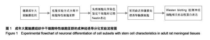
Neuronal differentiation of cell subsets with stem cell characteristics in adult rat meningeal tissues
Liu De1, Li Xiang-ming2, Gu Xi-juan3
- 1Liaocheng Third People’s Hospital, Liaocheng 252000, Shandong Province, China; 2Second Hospital of Shandong University, Jinan 250033, Shandong Province, China; 3Liaocheng University Hospital, Liaocheng 252000, Shandong Province, China
-
Received:2015-10-30Online:2015-12-03Published:2015-12-03 -
Contact:Gu Xi-juan, Associate chief physician, Liaocheng University Hospital, Liaocheng 252000, Shandong Province, China -
About author:Liu De, Associate chief physician, Liaocheng Third People’s Hospital, Liaocheng 252000, Shandong Province, China -
Supported by:the Scientific Tackle Key Project of Shandong Province, No. 2012GG10001020; the Natural Science Foundation of Shandong Province, No. ZR2011HQ045
CLC Number:
Cite this article
Liu De, Li Xiang-ming, Gu Xi-juan. Neuronal differentiation of cell subsets with stem cell characteristics in adult rat meningeal tissues[J]. Chinese Journal of Tissue Engineering Research, 2015, 19(50): 8172-8176.
share this article
"


"
"

"
| [1] 孙晓飞,金光辉,谢杨,等.神经干细胞移植治疗脊髓损伤的研究进展[J].中国矫形外科杂志,2015,23(18):1680-1682.
[2] 杨万章,吴芳,张敏,等.脐血源神经干细胞移植治疗神经系统疾病临床总结和分析[J].中西医结合心脑血管病杂志,2009,7(3): 287-290.
[3] 陈东,孟晓婷,刘佳梅,等.鹿茸多肽对胎大鼠脑神经干细胞体外诱导分化的实验研究[J].解剖学报,2004,35(3):240-243.
[4] Menendez L, Yatskievych TA, Antin PB, et al. Wnt signaling and a Smad pathway blockade direct the differentiation of human pluripotent stem cells to multipotent neural crest cells. Proc Natl Acad Sci U S A. 2011;108(48):19240-19245.
[5] Li YC, Liao YT, Chang HH, et al. Covalent bonding of GYIGSR to EVAL membrane surface to improve migration and adhesion of cultured neural stem/precursor cells. Colloids Surf B Biointerfaces. 2013;102:53-62.
[6] 张宝华,仇福成,董慈,等.脑脊液途径移植神经干细胞治疗中枢神经系统疾病的研究现状[J].中国组织工程研究,2014,18(6): 974-978.
[7] 吴家华,罗焕敏.神经干细胞移植治疗中枢神经系统疾病可行性研究[J].中国老年学杂志,2005,25(1):110-112.
[8] 温秀杰,刘鲁川,王尧,等.碱性成纤维生长因子和表皮生长因子对脊髓神经干细胞增殖与分化的影响[J].中国临床神经科学,2014, 22(2):121-125.
[9] 闫峰,张越林,刘宁,等.全骨髓贴壁法分离骨髓间充质干细胞并诱导其定向神经元样细胞的分化[J].细胞与分子免疫学杂志,2014, 30(11):1162-1165.
[10] 牛平,王耀山,吕永利,等.神经营养因子对体外培养多巴胺能神经元营养作用比较研究[J].中国医科大学学报,2001,30(6):408-410.
[11] 郭洪波,邹飞.大鼠海马神经干细胞的分离、培养及鉴定[J].第一军医大学学报,2004,24(10):1143-1146.
[12] 谭新杰,胡长林,蔡文琴,等.新生大鼠海马神经干细胞的分离、培养、分化和鉴定[J].国际脑血管病杂志,2006,14(2):107-109.
[13] 丁道芳,邢三丽,周鸣鸣,等.大鼠胚胎神经干细胞单克隆化及单层化培养和鉴定[J].生物化学与生物物理进展,2009,36(1):72-76.
[14] 史冬梅,周畅,谢佐平,等.神经干细胞有丝分裂过程中nestin表达变化[J].神经解剖学杂志,2003,19(2):119-123.
[15] 林旭妍,林绿标,许益民,等.神经膜细胞促进神经干细胞存活及分化的实验研究[J].中华脑科疾病与康复杂志:电子版,2012,2(5): 24-27.
[16] 杨忠旭,栾国明.神经干细胞的定向分化调控及移植治疗颞叶癫痫[J].立体定向和功能性神经外科杂志,2003,16(2):105-107.
[17] Heng BC, Saxena P, Fussenegger M. Heterogeneity of baseline neural marker expression by undifferentiated mesenchymal stem cells may be correlated to donor age. J Biotechnol. 2014;174:29-33.
[18] Caprini A, Silva D, Zanoni I, et al. A novel bioactive peptide: assessing its activity over murine neural stem cells and its potential for neural tissue engineering. N Biotechnol. 2013; 30(5):552-562.
[19] 陈娟,李娟,谭军,等.腺苷预处理对局灶性脑缺血再灌注损伤大鼠脑组织中内源性神经干细胞增殖的影响[J].郑州大学学报:医学版,2014,49(5):645-647,648.
[20] 许锋,姜红,周春清,等.促红细胞生成素对新生鼠缺氧缺血性脑损伤后神经干细胞的影响[J].中国新生儿科杂志, 2010, 25(2): 94-97.
[21] 唐蔚东,叶建亚,李彩格,等.巢蛋白和性别决定基因高迁移率组蛋白阳性细胞在成年大鼠穹隆下器的表达[J].解剖学杂志,2014, 37(2):202-204,220.
[22] 王婷.柔脑膜细胞具有神经干细胞特性的实验研究[D].西安:解放军第四军医大学,2008.
[23] Zou Z, Liu T, Li J, et al. Biocompatibility of functionalized designer self-assembling nanofiber scaffolds containing FRM motif for neural stem cells. J Biomed Mater Res A. 2014; 102(5): 1286-1293.
[24] 顾繁,高建华,鲁峰.hASCs的单克隆培养及干细胞相关标志物表达的实验研究[J].南方医科大学学报,2008,28(6):1067-1069,1075.
[25] 丛琳,朱晓峰.FGF-2,NT-3对海马表皮生长因子反应性神经干细胞分化为神经元的诱导作用[J].神经损伤与功能重建,2006, 1(1): 25-27,37.
[26] 李晓波,丁新生,潘凤华,等.碱性成纤维细胞生长因子、表皮生长因子对脑缺血大鼠内源性神经干细胞增殖的影响[J].临床神经病学杂志,2006,19(3):188-190.
[27] 段淑荣,王慧慧,戚基萍,等.脑缺血后碱性成纤维细胞生长因子、表皮生长因子对内源性神经干细胞增殖的影响[J].中华医学杂志,2008,88(47):3337-3341.
[28] 向云,燕铁斌,金冬梅,等.低频电刺激对急性脑梗死大鼠脑bFGF、EGF表达和内源性神经干细胞增殖的影响[J].中华物理医学与康复杂志,2010,32(12):881-886.
[29] Khosravizadeh Z, Razavi S, Bahramian H, et al. The beneficial effect of encapsulated human adipose-derived stem cells in alginate hydrogel on neural differentiation. J Biomed Mater Res B Appl Biomater. 2014;102(4):749-755.
[30] Lv Y, Zhao P, Chen G, et al. Effects of low-intensity pulsed ultrasound on cell viability, proliferation and neural differentiation of induced pluripotent stem cells-derived neural crest stem cells. Biotechnol Lett. 2013;35(12):2201-2212.
[31] Zhang Y, Guo Y, Wu B, et al. Ligand-mediated endocytosis of nanoparticles in neural stem cells: implications for cellular magnetic resonance imaging. Biotechnol Lett. 2013;35(12): 1997-2004.
[32] 陈东风,杜少辉,李伊为,等.龟板对局灶性脑缺血再灌注后Nestin表达的影响[J].解剖学杂志,2002,25(4):315-319.
[33] 程龙,朱培纯,司银楚,等.三七总皂甙对去大脑皮层血管后成年大鼠前脑侧脑室室管膜下层Nestin和bFGF表达的作用[J].北京中医药大学学报,2003,26(3):18-20.
[34] 王军,范亚珍,陈海,等.脐血单个核细胞诱导的神经干细胞中Foxg1和Nestin基因的表达及其相互关系[J].实用儿科临床杂志,2010,25(11):848-850,868.
[35] Ravindran G, Devaraj H.Prognostic significance of neural stem cell markers, Nestin and Musashi-1, in oral squamous cell carcinoma: expression pattern of Nestin in the precancerous stages of oral squamous epithelium.Clin Oral Investig. 2015;19(6):1251-1260.
[36] Di CG, Xiang AP, Jia L,et al. Involvement of extracellular factors in maintaining self-renewal of neural stem cell by nestin. Neuroreport. 2014;25(10):782-787.
[37] Miao HC, Wu F, Ding J, et al. Effects of electroacupuncture intervention combined with polysaccharide of Gastrodia elata Blume on expression of nestin and cytokines of neural stem cells in the dentate gyrus of cerebral ischemia rats. Zhen Ci Yan Jiu. 2014;39(1):40-45.
[38] Gao WL, Tian F, Zhang SQ, et al.Epidermal growth factor increases the expression of Nestin in rat reactive astrocytes through the Ras-Raf-ERK pathway. Neurosci Lett. 2014; 562:54-59.
[39] Tomita T, Akimoto J, Haraoka J, et al. Clinicopathological significance of expression of nestin, a neural stem/progenitor cell marker, in human glioma tissue. Brain Tumor Pathol. 2014;31(3):162-171.
[40] Yang P, Jin H, Xiao X,et al.Ventricular and subventricular zones under the frontal cortex of human fetus: development and distribution of nestin-positive cells.Nan Fang Yi Ke Da Xue Xue Bao. 2013;33(5):708-714.
[41] Wu MD, Montgomery SL, Rivera-Escalera F, et al.Sustained IL-1β expression impairs adult hippocampal neurogenesis independent of IL-1 signaling in nestin+ neural precursor cells. Brain Behav Immun. 2013;32:9-18.
[42] Shin YJ, Kim HL, Park JM, et al. Characterization of nestin expression and vessel association in the ischemic core following focal cerebral ischemia in rats. Cell Tissue Res. 2013;351(3):383-395. |
| [1] | Jiang Tao, Ma Lei, Li Zhiqiang, Shou Xi, Duan Mingjun, Wu Shuo, Ma Chuang, Wei Qin. Platelet-derived growth factor BB induces bone marrow mesenchymal stem cells to differentiate into vascular endothelial cells [J]. Chinese Journal of Tissue Engineering Research, 2021, 25(25): 3937-3942. |
| [2] | Chen Yang, Huang Denggao, Gao Yuanhui, Wang Shunlan, Cao Hui, Zheng Linlin, He Haowei, Luo Siqin, Xiao Jingchuan, Zhang Yingai, Zhang Shufang. Low-intensity pulsed ultrasound promotes the proliferation and adhesion of human adipose-derived mesenchymal stem cells [J]. Chinese Journal of Tissue Engineering Research, 2021, 25(25): 3949-3955. |
| [3] | Zhang Lishu, Liu Anqi, He Xiaoning, Jin Yan, Li Bei, Jin Fang. Alpl gene affects the therapeutic effect of bone marrow mesenchymal stem cells on ulcerative colitis [J]. Chinese Journal of Tissue Engineering Research, 2021, 25(25): 3970-3975. |
| [4] | Ruan Guangping, Yao Xiang, Liu-Gao Miyang, Cai Xuemin, Li Zian, Pang Rongqing, Wang Jinxiang, Pan Xinghua. Umbilical cord mesenchymal stem cell transplantation for traumatic systemic inflammatory response syndrome in tree shrews [J]. Chinese Journal of Tissue Engineering Research, 2021, 25(25): 3994-4000. |
| [5] | Mo Jianling, He Shaoru, Feng Bowen, Jian Minqiao, Zhang Xiaohui, Liu Caisheng, Liang Yijing, Liu Yumei, Chen Liang, Zhou Haiyu, Liu Yanhui. Forming prevascularized cell sheets and the expression of angiogenesis-related factors [J]. Chinese Journal of Tissue Engineering Research, 2021, 25(22): 3479-3486. |
| [6] | Chen Lei, Zheng Rui, Jie Yongsheng, Qi Hui, Sun Lei, Shu Xiong. In vitro evaluation of adipose-derived stromal vascular fraction combined with osteochondral integrated scaffold [J]. Chinese Journal of Tissue Engineering Research, 2021, 25(22): 3487-3492. |
| [7] | Wei Qin, Zhang Xue, Ma Lei, Li Zhiqiang, Shou Xi, Duan Mingjun, Wu Shuo, Jia Qiyu, Ma Chuang. Platelet-derived growth factor-BB induces the differentiation of rat bone marrow mesenchymal stem cells into osteoblasts [J]. Chinese Journal of Tissue Engineering Research, 2021, 25(19): 2953-2957. |
| [8] | Chen Xiao, Guo Zhi, Chen Lina, Liu Xuanyong, Zhang Yihuizhi, Li Xumian, Wang Yueqiao, Wei Liya, Xie Jing, Lin Li. Factors affecting the mobilization and collection of autologous peripheral blood hematopoietic stem cells [J]. Chinese Journal of Tissue Engineering Research, 2021, 25(19): 2958-2962. |
| [9] | Guo Zhibin, Wu Chunfang, Liu Zihong, Zhang Yuying, Chi Bojing, Wang Bao, Ma Chao, Zhang Guobin, Tian Faming. Simvastatin stimulates osteogenic differentiation of bone marrow mesenchymal stem cells [J]. Chinese Journal of Tissue Engineering Research, 2021, 25(19): 2963-2968. |
| [10] | Li Congcong, Yao Nan, Huang Dane, Song Min, Peng Sha, Li Anan, Lu Chao, Liu Wengang. Identification and chondrogenic differentiation of human infrapatellar fat pad derived stem cells [J]. Chinese Journal of Tissue Engineering Research, 2021, 25(19): 2976-2981. |
| [11] | Gao Yuanhui, Xiang Yang, Cao Hui, Wang Shunlan, Zheng Linlin, He Haowei, Zhang Yingai, Zhang Shufang, Huang Denggao. Comparison of biological characteristics of adipose derived mesenchymal stem cells in Wuzhishan inbreed miniature pigs aged two different months [J]. Chinese Journal of Tissue Engineering Research, 2021, 25(19): 2988-2993. |
| [12] | Cao Yang, Zhang Junping, Peng Li, Ding Yi, Li Guanghui. Isolation and culture of rabbit aortic endothelial cells and biological characteristics [J]. Chinese Journal of Tissue Engineering Research, 2021, 25(19): 3000-3003. |
| [13] | Dai Min, Wang Shuai, Zhang Nini, Huang Guilin, Yu Limei, Hu Xiaohua, Yi Jie, Yao Li, Zhang Ligang. Biological characteristics of hypoxic preconditioned human amniotic mesenchymal stem cells [J]. Chinese Journal of Tissue Engineering Research, 2021, 25(19): 3004-3008. |
| [14] | Qin Yanchun, Rong Zhen, Jiang Ruiyuan, Fu Bin, Hong Xiaohua, Mo Chunmei. Chinese medicine compound preparation inhibits proliferation of CD133+ liver cancer stem cells and the expression of stemness transcription factors [J]. Chinese Journal of Tissue Engineering Research, 2021, 25(19): 3016-3023. |
| [15] | Dai Yaling, Chen Lewen, He Xiaojun, Lin Huawei, Jia Weiwei, Chen Lidian, Tao Jing, Liu Weilin. Construction of miR-146b overexpression lentiviral vector and the effect on the proliferation of hippocampal neural stem cells [J]. Chinese Journal of Tissue Engineering Research, 2021, 25(19): 3024-3030. |
| Viewed | ||||||
|
Full text |
|
|||||
|
Abstract |
|
|||||