Chinese Journal of Tissue Engineering Research ›› 2015, Vol. 19 ›› Issue (50): 8161-8166.doi: 10.3969/j.issn.2095-4344.2015.50.022
Previous Articles Next Articles
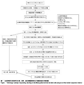
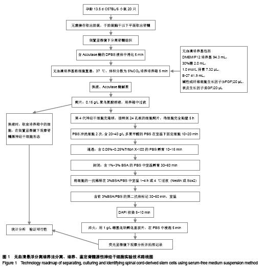
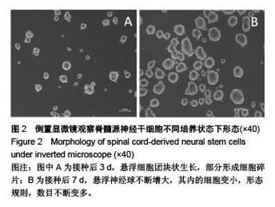
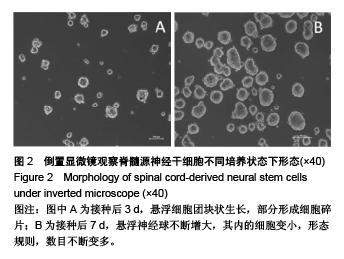
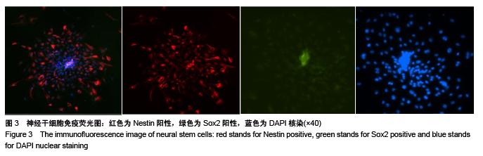
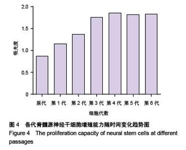
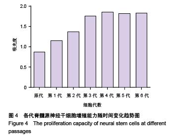
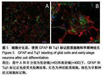
Spinal cord-derived neural stem cells cultured by serum-free suspension method: separation, cultivation and identification
Li Jun, Xu Da-bo, Fu Qiang, Liu Yan-bin
- Department of Orthopedic Surgery, the Affiliated Changhai Hospital of the Second Military Medical University, Shanghai 200433, China
-
Received:2015-10-26Online:2015-12-03Published:2015-12-03 -
Contact:Fu Qiang, Associate chief physician, Associate professor, Department of Orthopedic Surgery, the Affiliated Changhai Hospital of the Second Military Medical University, Shanghai 200433, China -
About author:Li Jun, Studying for master’s degree, Department of Orthopedic Surgery, the Affiliated Changhai Hospital of the Second Military Medical University, Shanghai 200433, China -
Supported by:the National Natural Science Foundation of China, No. 31371011
CLC Number:
Cite this article
Li Jun, Xu Da-bo, Fu Qiang, Liu Yan-bin. Spinal cord-derived neural stem cells cultured by serum-free suspension method: separation, cultivation and identification[J]. Chinese Journal of Tissue Engineering Research, 2015, 19(50): 8161-8166.
share this article

"

"
"

"
"

"
| [1] Ahmed S.The culture of neural stem cells.J Cell Biochem. 2009;106(1):1-6.
[2] Lee GY, Kim JA, Oh IH. Stem cell niche as a prognostic factor in leukemia. BMB Rep. 2015;48(8):427-428.
[3] Allazetta S, Lutolf MP. Stem cell niche engineering through droplet microfluidics. Curr Opin Biotechnol. 2015;35:86-93.
[4] DeCarolis NA, Kirby ED, Wyss-Coray T, et al. The Role of the Microenvironmental Niche in Declining Stem-Cell Functions Associated with Biological Aging.Cold Spring Harb Perspect Med. 2015;5(12). pii: a025874.
[5] Polisetti N, Zenkel M, Menzel-Severing J, et al. Cell Adhesion Molecules and Stem Cell-Niche-Interactions in the Limbal Stem Cell Niche.Stem Cells. 2015 Sep 9. doi: 10.1002/stem. 2191. [Epub ahead of print]
[6] Feng JF, Liu J, Zhang XZ, et al. Guided migration of neural stem cells derived from human embryonic stem cells by an electric field. Stem Cells. 2012;30(2):349-355.
[7] Li Q, Brus-Ramer M, Martin JH, et al. Electrical stimulation of the medullary pyramid promotes proliferation and differentiation of oligodendrocyte progenitor cells in the corticospinal tract of the adult rat. Neurosci Lett. 2010;479(2): 128-133.
[8] Koppes AN, Nordberg AL, Paolillo GM, et al. Electrical stimulation of schwann cells promotes sustained increases in neurite outgrowth. Tissue Eng Part A. 2014;20(3-4):494-506.
[9] Adams DS, Levin M. Endogenous voltage gradients as mediators of cell-cell communication: strategies for investigating bioelectrical signals during pattern formation. Cell Tissue Res. 2013;352(1):95-122.
[10] Sütterlin P, Williams EJ, Chambers D, et al. The molecular basis of the cooperation between EGF, FGF and eCB receptors in the regulation of neural stem cell function. Mol Cell Neurosci. 2013;52:20-30.
[11] Dupin E, Sommer L. Neural crest progenitors and stem cells: from early development to adulthood. Dev Biol. 2012;366(1): 83-95.
[12] Stemple DL, Anderson DJ. Isolation of a stem cell for neurons and glia from the mammalian neural crest. Cell. 1992; 71(6): 973-985.
[13] Mokrý J, Karbanová J. Foetal mouse neural stem cells give rise to ependymal cells in vitro. Folia Biol (Praha). 2006;52(5): 149-155.
[14] Deleyrolle LP, Reynolds BA. Identifying and enumerating neural stem cells: application to aging and cancer. Prog Brain Res. 2009;175:43-51.
[15] Zhang P, Wu X, Hu C, et al. Rho kinase inhibitor Y-27632 and Accutase dramatically increase mouse embryonic stem cell derivation. In Vitro Cell Dev Biol Anim. 2012;48(1):30-36.
[16] Bajpai R, Lesperance J, Kim M, et al. Efficient propagation of single cells Accutase-dissociated human embryonic stem cells. Mol Reprod Dev. 2008;75(5):818-827.
[17] Taha MF, Javeri A, Kheirkhah O, et al. Neural differentiation of mouse embryonic and mesenchymal stem cells in a simple medium containing synthetic serum replacement. J Biotechnol. 2014;172:1-10.
[18] Chojnacki A, Weiss S. Production of neurons, astrocytes and oligodendrocytes from mammalian CNS stem cells. Nat Protoc. 2008;3(6):935-940.
[19] Remaud S, López-Juárez SA, Bolcato-Bellemin AL, et al. Inhibition of Sox2 Expression in the Adult Neural Stem Cell Niche In Vivo by Monocationic-based siRNA Delivery. Mol Ther Nucleic Acids. 2013;2:e89.
[20] Kim SM, Flaßkamp H, Hermann A, et al. Direct conversion of mouse fibroblasts into induced neural stem cells. Nat Protoc. 2014;9(4):871-881.
[21] De Filippis L, Binda E. Concise review: self-renewal in the central nervous system: neural stem cells from embryo to adult. Stem Cells Transl Med. 2012;1(4):298-308.
[22] Yaqubi M, Mohammadnia A, Fallahi H. Predicting involvement of polycomb repressive complex 2 in direct conversion of mouse fibroblasts into induced neural stem cells. Stem Cell Res Ther. 2015;6:42.
[23] Nout YS, Rosenzweig ES, Brock JH, et al. Animal models of neurologic disorders: a nonhuman primate model of spinal cord injury. Neurotherapeutics. 2012;9(2):380-392.
[24] Conover JC, Notti RQ. The neural stem cell niche. Cell Tissue Res. 2008;331(1):211-224.
[25] Haubenwallner S, Katschnig M, Fasching U, et al. Effects of the polymeric niche on neural stem cell characteristics during primary culturing. J Mater Sci Mater Med. 2014;25(5): 1339-1355.
[26] Kerever A, Mercier F, Nonaka R, et al. Perlecan is required for FGF-2 signaling in the neural stem cell niche. Stem Cell Res. 2014;12(2):492-505.
[27] Belmadani A, Tran PB, Ren D, et al. Chemokines regulate the migration of neural progenitors to sites of neuroinflammation. J Neurosci. 2006;26(12):3182-3191.
[28] Illes S, Theiss S, Hartung HP, et al. Niche-dependent development of functional neuronal networks from embryonic stem cell-derived neural populations. BMC Neurosci. 2009; 10:93.
[29] Garcion E, Halilagic A, Faissner A, et al. Generation of an environmental niche for neural stem cell development by the extracellular matrix molecule tenascin C. Development. 2004; 131(14):3423-3432.
[30] Musienko P, van den Brand R, Maerzendorfer O, et al. Combinatory electrical and pharmacological neuroprosthetic interfaces to regain motor function after spinal cord injury. IEEE Trans Biomed Eng. 2009;56(11 Pt 2):2707-2711. |
| [1] | Jiang Tao, Ma Lei, Li Zhiqiang, Shou Xi, Duan Mingjun, Wu Shuo, Ma Chuang, Wei Qin. Platelet-derived growth factor BB induces bone marrow mesenchymal stem cells to differentiate into vascular endothelial cells [J]. Chinese Journal of Tissue Engineering Research, 2021, 25(25): 3937-3942. |
| [2] | Chen Yang, Huang Denggao, Gao Yuanhui, Wang Shunlan, Cao Hui, Zheng Linlin, He Haowei, Luo Siqin, Xiao Jingchuan, Zhang Yingai, Zhang Shufang. Low-intensity pulsed ultrasound promotes the proliferation and adhesion of human adipose-derived mesenchymal stem cells [J]. Chinese Journal of Tissue Engineering Research, 2021, 25(25): 3949-3955. |
| [3] | Zhang Lishu, Liu Anqi, He Xiaoning, Jin Yan, Li Bei, Jin Fang. Alpl gene affects the therapeutic effect of bone marrow mesenchymal stem cells on ulcerative colitis [J]. Chinese Journal of Tissue Engineering Research, 2021, 25(25): 3970-3975. |
| [4] | Ruan Guangping, Yao Xiang, Liu-Gao Miyang, Cai Xuemin, Li Zian, Pang Rongqing, Wang Jinxiang, Pan Xinghua. Umbilical cord mesenchymal stem cell transplantation for traumatic systemic inflammatory response syndrome in tree shrews [J]. Chinese Journal of Tissue Engineering Research, 2021, 25(25): 3994-4000. |
| [5] | Mo Jianling, He Shaoru, Feng Bowen, Jian Minqiao, Zhang Xiaohui, Liu Caisheng, Liang Yijing, Liu Yumei, Chen Liang, Zhou Haiyu, Liu Yanhui. Forming prevascularized cell sheets and the expression of angiogenesis-related factors [J]. Chinese Journal of Tissue Engineering Research, 2021, 25(22): 3479-3486. |
| [6] | Chen Lei, Zheng Rui, Jie Yongsheng, Qi Hui, Sun Lei, Shu Xiong. In vitro evaluation of adipose-derived stromal vascular fraction combined with osteochondral integrated scaffold [J]. Chinese Journal of Tissue Engineering Research, 2021, 25(22): 3487-3492. |
| [7] | Wei Qin, Zhang Xue, Ma Lei, Li Zhiqiang, Shou Xi, Duan Mingjun, Wu Shuo, Jia Qiyu, Ma Chuang. Platelet-derived growth factor-BB induces the differentiation of rat bone marrow mesenchymal stem cells into osteoblasts [J]. Chinese Journal of Tissue Engineering Research, 2021, 25(19): 2953-2957. |
| [8] | Chen Xiao, Guo Zhi, Chen Lina, Liu Xuanyong, Zhang Yihuizhi, Li Xumian, Wang Yueqiao, Wei Liya, Xie Jing, Lin Li. Factors affecting the mobilization and collection of autologous peripheral blood hematopoietic stem cells [J]. Chinese Journal of Tissue Engineering Research, 2021, 25(19): 2958-2962. |
| [9] | Guo Zhibin, Wu Chunfang, Liu Zihong, Zhang Yuying, Chi Bojing, Wang Bao, Ma Chao, Zhang Guobin, Tian Faming. Simvastatin stimulates osteogenic differentiation of bone marrow mesenchymal stem cells [J]. Chinese Journal of Tissue Engineering Research, 2021, 25(19): 2963-2968. |
| [10] | Li Congcong, Yao Nan, Huang Dane, Song Min, Peng Sha, Li Anan, Lu Chao, Liu Wengang. Identification and chondrogenic differentiation of human infrapatellar fat pad derived stem cells [J]. Chinese Journal of Tissue Engineering Research, 2021, 25(19): 2976-2981. |
| [11] | Gao Yuanhui, Xiang Yang, Cao Hui, Wang Shunlan, Zheng Linlin, He Haowei, Zhang Yingai, Zhang Shufang, Huang Denggao. Comparison of biological characteristics of adipose derived mesenchymal stem cells in Wuzhishan inbreed miniature pigs aged two different months [J]. Chinese Journal of Tissue Engineering Research, 2021, 25(19): 2988-2993. |
| [12] | Cao Yang, Zhang Junping, Peng Li, Ding Yi, Li Guanghui. Isolation and culture of rabbit aortic endothelial cells and biological characteristics [J]. Chinese Journal of Tissue Engineering Research, 2021, 25(19): 3000-3003. |
| [13] | Dai Min, Wang Shuai, Zhang Nini, Huang Guilin, Yu Limei, Hu Xiaohua, Yi Jie, Yao Li, Zhang Ligang. Biological characteristics of hypoxic preconditioned human amniotic mesenchymal stem cells [J]. Chinese Journal of Tissue Engineering Research, 2021, 25(19): 3004-3008. |
| [14] | Qin Yanchun, Rong Zhen, Jiang Ruiyuan, Fu Bin, Hong Xiaohua, Mo Chunmei. Chinese medicine compound preparation inhibits proliferation of CD133+ liver cancer stem cells and the expression of stemness transcription factors [J]. Chinese Journal of Tissue Engineering Research, 2021, 25(19): 3016-3023. |
| [15] | Dai Yaling, Chen Lewen, He Xiaojun, Lin Huawei, Jia Weiwei, Chen Lidian, Tao Jing, Liu Weilin. Construction of miR-146b overexpression lentiviral vector and the effect on the proliferation of hippocampal neural stem cells [J]. Chinese Journal of Tissue Engineering Research, 2021, 25(19): 3024-3030. |
| Viewed | ||||||
|
Full text |
|
|||||
|
Abstract |
|
|||||