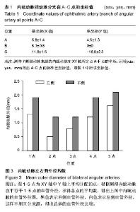
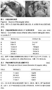
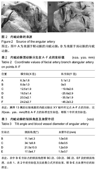
| [1] Rokenes HK,Bretteville G,Lovdal O,et al.The nasolabial skin flap in intraoral reconstruction.ORL J Otorhinolaryngol Relat Spec. 1991;53(6):346-348.
[2] 邵英,张舵,自然,等.侧鼻动脉为蒂的鼻唇沟皮瓣修复鼻缺损[J].中国修复重建外科杂志,2010,24(5):552-555.
[3] Napolitano M,Mast BA.The nasolabial flap revisited as an adjunct to floor-of mouth reconstruction.Ann Plast Surg. 2001; 46(3):265-268.
[4] 胡小华,黄桂林,张霓霓,等.鼻唇沟岛状皮瓣在口底组织缺损修复重建中的应用[J].中国修复重建外科杂志,2014,28(6):710-713.
[5] Yang HM,Lee JG,Hu KS,et al. New anatomical insights of the course and branching patterns of the facial artery: clinical implications regarding injectable treatments to the nasolabial fold and nasojugal groove. Plast Reconstr Surg. 2014.
[6] Mitz V,Peyronie M.The superficai muscul oapneurotic system (SMAS)in the parotid and cheek ates.Plast Reconstr Surg. 1976;58(1):80.
[7] 熊绍虎,许达传,程新德,等.面部真皮下血管网皮瓣血供的解剖学基础[J].中国临床解剖学杂志,2000,18(4):330-332.
[8] Fabrizio T, Savani A, Sanna M, et al. The retroangular flap for nasal reconstruction.Plast Reconstr Surg. 1996;97(2):431- 435.
[9] 吴晓勇,熊猛.面动脉及其分支的解剖及临床价值[J].中国美容医学2013,22(5):523-525.
[10] 谢义德,黄拔瑞,郭志辉.鼻唇沟皮瓣在面部整形中的应用[J].中华医学美容杂志,1997,6(3): 100-101.
[11] Guero S, Bastian D, Lassau JP, et al. Anatomical basis of a new nasolabial island flap. Surg Radial Anat. 1991;13(3):265- 270.
[12] Senem E, Figen G. Anatomy of the Supraorbital Region and the Evaluation of it for the Reconstruction of Facial Defects. J Craniofac Surg. 2007;18(1):104-112.
[13] Bouhanna A, Bruant-Rodier C, Himy S,et al. Reconstruction of the nasal alar defect with the superiorly based nasolabial flap describe by Burget: report of seven cases. Ann Chir Plast Esthet. 2007;24(3):35-38.
[14] 陈日亭.颌面颈手术解剖[M].北京:人民卫生出版社,1994:99.
[15] 吴镝,王新刚,黄晨煜,等.面动脉的解剖学研究及其临床意义[J].组织工程与重建外科杂志,2006,2(5):256-258.
[16] 郝鹏.鼻部血管的应用解剖及临床意义[D].山东大学,2011.
[17] Nanjan NS. An anatomical study of the facial artery. Ann Plast Surg. 1988;88:39-50.
[18] 江桂华,颜剑豪,林楚岚,等. 多层螺旋CT血管成像对面动脉的解剖学研究[J].南方医科大学学报,2008,28(3):457-459.
[19] 刘玉广,张先东.面动脉的彩色多普勒解剖分型及临床意义[J].医学影像学杂志,2013,23(8):1180-1182.
[20] Tan O,Atik B, Ergen D. The Retroangular Flap Revisited. Dermatol Surg. 2007;33(11):1343-1349.
[21] 沈倩云,章一新,李靖,等.鼻唇沟穿支皮瓣在鼻翼修复中的应用[J].中国美容整形外科杂志,2010,21(1):6-14. |