Chinese Journal of Tissue Engineering Research ›› 2015, Vol. 19 ›› Issue (7): 1104-1111.doi: 10.3969/j.issn.2095-4344.2015.07.022
Previous Articles Next Articles
A systematic review of 585 nm pulsed dye laser for repair of pathological scars
Song Li, Ye Jun-ru, Lu Mao, Peng Ke, Shen Yue-li, Zhu Jiang-ting
- Department of Dermatology, First Affiliated Hospital of Chengdu Medical College, Chengdu 610500, Sichuan Province, China
-
Online:2015-02-12Published:2015-02-12 -
About author:Song Li, Master, Attending physician, Department of Dermatology, First Affiliated Hospital of Chengdu Medical College, Chengdu 610500, Sichuan Province, China -
Supported by:the Project of Sichuan Provincial Health Bureau in 2012, No. 120480
CLC Number:
Cite this article
Song Li, Ye Jun-ru, Lu Mao, Peng Ke, Shen Yue-li, Zhu Jiang-ting. A systematic review of 585 nm pulsed dye laser for repair of pathological scars[J]. Chinese Journal of Tissue Engineering Research, 2015, 19(7): 1104-1111.
share this article
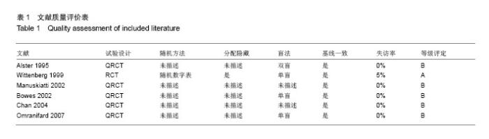
2.1 文献检索结果 按照设计的检索式在CCTR中检索出49篇,MEDLINE中121篇,Embase中48篇,中国期刊全文数据库和中文科技期刊全文数据库共60篇,共294篇。通过阅读题目和摘要,筛选出可能相关的共18篇。找出全文并仔细阅读后,最终纳入6篇[11-16],全部为英文文献,排除11 篇[17-27]。排除的11篇中4篇治疗组为激光加其他治 疗[17-20],1篇为CCT[21],3篇对象为手术后正常瘢痕[22-24],1篇为烧伤后正常瘢痕[25],1篇为痤疮瘢痕[26],1篇脉冲染料激光采用的是595 nm波长[27]。 手工检索及附加检索无补充。 2.2 纳入研究一般特征 2.2.1 样本量 共纳入6个研究[11-16],199例患者,208个瘢痕。试验样本量从10到120个瘢痕不等。 2.2.2 研究对象特点 纳入的研究对象包括增生性瘢痕及瘢痕疙瘩患者,年龄及性别无明确限制。其中6个研究均是将同一个瘢痕分为几部分,分别用于治疗组及对照组。 2.2.3 干预措施特点 纳入的研究按不同的对照组分类主要有以下几组:① 585 nm脉冲激光VS空白对照(3个研究[11,13,15])。②585 nm脉冲激光VS 532 nm倍频Nd:YAG激光(Q开关及可变脉冲模式)(1个研究[14])。③585 nm脉冲激光VS曲安奈德皮损内注射(1个研究[16])。④585 nm脉冲激光VS硅凝胶膜(1个研究[12])。⑤585 nm脉冲激光VS饵激光(1个研究[16])。 治疗措施均采用585 nm脉冲激光,在治疗组中,脉宽为450-1 500 ms,光斑大小5-10 mm,能量密度3.5-9 J/cm2,治疗间隔为4-8周,治疗次数为2-12次。 2.2.4 疗效判定 ①主要指标:温哥华烧伤瘢痕评价量表、红斑、瘢痕大小,包括厚度、体积、长度、宽度等,血流、柔韧度、弹性。②次要指标:所有研究均包括了次要指标的测量,主要有:患者症状、患者主观评价等。③治疗时间:所有研究治疗时间为22周至2年。1篇研究描述了失访率为5%[11],其余研究均报道失访率为0%。④不良反应:4个研究报告了不良反应,其中包括术中疼痛、紫癜、色素沉着、水疱等[12-15]。其中1个研究报道无不良反应[13]。另2个研究未描述不良反应[11,16]。 2.3 纳入研究质量 2.3.1 随机方法 文章纳入的6个研究中,包括6篇RCT[11-16],1篇采用计算机生成随机数字表的方法进行随机分配[12]。另有5篇QRCT[11,13-16],未描述随机方法。1个研究[12]进行分配隐藏,其他没有(见表1)。"

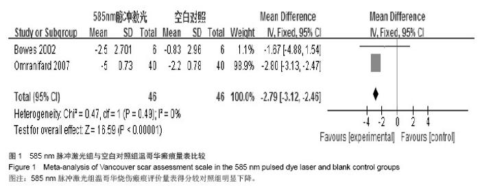
2.3.2 盲法 其中有1篇研究采取了双盲[11],3篇研究对评价者采用了盲法[12,14,16],2篇研究不清楚是否采用盲法[13,15]。 2.3.3 基线一致 所有的研究均描述了试验组与对照组的基线情况,具有可比性。 2.3.4 随访情况 全部研究进行了随访,随访时间为22周至2年。只有1个研究失访率5%[12],其他报道的失访率为0%。该研究未描述是否进行意向性处理(ITT)分析。 2.4 统计结果分析 2.4.1 585 nm脉冲激光与无治疗相比 共有5篇研究比较了585 nm脉冲激光组与无治疗组主要指标和次要指标[11-15]。 主要指标: 温哥华量表:其中有2个研究有关于温哥华烧伤瘢痕评价量表的结果(图1)[14,16],均显示温哥华量表得分有明显下降,与对照组结果相比有统计学意义。"

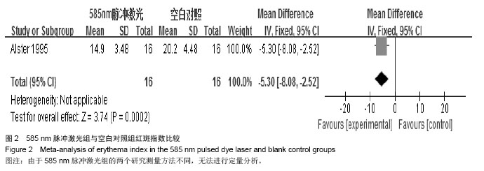
红斑:2个研究比较了红斑消退情况[11,13],其中1个研究显示在32周时红斑明显减退[13],P=0.02,但585 nm脉冲激光组与对照组比较,差异无显著性意义,另1个研究显示在1次及2次治疗结束示时红斑明显减退(图2)[11],与对照组相比差异有显著性意义,由于两个研究测量方法不同,且第1个研究未报道原始数据,无法进行定量分析。"

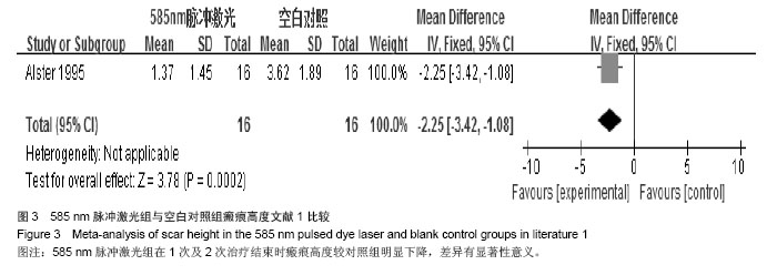
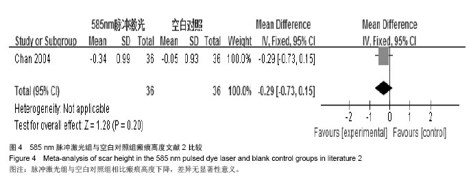
大小:包括高度、长度、宽度、体积等。4个研究比较了瘢痕大小的改变情况[11,12,13,15],其中3个研究描述了瘢痕的高度[11,13,15]。1个研究显示在1次及2次治疗结束示时瘢痕高度明显下降(图3)[11],与对照组相比差异有显著性意义;另1个研究显示在32周时瘢痕高度明显下降[13],与对照组相比差异有显著性意义,(P=0.005)但该研究未报道原始数据;还有1个研究[15]显示585 nm脉冲激光组与空白对照组相比高度下降,差异无显著性意义(P=0.20)[WMD=-0.29,95% CI(-0.73,0.15)](图4)。但由于这2个研究未报道完整的原始数据[11,15],无法进行数据合并,只有分别研究。1个研究描述了瘢痕的体积[12],40周的研究结果示585 nm组相对于空白对照组在瘢痕体积改变方面差异无显著性意义(P=0.13)。"

"
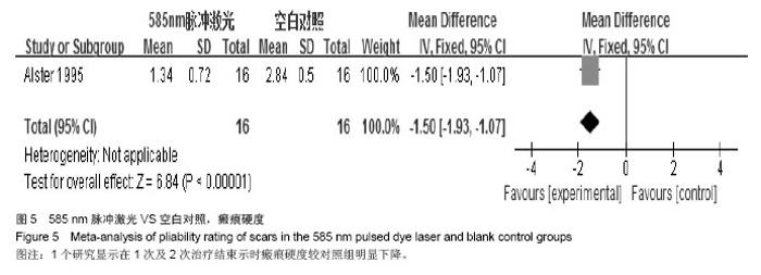
弹性或硬度:4个研究比较了瘢痕弹性或硬度的改变情况[11-13,15],其中2个研究描述了瘢痕的硬度[11,13],1个研究显示在1次(P=0.000 7)及2次治疗(P=0.000 4)结束示时瘢痕硬度明显下降[11],与对照组相比差异有显著性意义(图5)。而另外一个研究显示585 nm脉冲激光组在治疗32周后与基线相比瘢痕硬度改变[13],差异无显著性意义(P=0.02),而对照组24周时硬度改变与基线相比差异有显著性意义(P=0.046),但该差异未保持到32周,该研究无原始数据,不能与之前研究进行合并。其中2个研究描述了瘢痕的弹性[12,15],1个研究描述弹性变化治疗组与对照组间差异无显著性意义(P=0.76)[12],该研究未见原始研究数据。另1个研究用皮肤弹性测量仪进行弹性变化测量[15],只有1次测量弹性变化治疗组与对照组间差异有显著性意义,其他4次测量差异均无显著性意义,这2个研究因为缺乏原始数据不能合并。"

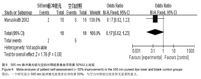
次要指标:患者自我评价:2个研究报道了患者对治疗的主观评 价[13,15],其中一个研究显示比较患者自评改善50%及以上的比例[13],治疗组与空白对照组差异无显著性意义(图6)。而另一个研究显示治疗组在痒(P=0.007)、疼痛(P =0.001)、敏感(P=0.018)、大小改变(P =0.023)方面患者的主观评有明显好转[15],差异有显著性意义,而在针刺感(P=0.201)及颜色改变(P=0.122)方面患者评价的明显好转率两组间差异无显著性意义,但总的评分治疗组优于空白对照组,两组间差异有显著性意义(P=0.003)。 患者症状:1个研究报道了患者的痒痛评分[12],数据显示585 nm脉冲激光组与空白对照组在改善患者痒痛症状方面差异无显著性意义(P=0.99)。 2.4.2 585 nm脉冲激光与硅凝胶膜组相比 共有1篇研究比较了585 nm脉冲激光组与无治疗组主要指标和次要指标[12]。 主要指标:①红斑:该研究显示585 nm组与硅凝胶膜组在改变瘢痕红斑(血流)方面[12],差异无显著性意义(P=0.73)。②弹性:该研究显示585 nm组与硅凝胶膜组在改变瘢痕弹性方面[12],差异无显著性意义。③体积:该研究显示585 nm组与硅凝胶膜组在改变瘢痕弹性方面[12],差异无显著性意义(P=0.85)。 次要指标:患者症状:1个研究报道了患者的痒痛评分[12],数据显示585 nm脉冲激光组硅凝胶膜组在改善患者痒痛症状方面差异无显著性意义(P=0.53)。 2.4.3 585 nm脉冲激光与532 nm倍频Nd:YAG激光(Q开关及可变脉冲模式)组相比 共有1篇研究比较了585 nm脉冲激光组与532 nm倍频Nd:YAG激光[14],该研究将532 nm倍频Nd:YAG激光分为Q开关及可变脉冲模式。 主要指标温哥华量表:该研究比较了585 nm脉冲激光及532 nm倍频Nd:YAG激光温哥华烧伤瘢痕评价量表的结果[14],均显示温哥华量表得分有明显下降,但与Q开关及可变脉冲模式组相比结果相比无统计学意义。 次要指标患者自我评价:1个研究报道了患者对治疗的主观评价[14],该研究显示参试的6人中的5人都认为Q开关532 nm倍频Nd:YAG激光从总体来讲是最好的治疗选择。 2.4.4 585 nm脉冲激光与曲安奈德皮损内注射组相比 共有2篇研究比较了585 nm脉冲激光组与曲安奈德皮损内注射[13,16]。 主要指标:①温哥华量表:其中有1个研究有关于温哥华烧伤瘢痕评价量表的结果[16],显示两组温哥华量表得分与治疗之前比有明显下降,但激光组与曲安奈德皮损内注射组相比差异无显著性意义。②红斑:该研究比较了585 nm脉冲激光及曲安奈德皮损内注射[13],结果显示两组在改变红斑方面,差异无无显著性意义。③硬度:该研究比较了585 nm脉冲激光及曲安奈德皮损内注射[13],结果显示两组在改变瘢痕硬度方面,差异无显著性意义。④体积:该研究比较了585 nm脉冲激光及曲安奈德皮损内注射治疗病理性瘢痕[13],随访至32周时曲安奈德皮损内注射组在改变瘢痕体积方面有更能减小瘢痕体积的趋势,但两组数据差异无显著性意义。 "

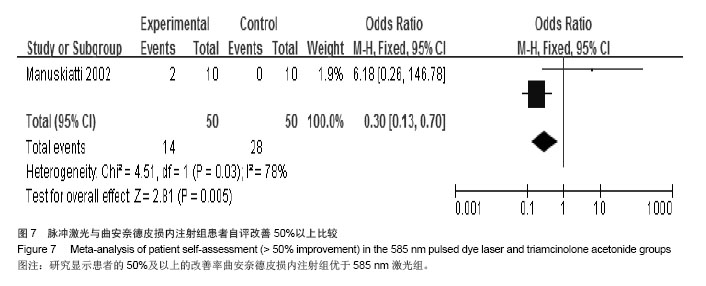
次要指标:患者自我评价:2个研究均报道了患者对治疗的主观评价[13,16],该研究显示患者的50%及以上的改善率曲安奈德皮损内注射组优于585 nm激光组,两组差异有显著性意义(图7)。 2.4.5 585 nm脉冲激光与铒激光(1个研究[16]) 共有1篇研究比较了585 nm脉冲激光组与铒激光[16]。 主要指标温哥华量表:其中有1个研究有关于温哥华烧伤瘢痕评价量表的结果[16],显示两组温哥华量表得分与治疗之前比有明显下降,但激光组与铒激光组相比差异无显著性意义。"
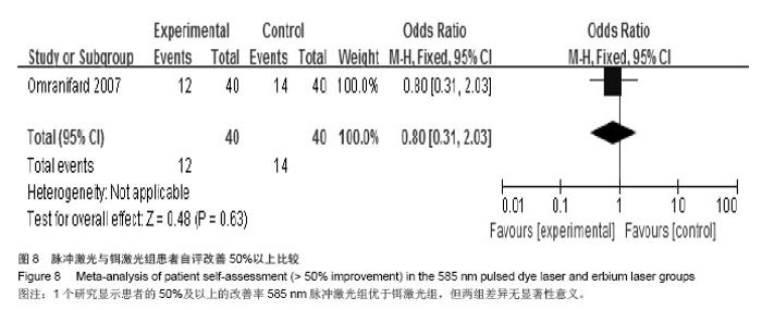
次要指标患者自我评价:1个研究报道了患者对治疗的主观评价[16],该研究显示患者的50%及以上的改善率585 nm脉冲激光组优于铒激光组,但两组差异无显著性意义(图8)。 2.5 不良反应 本课题共纳入使用585 nm脉冲激光治疗的病理性瘢痕208个,其中发生水疱的1例[15],部分患者有轻度紫癜及暂时的色素沉着[14],其他报道的不良反应主要是术中疼痛[12,14],1个研究报道了1例患者因无法忍受疼痛终止治疗[12]。所有研究未报道较严重的不良反应。多数不良反应在术后给予护理及对症处理后均可缓解,大多数研究显示下次治疗时降低治疗剂量均可避免再次出现不良反应。"

| [1]徐阳.增生性瘢痕的激光治疗进展[J].中国美容医学,2014,23(5): 420-424. [2]Tanaka A, Hatoko M, Tada H, et al. Expression of p53 family in scars. J Dermatol Science.2004;34:17-24. [3]Amadeu TP,Braune AS,Porto LC,et al.Fibrillin-1 and elastin are differentially expressed in hypertrophic scars and keloids. Wound Repair Regen. 2004;12:169-174. [4]蔡景龙.激光治疗瘢痕的新变化[J].中国美容整形外科杂志, 2013,24(11):645-647 [5]Liu A,Moy RL,Ozog DM.Current methods employed in the prevention andminimizatioof surgical scars. Dermatol Surg. 2011;37(12):1740-17446 [6]郭君.增生性瘢痕的激光治疗进展[J].中国美容医学2012;21(5): 878-880. [7]Alester TS.Improvement of erythematous and hypertrophic scars by 585nmflashlamp-pumpedpulseddyelaser. Ann Plast Surg.1994;32(2):186-190. [8]邹彦芬,于波,钟绮丽等.585nm/1064nm双波长复合激光预防手术后瘢痕增生的自体对照观察[J].中国皮肤性病学杂志, 2014, 28(4):372-374. [9]何传果,李栋梁,李大铁.不同治疗周期染料激光对兔耳增生性瘢痕硬度的影响[J].中国美容整形外科杂志,2011,22(11):697-699. [10]Asilian A,Darougheh A,Shariati F. Newcombinationoftriamcinolone,5-Fluorouracil,and pulsed-dye laser for treatment of keliod and hypertrophic scars,Dermatol surg.2006;32(7):907-915. [11]Alster TS, Williams CM. Treatment of keliod sternotomy scars with585nmflashlamp-pumpedplulsed-dyelaser.Lancet.1995; 345:1198-200. [12]Gregory P,Wittenberg MD.Prospective,Single-blind, Randomized,Controlled Study to Assess the efficacy of the 585-nm Flashlamp-Pumed plused-dye Laser and silicone Gel Sheeting in Hypertrophic Scar Treatment.Arch Dermatol. 1999;135(9):1049. [13]Manuskiatti W, Fitzpatrick RE. Treatment Pesponse of keliodal and Hypertrophic Sternotomy Scars.Arch Dermatol 2002;138(9):1149. [14]Bowes LE, Nouri K, Berman B,et al.Treatment of Pigmented Hypertrophic Scars with the 585nm Pulsed Dye Laser and the 532nm Frequncy-Doubled Nd:YAG Laser in the Q-Switched and Variable Pulsed Modes:A Comparative Study,Dermatol Surg .2002;28:714-719. [15]Chan HH, Wong DS, Ho WS, et al. The use of Pulsed Dye Laser for the Preventiong and Trentment of Hypertrophic scars in Chinese Persons.Dermatol Surg 2004;30:987-994. [16]Omranifard M.Comparing the effects of conventional method,plused dye laser and erbium laser for the treatment of hyperteophic scars in Iranian petients.JRMS. 2007;12(6): 277-281. [17]陈文革,喻楠,王建军,等.脉冲染料激光联合外用咪喹莫特治疗病理性瘢痕的疗效观察[J].中国皮肤性病学杂志.2014;28(4):369-371 [18]倪小丽,车敦发,张添.585nm脉冲染料激光联合外用硅凝胶膜早期治疗增生性瘢痕的疗效观察[J].中国美容医学, 2013,22(5): 550-552. [19]Ali Aslian MD,AfshinDaroughen MD. New Combination of Triaciolone,5-Fluororacil,and Pulsed-Dye Laser for Treatment of Keliod and Hypertrophic Scars.Dermatol Surg. 2006;32(7): 907-915. [20]宋金荣,翁伟丽,李勤等.脉冲染料激光联合点阵铒激光治疗增生性瘢痕的临床体会[J].中国美容医学,2012,21(7):1183-1184. [21]Paquet P, Hermanns JF, Pierard GE. Effect of the 585nm flashlamp-pumped pulsed dye laser for the treatment of keloids. Dermatologic surgery.2001;27(2):171-174. [22]Nouri K, Elsaie ML, Vejjabhinanta V,et al.Comparison of the effects of short-and long-pulse durations when using a 585nm pulsed dye laser in the treatment of new surgical scars.Lasers Med Sci.2010;25:121-126. [23]Nouri K, Rivas MP, Stevens M,et al.Comparision of the effectiveness of the pulsed dye laser 585nm versus 595nm in the treatment of new suigical scars.Lasers Med Sci. 2009;24: 801-810. [24]Davari P, Gorouhi F, Hashemi P, et al.Pulsed dye laser treatment with different onset times for new suigical scars:a single-blind randomized controlled trial.Lasers Med Sci. 2012; 27:1095-1098. [25]Hultman CS, Edkins RE, Wu C, et al.Prospective,Before-After Cohort Study to Assess the Efficacy of Laser Therapy on Hypertrophic Burn Scars.Annals of Plastic Surgery. 2013; 70(5):521-525. [26]Lee DH, Choi YS, Min SU.Comparision of a 585nm pulsed dye laser and a 1064-nm Nd :YAG laser for the treatment of acne scars:A randomized split-face clinical study.Dermatology Surgery.2009;11(2):801-807. [27]钟姗,梅雪岭,赵俊英.不同脉宽595nm染料激光治疗增生性瘢痕和瘢痕疙瘩的疗效.首都医科大学学报,2013,34(5):759-765 [28]何黎,刘玮.皮肤美容学[M].北京:人民卫生出版社,2013:34-35. [29]刘玮,赖维,王学民.中国城市女性人群皮肤类型调查及相关研究[J].临床皮肤科杂志,2005,34(7):420-423. [30]仲少敏,赵俊郁,朱学骏.皮肤光反应类型与紫外线诱导黑化反应相关性分析[J].中国皮肤性病学杂志,2013,27(12):1208-1210. |
| [1] | Ruan Guangping, Yao Xiang, Cai Xuemin, Li Zian, Pang Rongqing, Pan Xinghua. Effect of umbilical cord mesenchymal stem cell transplantation for treating systemic lupus erythematosus in a tree shrew model [J]. Chinese Journal of Tissue Engineering Research, 2021, 25(1): 90-95. |
| [2] | Zhou Yanxing1, Peng Xinsheng2, Hou Gan1, Li Jiangbin1, Zhang Hua1, Zhou Zhikun2, Zhou Yanfang3 . Inhibitory effect of capsaicin on fibroblast proliferation and its molecular mechanism [J]. Chinese Journal of Tissue Engineering Research, 2019, 23(7): 1018-1022. |
| [3] | Ma Linjie, Xue Wentao, Tan Jupeng. Induced pluripotent stem cell transplantation for systemic lupus erythematosus in a mouse model [J]. Chinese Journal of Tissue Engineering Research, 2019, 23(33): 5286-5292. |
| [4] | Fan Siqi1, Zeng Ping2, Zhou Yi3, Qin Gang2, Liao Xiaobo2, He Kaiyi2. Screening differentially expressed proteins in the serum of patients with systemic lupus erythematosus combined with osteonecrosis of the femoral head by iTRAQ technology [J]. Chinese Journal of Tissue Engineering Research, 2019, 23(3): 476-481. |
| [5] | Ye Ling, Zhu Jing, He Chengsong. Intervention with autologous adipose-derived mesenchymal stem cells for immune function in mice with systemic lupus erythematosus [J]. Chinese Journal of Tissue Engineering Research, 2019, 23(17): 2696-2702. |
| [6] | Li Xiang, Wu Zhixian, Liu Hongwei, Liang Jie, Mo Zizeng. Molecular mechanism underlying the effect of adipose-derived stem cells on the proliferation of keloid fibroblasts [J]. Chinese Journal of Tissue Engineering Research, 2019, 23(1): 61-67. |
| [7] | Jing Ya-jun 1, Zhang Lei1, Zhang Shao-qun1, Liao Li-qing1, Yuan Shi-guo2, Li Yi-kai1. Pathological changes of the skeletal muscle after local loosening therapies [J]. Chinese Journal of Tissue Engineering Research, 2018, 22(4): 535-541. |
| [8] | Ge Wenjia, Ma Xiaorong, Ouyang Tianxiang . Aminolevulinic acid photodynamic therapy can significantly inhibit the growth of endothelial cells and induce cell apoptosis [J]. Chinese Journal of Tissue Engineering Research, 2018, 22(36): 5840-5845. |
| [9] | Lv Ying1, An Mei-wen1, Hou Chun-sheng2. Finite element analysis of skin closure stress in different directions [J]. Chinese Journal of Tissue Engineering Research, 2017, 21(4): 609-614. |
| [10] | Wu Zi-han1, Li Gao-feng2. Correlation of angiopoietin-1 with angiogenesis during scar formation [J]. Chinese Journal of Tissue Engineering Research, 2017, 21(32): 5158-5163. |
| [11] | Wang Ying-cui, Pan Xing-hua . Immunoregulatory mechanism of mesenchymal stem cells on the T and B cells in systemic lupus erythematosus [J]. Chinese Journal of Tissue Engineering Research, 2017, 21(29): 4734-4741. |
| [12] | Liu Ying-wei, Zhang Wan-li, Chi Cheng-tao, Xu Qing-yu, Lu De-zhi. Effects of hyaluronic acid on scar formation in the acellular nerve allograft [J]. Chinese Journal of Tissue Engineering Research, 2016, 20(42): 6317-6323. |
| [13] | Fan Xu-hui, Yang Bo, Hu Xiang, Guan Fang-xia. Reactive hyperplasia of glial cells induced by spinal cord injury in a rat model [J]. Chinese Journal of Tissue Engineering Research, 2016, 20(40): 6001-6006. |
| [14] | Ding Chao, Sun Qiang, Tang Cheng. Comparison of 3.0T MRI and SPECT-CT in the diagnosis of osteoporotic vertebral compression fractures [J]. Chinese Journal of Tissue Engineering Research, 2016, 20(39): 5885-5891. |
| [15] | Li Jian-feng, Yan Jin-yu, Xia Run-fu, Zhang Xu, Tan Xiao-hui, Guan Jian, Ye Zhen, Zhang Shu-lian. Glial scar formation and astrocyte role in spinal cord injury [J]. Chinese Journal of Tissue Engineering Research, 2016, 20(37): 5609-5616. |
| Viewed | ||||||
|
Full text |
|
|||||
|
Abstract |
|
|||||