Chinese Journal of Tissue Engineering Research ›› 2014, Vol. 18 ›› Issue (53): 8634-8640.doi: 10.3969/j.issn.2095-4344.2014.53.020
Previous Articles Next Articles
Three dimensional test of lumbar canal volume in degenerative spondylolisthesis patients under physiological load
Xu Hong-da1, 2, Miao Jun2, Xia Qun2
- 1Graduate School of Tianjin Medical University, Tianjin 300070, China
2Department of Spinal Surgery, Tianjin Hospital, Tianjin 300211, China
-
Revised:2014-11-02Online:2014-12-24Published:2014-12-24 -
About author:Xu Hong-da, Studying for master’s degree, Graduate School of Tianjin Medical University, Tianjin 300070, China; Department of Spinal Surgery, Tianjin Hospital, Tianjin 300211, China -
Supported by:the National Natural Science Foundation of China, No. 81371992.
CLC Number:
Cite this article
Xu Hong-da, Miao Jun, Xia Qun . Three dimensional test of lumbar canal volume in degenerative spondylolisthesis patients under physiological load[J]. Chinese Journal of Tissue Engineering Research, 2014, 18(53): 8634-8640.
share this article

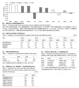
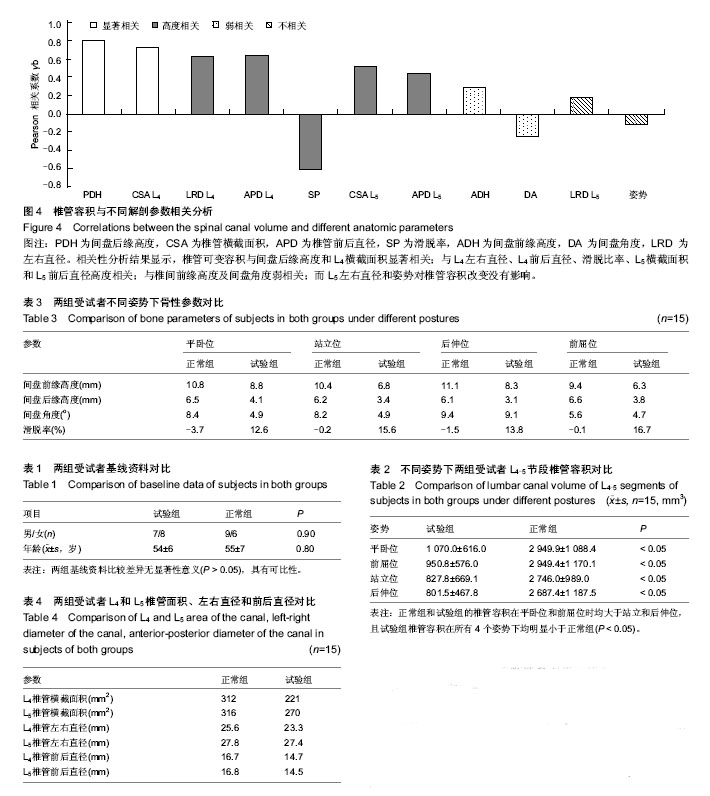
2.1 参与者数量分析 按意向性处理,共纳入试验组L4退变滑脱患者15例,正常组15例,全部进入结果分析,无脱落。 2.2 基线资料比较 两组基线资料比较差异无显著性意义(P > 0.05),具有可比性,见表1。 2.3 L4-5节段椎管容积测量 腰椎退变滑脱患者椎管容积(809.9-1 070.0 mm3)在4个姿势下均明显小于正常人 (2 687.4-2 949.9 mm3,P < 0.05)。 同时两组受试者椎管容积随着姿势改变出现相同的规律,即椎管容积在平卧位和前屈位均分别大于站立位和后伸位,见表2。 2.4 影响L4-5滑脱节段椎管容积的解剖因素 测量了可能影响椎管容积的相关参数(表3,4)。椎管可变容积与间盘后缘高度(γb=0.80,P=0.00)和L4横截面积(γb=0.74, P显著相关;与L4左右直径(γb=0.67,P=0.00)、L4前后直径(γb=0.61,P=0.00)、滑脱比率(γb=-0.61, P、L5横截面积(γb=0.57,P=0.00)和L5前后直径(γb=0.47,P=0.00)高度相关;与椎间前缘高度(γb=0.28, P及间盘角度(γb=-0.24,P=0.02)弱相关;而L5左右直径(γb=0.18,P=0.06)和姿势(γb=-0.09,P=0.23)对椎管容积改变没有影响(图4)。=0.00)=0.00)=0.00)"

| [1] Smorgick Y, Mirovsky Y, Fischgrund JS, et al. Radiographic predisposing factors for degenerative spondylolisthesis. Orthopedics. 2014;37(3): e260-264. [2] Kepler CK, Hilibrand AS, Sayadipour A, et al. Clinical and Radiographic Degenerative Spondylolisthesis (CARDS) Classification. Spine J. 2014. Epub ahead of print. [3] Sengupta DK, Herkowitz HN. Degenerative spondylolisthesis: review of current trends and controversies. Spine (Phila Pa 1976). 2005;30(6 Suppl): S71-81. [4] Dai LY, Xu YK, Zhang WM, et al. The effect of flexion-extension motion of the lumbar spine on the capacity of the spinal canal. An experimental study. Spine (Phila Pa 1976). 1989;14(5): 523-525. [5] Willen J, Danielson B, Gaulitz A, et al. Dynamic effects on the lumbar spinal canal: axially loaded CT-myelography and MRI in patients with sciatica and/or neurogenic claudication. Spine (Phila Pa 1976). 1997;22(24): 2968-2976. [6] Mauch F, Jung C, Huth J, et al. Changes in the lumbar spine of athletes from supine to the true-standing position in magnetic resonance imaging. Spine (Phila Pa 1976). 2010;35(9):1002-1007. [7] Madsen R, Jensen TS, Pope M, et al. The effect of body position and axial load on spinal canal morphology: an MRI study of central spinal stenosis. Spine (Phila Pa 1976). 2008;33(1): 61-67. [8] Danielson B, Willen J. Axially loaded magnetic resonance image of the lumbar spine in asymptomatic individuals. Spine (Phila Pa 1976). 2001;26(23):2601-2606. [9] Hirasawa Y, Bashir WA, Smith FW, et al. Postural changes of the dural sac in the lumbar spines of asymptomatic individuals using positional stand-up magnetic resonance imaging. Spine (Phila Pa 1976). 2007;32(4): E136-140. [10] Eubanks BA, Cann CE, Brant-Zawadzki M. CT measurement of the diameter of spinal and other bony canals: effects of section angle and thickness. Radiology. 1985;157(1): 243-246. [11] Bayley JC, Kruger DM, Schlegel JM. Does the angle of the computed tomographic scan change spinal canal measurements? Spine (Phila Pa 1976). 1991;16(10 Suppl): S526-529. [12] 夏群, Wang SB, Li GA.腰椎椎体间旋转中心的在体研究[J]. 中华骨科杂志,2010,30(4):325-329. [13] Bai JQ, Hu YC, Du LQ, et al. Assessing validation of dual fluoroscopic image matching method for measurement of in vivo spine kinematics. Chin Med J (Engl). 2011;124(11): 1689-1694. [14] Niggemann P, Kuchta J, Grosskurth D, et al. Spondylolysis and isthmic spondylolisthesis: impact of vertebral hypoplasia on the use of the Meyerding classification. Br J Radiol. 2012;85(1012):358-362. [15] Hong SW, Lee HY, Kim KH, et al. Interspinous ligamentoplasty in the treatment of degenerative spondylolisthesis: midterm clinical results. J Neurosurg Spine. 2010;13(1):27-35. [16] Wan Z, Wang S, Kozanek M, et al. Biomechanical evaluation of the X-Stop device for surgical treatment of lumbar spinal stenosis. J Spinal Disord Tech. 2012;25(7):374-378. [17] Mannion AF, Pittet V, Steiger F, et al. Development of appropriateness criteria for the surgical treatment of symptomatic lumbar degenerative spondylolisthesis (LDS). Eur Spine J. 2014. Epub ahead of print. [18] Audat ZM, Darwish FT, Al Barbarawi MM, et al. Surgical management of low grade isthmic spondylolisthesis; a randomized controlled study of the surgical fixation with and without reduction. Scoliosis. 2011; 6(1):14-21. [19] Steiger F, Becker HJ, Standaert CJ, et al. Surgery in lumbar degenerative spondylolisthesis: indications, outcomes and complications. A systematic review. Eur Spine J. 2014;23(5): 945-973. [20] Roussouly P, Gollogly S, Berthonnaud E, et al. Sagittal alignment of the spine and pelvis in the presence of L5-s1 isthmic lysis and low-grade spondylolisthesis. Spine (Phila Pa 1976). 2006; 31(21):2484-2490. [21] Labelle H, Roussouly P, Chopin D, et al. Spino-pelvic alignment after surgical correction for developmental spondylolisthesis. Eur Spine J. 2008; 17(9):1170-1176. [22] Yan DL, Pei FX, Li J, et al. Comparative study of PILF and TLIF treatment in adult degenerative spondylolisthesis. Eur Spine J. 2008;17(10):1311-1316. [23] Takahashi K, Kitahara H, Yamagata M, et al. Long-term results of anterior interbody fusion for treatment of degenerative spondylolisthesis. Spine (Phila Pa 1976). 1990; 15(11):1211-1215. [24] McAfee PC, DeVine JG, Chaput CD, et al. The indications for interbody fusion cages in the treatment of spondylolisthesis: analysis of 120 cases. Spine (Phila Pa 1976). 2005;30 (6 Suppl):S60-65. [25] Vamvanij V, Ferrara LA, Hai Y, et al. Quantitative changes in spinal canal dimensions using interbody distraction for spondylolisthesis. Spine (Phila Pa 1976). 2001;26(3):E13-18. [26] Infusa A, An HS, Glover JM, et al. The ideal amount of lumbar foraminal distraction for pedicle screw instrumentation. Spine (Phila Pa 1976). 1996;21(19):2218-2223. [27] Majid K, Fischgrund JS. Degenerative lumbar spondylolisthesis: trends in management. J Am Acad Orthop Surg. 2008;16(4):208-215. [28] Bassewitz H, Herkowitz H. Lumbar stenosis with spondylolisthesis: current concepts of surgical treatment. Clin Orthop Relat Res. 2001;(38):54-60. [29] Chang HS, Fujisawa N, Tsuchiya T, et al. Degenerative spondylolisthesis does not affect the outcome of unilateral laminotomy with bilateral decompression in patients with lumbar stenosis. Spine (Phila Pa 1976). 2014;39(5):400-408. [30] Chen IR, Wei TS. Disc height and lumbar index as independent predictors of degenerative spondylolisthesis in middle-aged women with low back pain. Spine. 2009;34: 1402-1409. [31] Kalichman L, Hunter DJ, Kim DH, et al. Association between disc degeneration and degenerative spondylolisthesis? Pilot study. J Back Musculoskelet Rehabil. 2009;22:21-25. [32] Chagnon A, Aubin CE, Villemure I.Biomechanical influence of disk properties on the load transfer of healthy and degenerated disks using a poroelastic finite element model. J Biomech Eng. 2010;132:136. [33] Toyone T, Ozawa T, Kamikawa K, et al.Facet joint orientation difference between cephalad and caudad portions: a possible cause of degenerative spondylolisthesis. Spine. 2009;34: 2259-2262. [34] Fujiwara A, Tamai K, An HS, et al.The relationship between disc degeneration, facet joint osteoarthritis, and stability of the degenerative lumbar spine. J Spinal Disord. 2009;13: 444-450. [35] Takayanagi K, Takahashi K, Yamagata M, et al.Using cineradiography for continuous dynamic-motion analysis of the lumbar spine. Spine. 2001;26:1858-1865. [36] Okawa A, Shinomiya K, Komori H, et al.Dynamic motion study of the whole lumbar spine by videofluoroscopy. Spine. 2001;23:1743-1749. |
| [1] | Yao Rubin, Wang Shiyong, Yang Kaishun. Minimally invasive transforaminal lumbar interbody fusion for treatment of single-segment lumbar spinal stenosis improves lumbar-pelvic balance [J]. Chinese Journal of Tissue Engineering Research, 2021, 25(9): 1387-1392. |
| [2] | Song Chengjie, Chang Hengrui, Shi Mingxin, Meng Xianzhong. Research progress in biomechanical stability of lateral lumbar interbody fusion [J]. Chinese Journal of Tissue Engineering Research, 2021, 25(6): 923-928. |
| [3] | Tian Yang, Tang Chao, Liao Yehui, Tang Qiang, Ma Fei, Zhong Dejun. Consistency and repeatability of CT and MRI in measurement of spinal canal area in patients with lumbar spinal stenosis [J]. Chinese Journal of Tissue Engineering Research, 2021, 25(24): 3882-3887. |
| [4] | Yang Qin, Zhou Honghai, Chen Longhao, Zhong Zhong, Xu Yigao, Huang Zhaozhi. Research status and development trend of pelvic reconstruction techniques: a bibliometric and visual analysis [J]. Chinese Journal of Tissue Engineering Research, 2021, 25(23): 3718-3724. |
| [5] | Zhao Hongshun, A Jiancuo, Wang Deyuan, Xu Zhihua, Gao Shunhong. Factors affecting the height of early intervertebral space after lumbar interbody fusion via lateral approach [J]. Chinese Journal of Tissue Engineering Research, 2021, 25(21): 3332-3336. |
| [6] | Zhang Chongfeng, Li Xianlin, Peng Weibing, Jia Hongsheng, Cai Lei. Treating lumbar disc herniation of blood stasis type with Chinese herbs, acupuncture, moxibustion, and massage: a Bayesian network Meta-analysis [J]. Chinese Journal of Tissue Engineering Research, 2021, 25(17): 2781-2788. |
| [7] |
Zhang Cong, Zhao Yan, Du Xiaoyu, Du Xinrui, Pang Tingjuan, Fu Yining, Zhang Hao, Zhang Buzhou, Li Xiaohe, Wang Lidong.
Biomechanical analysis of the lumbar spine and pelvis in adolescent
idiopathic scoliosis with lumbar major curve |
| [8] | Tuerhongjiang·Abuduresiti, Meng Xiangyu, Maihemuti·Yakufu, Wang Tiantang, Xieraili·Maimaiti, Dai Jifang, Wang Wei. Biomechanical advantages of percutaneous endoscopic lumbar discectomy for lumbar disc herniation [J]. Chinese Journal of Tissue Engineering Research, 2020, 24(36): 5768-5773. |
| [9] | Wang Jing, Xu Shuai, Liu Haiying. Hidden blood loss during posterior lumbar interbody fusion in lumbar spinal stenosis patients with and without rheumatoid arthritis [J]. Chinese Journal of Tissue Engineering Research, 2020, 24(33): 5307-5314. |
| [10] | Zhang Chunlin, Shang Lijie, Yan Xu, Cao Zhengming, Shao Chenglong, Feng Yang. Mid-long-term effect of only placed expandable interbody fusion cage in the treatment of lumbar spinal stenosis with vertebral instability using micro-endoscopic discectomy system [J]. Chinese Journal of Tissue Engineering Research, 2020, 24(3): 335-341. |
| [11] | Liu Jinyu, Ding Yu, Jiang Qiang, Cui Hongpeng, Lu Zhengcao. A finite element model of full endoscope lumbar fenestration and biomechanical characteristics [J]. Chinese Journal of Tissue Engineering Research, 2020, 24(27): 4291-4296. |
| [12] | Shen Canghai, Feng Yongjian, Song Yancheng, Liu Gang, Liu Zhiwei, Wang Ling, Dai Haiyang. Value of quantitative MRI T2WI parameters in predicting surgical outcome of thoracic ossification of the ligamentum flavum [J]. Chinese Journal of Tissue Engineering Research, 2020, 24(18): 2893-2899. |
| [13] |
Wen Yi, Su Feng, Liu Su, Zong Zhiguo, Zhang Xin, Ma Pengpeng, Li Yuexuan, Li Rui, Zhang Zhimin.
Establishment of finite element model of L4-5 and mechanical analysis of degenerative intervertebral discs
|
| [14] | Tian Jie, Ru Jiangying. Preservation of the spinous process ligament complex in expanded decompression of lumbar spinal canal: advantages and disadvantages [J]. Chinese Journal of Tissue Engineering Research, 2019, 23(8): 1228-1234. |
| [15] | Wang Liang, Li Lijun, Zhu Fuliang, Jiang Zhuyan, Wang Shuai, Ni Dongkui . Cortical bone trajectory screw versus pedicle screw fixation after posterior lumbar interbody fusion: a meta-analysis [J]. Chinese Journal of Tissue Engineering Research, 2019, 23(8): 1291-1298. |
| Viewed | ||||||
|
Full text |
|
|||||
|
Abstract |
|
|||||