Chinese Journal of Tissue Engineering Research ›› 2014, Vol. 18 ›› Issue (50): 8113-8121.doi: 10.3969/j.issn.2095-4344.2014.50.014
Previous Articles Next Articles
Effects of bone marrow mesenchymal stem cell transplantation on colon anastomotic healing
Feng Tao, Zhao Xin, Xu Xing-wei, Zheng Peng, Kao Xiao-ming, Ji Wu
- Affiliated Jinling Hospital of Nanjing University School of Medicine, Military Institute of General Surgery, Nanjing General Hospital of Nanjing Military Command, Nanjing 210002, Jiangsu Province, China
-
Received:2014-11-02Online:2014-12-03Published:2014-12-03 -
Contact:Ji Wu, M.D., Chief physician, Professor, Doctoral supervisor, Affiliated Jinling Hospital of Nanjing University School of Medicine, Military Institute of General Surgery, Nanjing General Hospital of Nanjing Military Command, Nanjing 210002, Jiangsu Province, China -
About author:Feng Tao, Studying for master’s degree, Affiliated Jinling Hospital of Nanjing University School of Medicine, Military Institute of General Surgery, Nanjing General Hospital of Nanjing Military Command, Nanjing 210002, Jiangsu Province, China -
Supported by:the Science and Technology Developmental Plan of Nanjing City, No. 201104027
CLC Number:
Cite this article
Feng Tao, Zhao Xin, Xu Xing-wei, Zheng Peng, Kao Xiao-ming, Ji Wu. Effects of bone marrow mesenchymal stem cell transplantation on colon anastomotic healing[J]. Chinese Journal of Tissue Engineering Research, 2014, 18(50): 8113-8121.
share this article
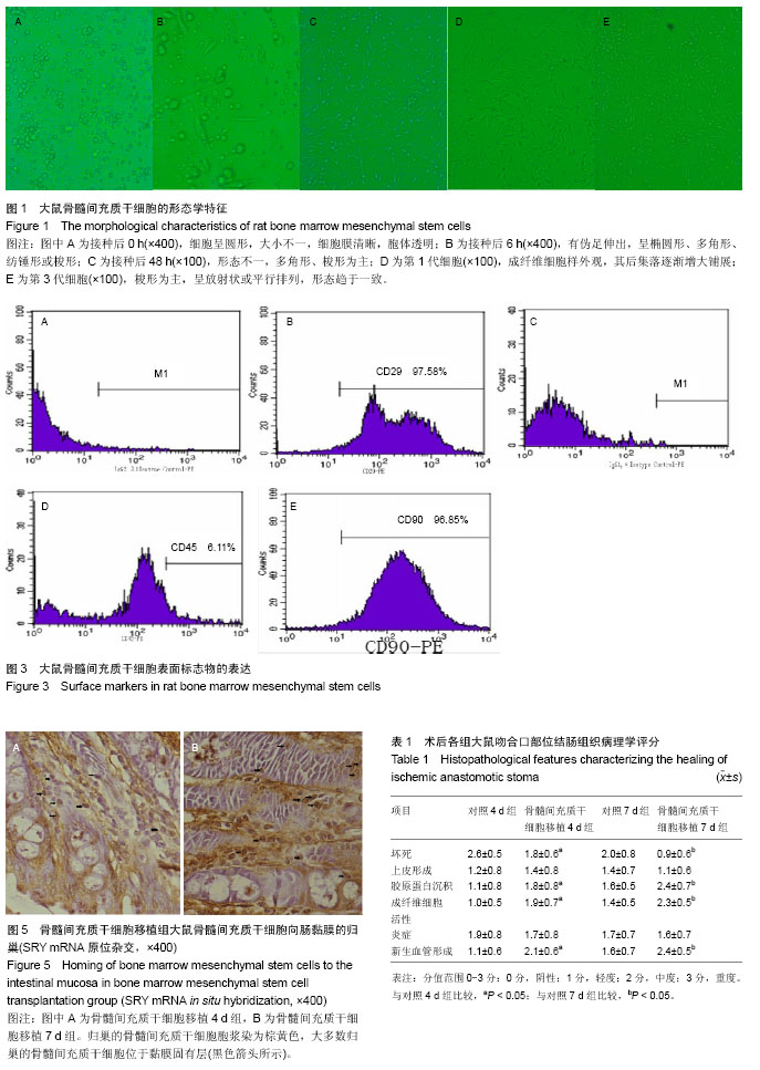
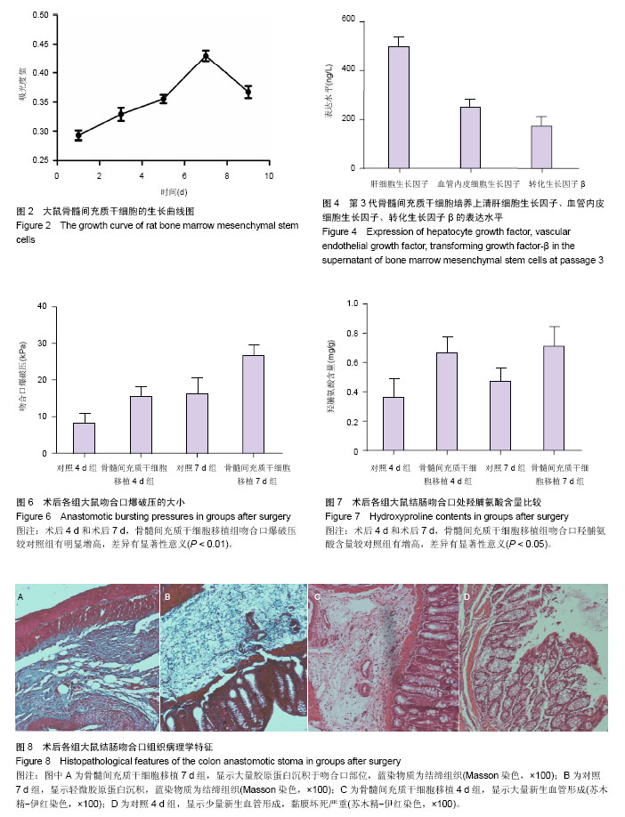
2.1 大鼠骨髓间充质干细胞的形态学特征 刚接种的细胞呈圆形,大小不一,细胞膜清晰,细胞体透明,悬浮于培养液中,见图1A。培养6-8 h后部分细胞贴附在塑料培养皿底,开始亦呈圆形,部分细胞有伪足伸出,呈椭圆形、多角形、纺锤形或梭形,见图1B。24 h后大部分细胞贴壁,48 h后细胞基本完成贴壁,进行首次半量换液,后每3 d全量换液,见图1C。红细胞等杂细胞随换液逐渐被清除,随后贴壁细胞开始分裂增殖,四五天后部分区域开始形成集落,细胞呈成纤维细胞样外观,其后集落逐渐增大铺展,10 d左右细胞基本汇合长满瓶底,即为第1代,见图1D。传代后,细胞贴壁迅速,形态以梭形为主,不再以集落的方式生长,细胞呈放射状或平行排列,形态趋于一致,见图1E,7 d即可传代1次。 2.2 骨髓间充质干细胞的活力以及增殖能力评估 锥虫蓝拒染实验显示骨髓间充质干细胞活力良好,细胞存活率为(95.69±2.17)%。CCK-8 法检测细胞增殖能力示显示骨髓间充质干细胞生长状态良好,增殖能力较强,培养第1,2天为相对潜伏期,接种第3天始细胞增殖活跃,步入对数增长期,第5-7天达到高峰,逐渐进入平台期,见图2。 2.3 骨髓间充质干细胞细胞表型鉴定 体外培养骨髓间充质干细胞常用的标志物有CD29、CD44、CD90、CD106等,本实验选取CD29-PE,CD90-PE作为骨髓间充质干细胞阳性对照。CD45在淋巴造血干细胞表面高度表达,选取CD45-PE作为骨髓间充质干细胞的阴性对照,同型对照分别为 IgG1, κ Isotype Control-PE,IgG2,λ1 Isotype Control-PE,结果显示骨髓间充质干细胞CD29、CD90阳性表达率为97.58%,96.85%,而CD45阳性表达率仅为6.11%,见图3。 2.4 细胞培养上清中肝细胞生长因子、血管内皮细胞生长因子、转化生长因子β水平 第3代骨髓间充质干细胞培养上清液肝细胞生长因子、血管内皮细胞生长因子、转化生长因子β水平分别为(498.42±39.78),(249.90±34.57),(172.28±39.57) ng/L,见图4。 2.5 骨髓间充质干细胞在肠黏膜归巢的检测 Y染色体示踪实验显示,在肠道黏膜固有层、肠黏膜上皮细胞层和肠黏膜下层均可见归巢的骨髓间充质干细胞,其中肠黏膜固有层居多。骨髓间充质干细胞移植4 d组和骨髓间充质干细胞移植7 d组细胞归巢率分别为(9.62±2.57)%、(11.69± 3.09)%,见图5。 2.6 吻合口爆破压的测定 术后第4天,骨髓间充质干细胞移植组吻合口压力较对照组明显增高[(15.4±2.77) kPa,(8.25±2.54) kPa,P < 0.01],同样,术后第7天,骨髓间充质干细胞移植组吻合口压力较对照组明显增高[(26.6± 2.94) kPa,(16.3±4.34) kPa,P < 0.01],见图6。 2.7 羟脯氨酸含量的测定 术后第4天,骨髓间充质干细胞移植组羟脯氨酸含量较对照组明显增高[(0.66±0.11) mg/g,(0.36±0.12) mg/g,P < 0.05],同样,术后第7天,骨髓间充质干细胞移植组羟脯氨酸含量较对照组明显增高[(0.71±0.13) mg/g,(0.47±0. 09) mg/g,P < 0.05],见图7。 2.8 组织病理学特征 骨髓间充质干细胞移植组黏膜坏死、新生血管形成、胶原蛋白沉积量及成纤维细胞活性评分与对照组有明显区别,差异有显著性意义(P < 0.05),骨髓间充质干细胞移植组上皮形成、炎症反应评分和对照组比较差异无显著性意义(P > 0.05),见图8,表1。"
"
| [1] Egger B, Inglin R, Zeeh J, et al. Insulin-like growth factor I and truncated keratinocyte growth factor accelerate healing of left-sided colonic anastomoses.Br J Surg. 2001;88(1): 90-98. [2] Thornton FJ, Barbul A.Healing in the gastrointestinal tract.Surg Clin North Am. 1997;77(3):549-573. [3] Mast BA.Healing in other tissues.Surg Clin North Am. 1997; 77(3):529-547. [4] Thompson SK, Chang EY, Jobe BA.Clinical review: Healing in gastrointestinal anastomoses, part I.Microsurgery. 2006;26(3): 131-136. [5] Enestvedt CK, Thompson SK, Chang EY,et al.Clinical review: Healing in gastrointestinal anastomoses, part II.Microsurgery. 2006;26(3):137-143. [6] Quaedackers JS, Beuk RJ, Bennet L,et al.An evaluation of methods for grading histologic injury following ischemia/ reperfusion of the small bowel.Transplant Proc. 2000;32(6): 1307-1310. [7] Yo LS, Consten EC, Quarles van Ufford HM,et al.Buttressing of the staple line in gastrointestinal anastomoses: overview of new technology designed to reduce perioperative complications. Dig Surg. 2006;23(5-6):283-291. [8] Pommergaard HC, Achiam MP, Rosenberg J.External coating of colonic anastomoses: a systematic review.Int J Colorectal Dis. 2012;27(10):1247-1258. [9] van der Ham AC, Kort WJ, Weijma IM,et al.Effect of antibiotics in fibrin sealant on healing colonic anastomoses in the rat.Br J Surg. 1992;79(6):525-528. [10] Nguyen NT, Nguyen CT, Stevens CM,et al.The efficacy of fibrin sealant in prevention of anastomotic leak after laparoscopic gastric bypass.J Surg Res. 2004;122(2): 218-224. [11] Binnebösel M, Junge K, Kaemmer DA,et al.Intraperitoneally applied gentamicin increases collagen content and mechanical stability of colon anastomosis in rats.Int J Colorectal Dis. 2009;24(4):433-440. [12] Junge K, Klinge U, Rosch R,et al.Improved collagen type I/III ratio at the interface of gentamicin-supplemented polyvinylidenfluoride mesh materials.Langenbecks Arch Surg. 2007;392(4):465-471. [13] Khanna A, Rossman J, Caty MG,et al.Beneficial effects of intraluminal nitroglycerin in intestinal ischemia-reperfusion injury in rats.J Surg Res. 2003;114(1):15-24. [14] 冯涛,考晓明,嵇武.干细胞的临床应用:缺少大动物模型和人体试验[J].中国组织工程研究, 2014,18(14):2269-2274. [15] Wu Y, Chen L, Scott PG,et al.Mesenchymal stem cells enhance wound healing through differentiation and angiogenesis.Stem Cells. 2007;25(10):2648-2659. [16] Hayashi Y, Tsuji S, Tsujii M,et al.Topical implantation of mesenchymal stem cells has beneficial effects on healing of experimental colitis in rats.J Pharmacol Exp Ther. 2008; 326(2):523-531. [17] Huang Y, Chang C, Zhang JW, et al. Bone marrow-derived mesenchymal stem cells increase dopamine synthesis in the injured striatum. Neural Regen Res. 2012; 7(34): 2653-2662. [18] Piao H, Youn TJ, Kwon JS,et al.Effects of bone marrow derived mesenchymal stem cells transplantation in acutely infarcting myocardium.Eur J Heart Fail. 2005;7(5):730-738. [19] Adas G, Arikan S, Karatepe O,et al.Mesenchymal stem cells improve the healing of ischemic colonic anastomoses (experimental study).Langenbecks Arch Surg. 2011;396(1): 115-126. [20] Rabau M, Eyal A, Kluger Y,et al.Bursting pressure in anastomotic healing in experimentally induced colitis in rats.Dis Colon Rectum. 1998;41(4):468-472. [21] Kingham TP, Pachter HL.Colonic anastomotic leak: risk factors, diagnosis, and treatment.J Am Coll Surg. 2009; 208(2):269-278. [22] Tadros T, Wobbes T, Hendriks T.Blood transfusion impairs the healing of experimental intestinal anastomoses.Ann Surg. 1992;215(3):276-281. [23] Guzel S, Sunamak O, AS A,et al.Effects of hyperbaric oxygen and Pgg-glucan on ischemic colon anastomosis.World J Gastroenterol. 2006;12(9):1421-1425. [24] Azevedo LA, Parra RS, Da Rocha JJ, et al.Hyperbaric oxygen on the healing of ischemic colonic anastomosis--an experimental study in rats.Undersea Hyperb Med. 2010; 37(6):405-411. [25] Cronin K, Jackson DS, Dunphy JE.Changing bursting strength and collagen content of the healing colon.Surg Gynecol Obstet. 1968;126(4):747-753. [26] Jiang H, Qu L, Li Y,et al.Bone marrow mesenchymal stem cells reduce intestinal ischemia/reperfusion injuries in rats.J Surg Res. 2011;168(1):127-134. [27] Gao J, Dennis JE, Muzic RF,et al.The dynamic in vivo distribution of bone marrow-derived mesenchymal stem cells after infusion.Cells Tissues Organs. 2001;169(1):12-20. [28] Voswinkel J, Francois S, Gorin NC,et al.Gastro-intestinal autoimmunity: preclinical experiences and successful therapy of fistulizing bowel diseases and gut Graft versus host disease by mesenchymal stromal cells.Immunol Res. 2013;56(2-3):241-248. [29] Forraz N, Wright KE, Jurga M,et al.Experimental therapies for repair of the central nervous system: stem cells and tissue engineering.J Tissue Eng Regen Med. 2013;7(7):523-536. [30] Adas G, Kemik O, Eryasar B,et al.Treatment of ischemic colonic anastomoses with systemic transplanted bone marrow derived mesenchymal stem cells.Eur Rev Med Pharmacol Sci. 2013;17(17):2275-2285. [31] Sémont A, François S, Mouiseddine M,et al.Mesenchymal stem cells increase self-renewal of small intestinal epithelium and accelerate structural recovery after radiation injury.Adv Exp Med Biol. 2006;585:19-30. [32] 高光周,李大伟,李欣,等. 同种异体大鼠骨髓间充质干细胞移植在肠缺血-再灌注损伤肠道中的定植及其分化效应[J].中国组织工程研究与临床康复, 2010, 14(23): 4262-4266. [33] Månsson P, Zhang XW, Jeppsson B,et al.Anastomotic healing in the rat colon: comparison between a radiological method, breaking strength and bursting pressure.Int J Colorectal Dis. 2002;17(6):420-425. [34] Tadauchi A, Narita Y, Kagami H,et al.Novel cell-based therapeutic strategy for ischemic colitis with use of bone marrow-derived mononuclear cells in rats.Dis Colon Rectum. 2009;52(8):1443-1451. [35] Gao F, He T, Wang H,et al.A promising strategy for the treatment of ischemic heart disease: Mesenchymal stem cell-mediated vascular endothelial growth factor gene transfer in rats.Can J Cardiol. 2007;23(11):891-898. [36] François S, Bensidhoum M, Mouiseddine M,et al.Local irradiation not only induces homing of human mesenchymal stem cells at exposed sites but promotes their widespread engraftment to multiple organs: a study of their quantitative distribution after irradiation damage.Stem Cells. 2006;24(4): 1020-1029. [37] Sémont A, Mouiseddine M, François A,et al.Mesenchymal stem cells improve small intestinal integrity through regulation of endogenous epithelial cell homeostasis.Cell Death Differ. 2010;17(6):952-961. [38] Shackleton M.Normal stem cells and cancer stem cells: similar and different.Semin Cancer Biol. 2010;20(2):85-92. [39] Peters BA, Diaz LA, Polyak K,et al.Contribution of bone marrow-derived endothelial cells to human tumor vasculature. Nat Med. 2005;11(3):261-262. [40] Potten CS, Booth C, Hargreaves D.The small intestine as a model for evaluating adult tissue stem cell drug targets.Cell Prolif. 2003;36(3):115-129. [41] Secko D.Do stem cells cause gastric cancer. CMAJ. 2005; 172(3):325-326. [42] Studeny M, Marini FC, Dembinski JL,et al.Mesenchymal stem cells: potential precursors for tumor stroma and targeted-delivery vehicles for anticancer agents.J Natl Cancer Inst. 2004;96(21):1593-1603 |
| [1] | Hou Jingying, Yu Menglei, Guo Tianzhu, Long Huibao, Wu Hao. Hypoxia preconditioning promotes bone marrow mesenchymal stem cells survival and vascularization through the activation of HIF-1α/MALAT1/VEGFA pathway [J]. Chinese Journal of Tissue Engineering Research, 2021, 25(7): 985-990. |
| [2] | Liang Xueqi, Guo Lijiao, Chen Hejie, Wu Jie, Sun Yaqi, Xing Zhikun, Zou Hailiang, Chen Xueling, Wu Xiangwei. Alveolar echinococcosis protoscolices inhibits the differentiation of bone marrow mesenchymal stem cells into fibroblasts [J]. Chinese Journal of Tissue Engineering Research, 2021, 25(7): 996-1001. |
| [3] | Geng Yao, Yin Zhiliang, Li Xingping, Xiao Dongqin, Hou Weiguang. Role of hsa-miRNA-223-3p in regulating osteogenic differentiation of human bone marrow mesenchymal stem cells [J]. Chinese Journal of Tissue Engineering Research, 2021, 25(7): 1008-1013. |
| [4] | Lun Zhigang, Jin Jing, Wang Tianyan, Li Aimin. Effect of peroxiredoxin 6 on proliferation and differentiation of bone marrow mesenchymal stem cells into neural lineage in vitro [J]. Chinese Journal of Tissue Engineering Research, 2021, 25(7): 1014-1018. |
| [5] | Zhu Xuefen, Huang Cheng, Ding Jian, Dai Yongping, Liu Yuanbing, Le Lixiang, Wang Liangliang, Yang Jiandong. Mechanism of bone marrow mesenchymal stem cells differentiation into functional neurons induced by glial cell line derived neurotrophic factor [J]. Chinese Journal of Tissue Engineering Research, 2021, 25(7): 1019-1025. |
| [6] | Pei Lili, Sun Guicai, Wang Di. Salvianolic acid B inhibits oxidative damage of bone marrow mesenchymal stem cells and promotes differentiation into cardiomyocytes [J]. Chinese Journal of Tissue Engineering Research, 2021, 25(7): 1032-1036. |
| [7] | Wang Shiqi, Zhang Jinsheng. Effects of Chinese medicine on proliferation, differentiation and aging of bone marrow mesenchymal stem cells regulating ischemia-hypoxia microenvironment [J]. Chinese Journal of Tissue Engineering Research, 2021, 25(7): 1129-1134. |
| [8] | Chen Junyi, Wang Ning, Peng Chengfei, Zhu Lunjing, Duan Jiangtao, Wang Ye, Bei Chaoyong. Decalcified bone matrix and lentivirus-mediated silencing of P75 neurotrophin receptor transfected bone marrow mesenchymal stem cells to construct tissue-engineered bone [J]. Chinese Journal of Tissue Engineering Research, 2021, 25(4): 510-515. |
| [9] | Jiang Tao, Ma Lei, Li Zhiqiang, Shou Xi, Duan Mingjun, Wu Shuo, Ma Chuang, Wei Qin. Platelet-derived growth factor BB induces bone marrow mesenchymal stem cells to differentiate into vascular endothelial cells [J]. Chinese Journal of Tissue Engineering Research, 2021, 25(25): 3937-3942. |
| [10] | Sun Jianwei, Yang Xinming, Zhang Ying. Effect of montelukast combined with bone marrow mesenchymal stem cell transplantation on spinal cord injury in rat models [J]. Chinese Journal of Tissue Engineering Research, 2021, 25(25): 3962-3969. |
| [11] | Zhang Lishu, Liu Anqi, He Xiaoning, Jin Yan, Li Bei, Jin Fang. Alpl gene affects the therapeutic effect of bone marrow mesenchymal stem cells on ulcerative colitis [J]. Chinese Journal of Tissue Engineering Research, 2021, 25(25): 3970-3975. |
| [12] | Hao Xiaona, Zhang Yingjie, Li Yuyun, Xu Tao. Bone marrow mesenchymal stem cells overexpressing prolyl oligopeptidase on the repair of liver fibrosis in rat models [J]. Chinese Journal of Tissue Engineering Research, 2021, 25(25): 3988-3993. |
| [13] | Yi Meizhi, Luo Guanghua, Xiao Yawen, Hu Rong, Chen Xiaolong, Zhao Heng. MRI findings of anatomical variations of the talus [J]. Chinese Journal of Tissue Engineering Research, 2021, 25(24): 3888-3893. |
| [14] | Mo Jianling, He Shaoru, Feng Bowen, Jian Minqiao, Zhang Xiaohui, Liu Caisheng, Liang Yijing, Liu Yumei, Chen Liang, Zhou Haiyu, Liu Yanhui. Forming prevascularized cell sheets and the expression of angiogenesis-related factors [J]. Chinese Journal of Tissue Engineering Research, 2021, 25(22): 3479-3486. |
| [15] | Wei Qin, Zhang Xue, Ma Lei, Li Zhiqiang, Shou Xi, Duan Mingjun, Wu Shuo, Jia Qiyu, Ma Chuang. Platelet-derived growth factor-BB induces the differentiation of rat bone marrow mesenchymal stem cells into osteoblasts [J]. Chinese Journal of Tissue Engineering Research, 2021, 25(19): 2953-2957. |
| Viewed | ||||||
|
Full text |
|
|||||
|
Abstract |
|
|||||