Chinese Journal of Tissue Engineering Research ›› 2014, Vol. 18 ›› Issue (33): 5282-5287.doi: 10.3969/j.issn.2095-4344.2014.33.006
Previous Articles Next Articles
Effects of Wallerian degeneration on biological characteristics and secretory function of Schwann cells in rats with sciatic nerve injury
Li Yue-zhen, Wu Geng, Wu Yang, Jin Xiu-dong, Zhang Ji-fei, Zhao Fu-sheng
- Mudanjiang Medical University, Mudanjiang 157011, Heilongjiang Province, China
-
Online:2014-08-13Published:2014-08-13 -
Contact:Zhao Fu-sheng, Master, Lecturer, Mudanjiang Medical University, Mudanjiang 157011, Heilongjiang Province, China -
About author:Li Yue-zhen, Master, Professor, Mudanjiang Medical University, Mudanjiang 157011, Heilongjiang Province, China -
Supported by:Scientific Research Project of Mudanjiang City, No. G2011s0018; Young Scholar Funds in Universities of Heilongjiang Province, No. 1253G061
CLC Number:
Cite this article
Li Yue-zhen, Wu Geng, Wu Yang, Jin Xiu-dong, Zhang Ji-fei, Zhao Fu-sheng. Effects of Wallerian degeneration on biological characteristics and secretory function of Schwann cells in rats with sciatic nerve injury[J]. Chinese Journal of Tissue Engineering Research, 2014, 18(33): 5282-5287.
share this article

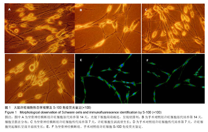
2.1 大鼠许旺细胞分离培养及鉴定 坐骨神经横断组,原代培养第5天,局部有许旺细胞从神经段边缘爬出;第7天可见典型许旺细胞,胞体细长呈极性生长;第14天细胞数量显著增多,呈线状排列,细胞端与端连成长串(图1A);手术对照组,许旺细胞增殖缓慢,在原代培养第14天时,神经段边缘可见少许三极或多极散在分布许旺细胞(图1B);传代培养第7天,坐骨神经横断组许旺细胞分裂增殖旺盛,呈涡流状排列或编织袋状生长(图1C)。手术对照组许旺细胞形态呈梭形,突起细长呈肩并肩状生长(图1D)。S-100免疫荧光鉴定,两组许旺细胞均呈阳性,胞体呈梭形,两端伸出纤长突起(图1E、F)。"
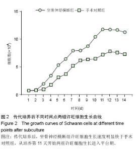
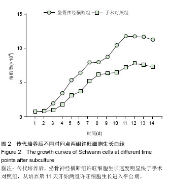
2.2 许旺细胞生长曲线 传代培养第3天,两组许旺细胞均进入对数增长期,随时间延长,许旺细胞生长速度逐渐加快,从培养第11天开始许旺细胞生长进入平台期。传代培养前1,2 d, 两组许旺细胞数比较差异均无显著性意义(P > 0.05);在培养第3,4,5,6,7,9,10,11,12,13,14天,坐骨神经横断组许旺细胞数明显多于手术对照组,差异均有显著性意义(P < 0.05),见图2。"
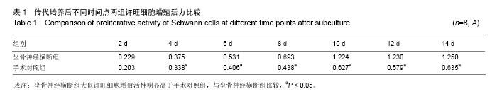
2.3 许旺细胞增殖活性检测 许旺细胞传代培养第2天,两组A值比较,差异无显著性意义(P > 0.05);传代培养4,6,8,10,12,14 d,坐骨神经横断组A值显著高于手术对照组,差异均有显著性意义(P < 0.05,表1)。"

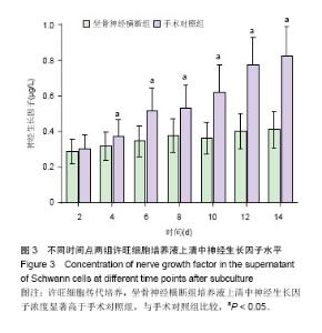
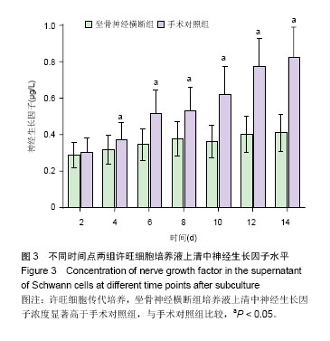
2.4 许旺细胞黏附能力检测 许旺细胞传代培养后 4 h,坐骨神经横断组吸光度(A)值为0.736±0.059,手术对照组吸光度(A)值为0.631±0.053,两组比较差异有显著性意义(P < 0.05),表明瓦勒变性可显著增加许旺细胞黏附能力。 2.5 神经生长因子浓度检测 许旺细胞传代培养第2天,坐骨神经横断组神经生长因子浓度与手术对照组比较,差异无显著性意义(P > 0.05);在传代培养4,6,8,10,12,14 d,坐骨神经横断组神经生长因子浓度均显著高于手术对照组,差异均有显著性意义(P < 0.05),见图3。"
| [1]Piirsoo M, Kaljas A, Tamm K, et al. Expression of NGF and GDNF family members and their receptors during peripheral nerve development and differentiation of Schwann cells in vitro. J Neurosci Lett.2010;469(1):135-140. [2]Ma Z, Wang J, Song F, et al. Critical period of axoglial signaling between Neuregulin-1 and BDNF required for early Schwann cell survival and differentiation. J Neurosci. 2011; 31(26): 9630-9640. [3]Bhatheja K, Field J. Schwann cells: origins and role in axonal main-tenance and regeneration. Int J Biochem Cell Biol. 2006; 38(12):1995-1999. [4]Tao HY, He B, Liu SQ, et al. Effect of carboxymethylated chitosan on the biosynthesis of NGF and activation of the Wnt/β-catenin signaling pathway in the proliferation of Schwann cells. Eur J Pharmacol, 2013;702(1-3):85-92. [5]Torigoe K, H ashimoto K, Lundborg G. A role of migratory Schwann cells in a conditioning effect of peripheral nerve regeneration. J Exp Neurol.1999;160(1):99-108. [6]Yao D, Li M, Shen D, et al. Expression changes and bioinformatic analysis of Wallerian degeneration after sciatic nerve injury in rat. Neurosci Bull,2013,29(3): 321-32. [7]Frostick SP, Yin Q, Kemp GJ. Schwann cells, neurotrophic factors, and peripheral nerve regeneration. Microsurgery. 1998;18(7): 397-405. [8]Sangsanoh P, Waleetorncheepsawat S, Suwantong O, et al. In vitro Bio-Compatibility of schwann cells on surfaces of biocompatible polymeric electrospun fibrous and solution-cast film scaffolds. Biomacromolecules.2007;8(5): 1587-1594. [9]Sinis N, Schaller HE, Becker ST, et al. Long nerve gaps limit the Regenerative potential of bioartificial nerve conduits filled with Schwann cells. Restor Neurol Neurosci.2007;25(2): 131-141. [10]Waller A. Experiments on the section of the glossopharyngeal and hypoglossal nerves of the frog, and observations of the alterations produced thereby in the structure of their primitive ?bres. Philos. Trans. R. Soc. London B, No. 140. (1850): 423-429. [11]赵富强,姜保国,张培训,等.鼠坐骨神经损伤后雪旺细胞增殖规律的免疫组织化学观察[J].中华外科杂志,2006,44(4): 268-270. [12]Li RH, Sliwkowski MX, Lo J, et al. Establishment of Schwann cell lines from normal adult and embryonic rat dorsal root ganglia. J Neurosci Methods.1996;67(1):57-69. [13]付国强,李学拥.兔坐骨神经电损伤后许旺细胞的增殖变化[J]. 中国临床康复,2005, 9(34):18-19. [14]Ide C, Tohyama K, Yokota R,et al. Schwann cell processes guide regeneration of peripheral axons. Brain Res.1983; 288(1-2):61-75. [15]Dong Z, Brennan A, Liu N,et al. Neu differentiation factor is a neuron-glia signal and regulates survival, proliferation, and maturation of rat Schwann cell precursors. Neuron. 1995;1 5(3):585-596. [16]Wang DY, Huang YC, Chiang H, et al. Microcontact printing of laminin on oxygen plasma activated substrates for the alignment and growth of Schwann cells. J Biomed Mater Res B Appl Biomater.2007;80(2):447-453. [17]Bunge RP, Bunge MB, Eldridge CF. Linkage between axonal ensheathment and basal lamina production by Schwann cells. Annu Rev Neurosci.1986;9:305-328. [18]Maisonpierre PC, Belluscio L, Friedman B, et al. NT-3, BDNF, and NGF in the developing rat nervous system: parallel as well as reciprocal patterns of expression. Neuron. 1990;5(4): 501-509. [19]Lindsay RM. Nerve growth factors (NGF, BDNF) enhance axonal regeneration but are not required for survival of adult sensory neurons. J Neurosci.1988;8(7): 2394-2405. [20]Shakhbazau A, Kawasoe J, Hoyng SA, et al.Early regenerative effects of NGF-transduced Schwann cells in peripheral nerve repair. Mol Cell Neurosci.2012;50(1): 103-112. [21]Shin YH, Lee SJ, Jung J.Extracellular ATP inhibits Schwann cell dedifferentiation and proliferation in an ex vivo model of Wallerian degeneration.Biochem Biophys Res Commun. 2013;430(2):852-857. [22]Somandin C, Gerber D, Pereira JA,et al.LITAF (SIMPLE) regulates Wallerian degeneration after injury but is not essential for peripheral nerve development and maintenance: implications for Charcot-Marie-Tooth disease. Glia. 2012; 60(10):1518-1528. [23]Gaudet AD, Popovich PG, Ramer MS. Wallerian degeneration: gaining perspective on inflammatory events after peripheral nerve injury.J Neuroinflammation. 2011;8:110. [24]Jung J, Cai W, Jang SY,et al.Transient lysosomal activation is essential for p75 nerve growth factor receptor expression in myelinated Schwann cells during Wallerian degeneration. Anat Cell Biol. 2011;44(1):41-49. [25]Jung J, Cai W, Lee HK, et al. Actin polymerization is essential for myelin sheath fragmentation during Wallerian degeneration. J Neurosci.2011;31(6):2009-2015. [26]Chaballe L, Close P, Sempels M,et al.Involvement of placental growth factor in Wallerian degeneration.Glia. 2011; 59(3):379-396. [27]Girolami EI, Bouhy D, Haber M, et al.Differential expression and potential role of SOCS1 and SOCS3 in Wallerian degeneration in injured peripheral nerve.Exp Neurol. 2010; 223(1):173-182. [28]Lee HK, Seo IA, Suh DJ, et al.Interleukin-6 is required for the early induction of glial fibrillary acidic protein in Schwann cells during Wallerian degeneration.J Neurochem. 2009;108(3): 776-786. [29]Keilhoff G, Schild L, Fansa H.Minocycline protects Schwann cells from ischemia-like injury and promotes axonal outgrowth in bioartificial nerve grafts lacking Wallerian degeneration.Exp Neurol. 2008;212(1):189-200. [30]刘璋寅.周围神经中许旺细胞特异性标记物[J].组织工程与重建外科杂志,2012,8(5):292-297. [31]Kubota A, Komiyama A, Matsumoto M, et al.Effect of macrophage suppression with silica on the proliferation of Schwann cells during Wallerian degeneration.Brain Res. 1998;802(1-2):254-258. [32]Jung J, Jo HW, Kwon H,et al.ATP Release through Lysosomal Exocytosis from Peripheral Nerves: The Effect of Lysosomal Exocytosis on Peripheral Nerve Degeneration and Regeneration after Nerve Injury.Biomed Res Int. 2014;2014: 936891. |
| [1] | Zeng Yanhua, Hao Yanlei. In vitro culture and purification of Schwann cells: a systematic review [J]. Chinese Journal of Tissue Engineering Research, 2021, 25(7): 1135-1141. |
| [2] | Luo Xuanxiang, Jing Li, Pan Bin, Feng Hu. Effect of mecobalamine combined with mouse nerve growth factor on nerve function recovery after cervical spondylotic myelopathy surgery [J]. Chinese Journal of Tissue Engineering Research, 2021, 25(5): 719-722. |
| [3] | Xie Yang, Lü Zhiyu, Zhang Shujiang, Long Ting, Li Zuoxiao. Effects of recombinant adeno-associated virus mediated nerve growth factor gene transfection on oligodendrocyte apoptosis and myelination in experimental autoimmune encephalomyelitis mice [J]. Chinese Journal of Tissue Engineering Research, 2021, 25(23): 3678-3683. |
| [4] | Yan Nan, Si Xiaofeng, Zeng Liang, Tian Wei, Shan Guangdong, Xiong Lishuo, Yang Weijie, Wang Zhengdong. Nerve growth factor interferes with proliferation and alpha-actin expression of skeletal muscle satellite cells in rats [J]. Chinese Journal of Tissue Engineering Research, 2021, 25(13): 2030-2035. |
| [5] | Wu Zhifeng, Luo Min. Biomechanical analysis of chemical acellular nerve allograft combined with bone marrow mesenchymal stem cell transplantation for repairing sciatic nerve injury [J]. Chinese Journal of Tissue Engineering Research, 2020, 24(7): 991-995. |
| [6] | Liu Guoming, Wang Qinfen, Lin Kefeng, Zhou Shiguo, Chen Zuxing, Lin Shishui. Whole body application of nerve growth factor promotes early healing of tibial shaft fracture and improves expression of bone morphogenetic protein-2 and vascular endothelial growth factor in rats [J]. Chinese Journal of Tissue Engineering Research, 2020, 24(29): 4680-4685. |
| [7] | Li Qinwen, Liang Jie, Wang Dongmei, Shang Zhenghui. Fibrotic changes in rat dorsal root ganglion following chronic sciatic nerve compression [J]. Chinese Journal of Tissue Engineering Research, 2020, 24(29): 4686-4691. |
| [8] | Gao Jianbo, Xia Bing, Li Shengyou, Yang Yujie, Ma Teng, Yu Peng, Luo Zhuojing, Huang Jinghui. Effect of nanoparticles carrying chondroitin sulfate ABC on the migration of Schwann cells in a magnetic field [J]. Chinese Journal of Tissue Engineering Research, 2020, 24(28): 4526-4532. |
| [9] |
Wang Ning, Chen Junyi, Zhu Lunjing, Duan Jiangtao, Wang Ye, Li Zhijun, Bei Chaoyong.
Lentivirus-mediated P75 neurotrophin receptor silencing combined with nerve growth factor overexpression promotes proliferation of rat bone marrow mesenchymal stem cells [J]. Chinese Journal of Tissue Engineering Research, 2020, 24(25): 3988-3993. |
| [10] | Zhu Yaqiong, Jin Zhuang, Wang Jing, Fang Jie, Li Chaochao, Hu Yongqiang, Tian Xiaoqi, Zhang Ying, Song Qing, Wang Yuexiang, Luo Yukun. Ultrasound-guided platelet-rich plasma injection for repair of sciatic nerve crush injury [J]. Chinese Journal of Tissue Engineering Research, 2020, 24(20): 3196-3201. |
| [11] | Luo Xuanxiang, Feng Hu, Jing Li, Pan Bin. Important roles of non-coding RNA in peripheral nerve repair [J]. Chinese Journal of Tissue Engineering Research, 2020, 24(14): 2271-2276. |
| [12] | Cao Peng, Wang Haonan, Tian Weifeng, Sun Naichao, Bai Jiangbo, Yu Kunlun, Tian Dehu. Human amniotic membrane repairs acute sciatic nerve injury in rat models [J]. Chinese Journal of Tissue Engineering Research, 2019, 23(7): 1046-1051. |
| [13] | Wu Bo, Li Xiangkui, Wang Hua, Wen Zhiyuan. Sustained-release properties and histocompatibility of ropivacaine-coated polyethylene glycol/polylactic acid microspheres implanted around the sciatic nerve [J]. Chinese Journal of Tissue Engineering Research, 2019, 23(34): 5486-5491. |
| [14] | Zong Qiang, Xu Yanan, Qu Tianyi, Li Lijun, Hai Miti·Abuduaini, Ni Dongkui. Magnetic verapamil nanoparticles promote peripheral nerve regeneration [J]. Chinese Journal of Tissue Engineering Research, 2019, 23(34): 5425-5429. |
| [15] | Wang Tong, Liu Yang, Zhu Yetao. Combined use of exogenous nerve growth factor and basic fibroblast growth factor promotes proliferation of endogenous brain cells in a rat model of severe traumatic brain injury [J]. Chinese Journal of Tissue Engineering Research, 2019, 23(25): 3998-4003. |
| Viewed | ||||||
|
Full text |
|
|||||
|
Abstract |
|
|||||