Chinese Journal of Tissue Engineering Research ›› 2013, Vol. 17 ›› Issue (42): 7341-7348.doi: 10.3969/j.issn.2095-4344.2013.42.001
The microstructure of antigen-extracted heterologous bone
Cui Jie, Li Zheng, He Hui-yu, Hu Yang
- First Affiliated Hospital of Xinjiang Medical University, Urumqi 830054, Xinjiang Uygur Autonomous Region, China
-
Received:2013-04-09Revised:2013-04-18Online:2013-10-15Published:2013-10-31 -
Contact:He Hui-yu, M.D., Professor, Chief physician, First Affiliated Hospital of Xinjiang Medical University, Urumqi 830054, Xinjiang Uygur Autonomous Region, China hehuiyu01@126.com -
About author:Cui Jie★, Master, First Affiliated Hospital of Xinjiang Medical University, Urumqi 830054, Xinjiang Uygur Autonomous Region, China cuijieme@163.com -
Supported by:the National Natural Science Foundation of China, No. 81060088*; Special Fund for Tissue Engineering, First Affiliated Hospital of Xinjiang Medical University, No. 2012ZZG02*
CLC Number:
Cite this article
Cui Jie, Li Zheng, He Hui-yu, Hu Yang. The microstructure of antigen-extracted heterologous bone[J]. Chinese Journal of Tissue Engineering Research, 2013, 17(42): 7341-7348.
share this article
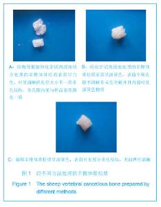
2.1 各组一般观察 见图1。"

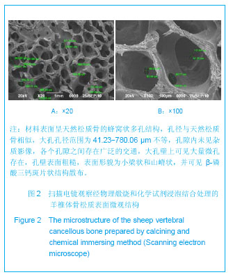
物理煅烧组样品表面呈白色,可见清晰的孔径大小不一的多孔结构,各孔隙内部与样品表面颜色一致;逆光观察可见样品透光,手指用力按压可将骨块压碎,碎片较规则。化学处理组样品表面呈淡黄色,表面个别孔隙不清晰并未完全敞开且内部可见深黄色物质;样品透光性较物理煅烧组差,手指用力按压无法将骨块压碎,但可使其发生形变。对照组样品呈深黄色,表面可见部分多孔结构,无前两组清晰;逆光观察透光差,并可见内部大量杂质手指用力按压无法将骨块压碎,但可使其发生少量形变,见图1。 2.2 支架孔隙率测定 分别将两种测定方法所得的3组平均孔隙率整理,设定检验水准α=0.05水准,结果见表1。"

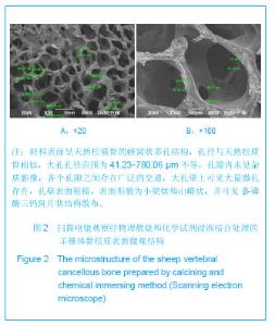
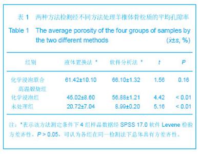
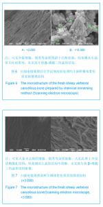
同一测定方法条件下,采用单因素方差分析检测得出结果:①液体置换法:组间比较F=27.89,P < 0.01,说明各组间孔隙率差异不全相同,再用Bonrerroni法行组间两两比较,各组间P < 0.01,说明液体置换法测定各组间孔隙率差异有显著性意义。②软件分析法:组间比较F=62.06,P < 0.01,说明各组间孔隙率差异不全相同,再用Bonrerroni法行组间两两比较,各组间P < 0.01,说明软件分析法测定各组间孔隙率差异有显著性意义。 同一分组条件下,采用配对t 检验比较两种测定孔隙率方法:①物理煅烧组:根据结果得出液体置换法与软件分析法测定孔隙率的差异无显著性意义(P=0.16)。②化学处理组、对照组:根据结果得出液体置换法与软件分析法测定孔隙率的差异有显著性意义(P < 0.01)。 2.3 支架超微形态结构观察 扫描电镜低放大倍数下观察(放大20-100倍):物理煅烧组材料表面呈天然松质骨的蜂窝状多孔结构,孔径与天然松质骨相似,大孔孔径范围为41.23-780.06 μm不等,孔隙内未见杂质影像,各个孔隙之间存在广泛的交通,大孔壁上可见大量微孔存在,孔壁表面粗糙,表面形貌为小梁状和山嵴状,并可见β-磷酸三钙斑片状结构散布,见图2。"
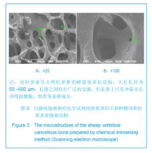
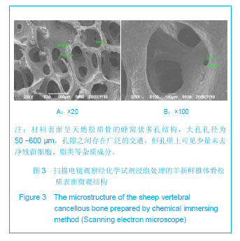
化学处理组表面与物理煅烧组相近,大孔孔径为50-600 μm,孔隙之间存在广泛的交通,但孔壁上可见少量未去净残留细胞、脂类等杂质成分,见图3。"
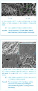
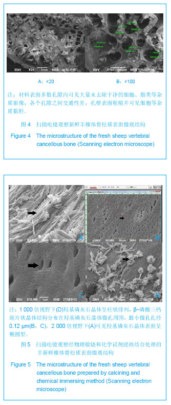
对照组材料表面多数孔隙内可见大量未去除干净的细胞、脂类等杂质影像,各个孔隙之间交通性差,孔壁表面粗糙并可见细胞等杂质黏附,见图4。 扫描电镜高放大倍数下观察(放大倍数1 000倍以上):物理煅烧组1 000倍视野下羟基磷灰石晶体呈柱状排列,β-磷酸三钙斑片状晶体结构分布在羟基磷灰石晶体微孔周围,最小微孔孔径0.12 μm,2 000倍视野下可见羟基磷灰石晶体表面呈椭圆型,见图5。"
化学处理组可见少量细胞、脂类等杂质残留于孔壁表面,羟基磷灰石晶体呈柱状排列,未见斑片状β-磷酸三钙晶体结构,见图6。对照组可见大量未去除的细胞、脂类等杂质影像,大孔孔壁上少见清晰微孔结构,羟基磷灰石晶体结构不清晰,未见斑片状β-磷酸三钙晶体结构影像,见图7。"

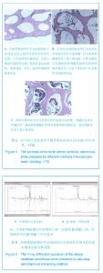
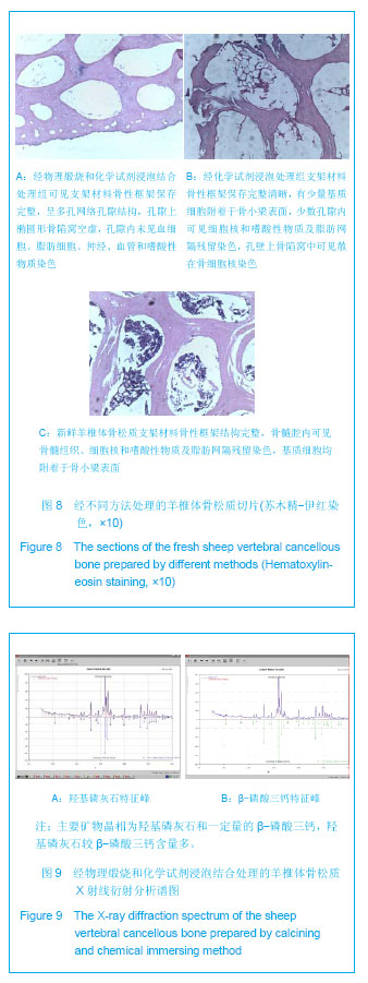
2.4 支架组织学观察 物理煅烧组切片经苏木精-伊红染色后可见支架材料骨性框架保存完整,呈多孔网络孔隙结构,孔隙上椭圆形骨陷窝空虚,孔隙内未见血细胞、脂肪细胞、神经、血管和嗜酸性物质染色,见图8A。化学处理组支架材料骨性框架保存完整清晰,有少量基质细胞附着于骨小梁表面,少数孔隙内可见细胞核和嗜酸性物质及脂肪网隔残留染色,孔壁上骨陷窝中可见散在骨细胞核染色,见图8B。对照组支架材料骨性框架结构完整,骨髓腔内可见骨髓组织、细胞核和嗜酸性物质及脂肪网隔残留染色,基质细胞均附着于骨小梁表面,见图8C。 2.5 支架X射线衍射分析 物理煅烧组样品衍射角2-Theta值特征峰尖锐清晰,与标准数据库特征峰值对比中得到主要矿物晶相为羟基磷灰石和一定量的β-磷酸三钙,分子式Ca5(PO4)3OH和Ca3(PO4)2,根据特征峰值偏移程度可见羟基磷灰石较β-磷酸三钙含量多,该结果亦在扫描电镜观测结果中得到证实,见图9。"
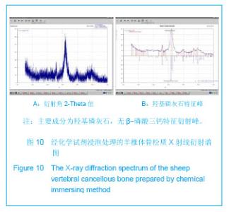
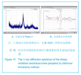
化学处理组样品衍射角2-Theta值特征衍射峰值清晰程度较物理煅烧组差,可见许多杂质衍射峰出现,其主要衍射特征峰与标准数据库特征衍射峰值对比中得到主要成分为羟基磷灰石,分子式Ca5(PO4)3OH,无β-磷酸三钙特征衍射峰,见图10。"
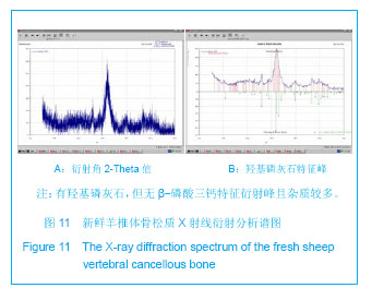
对照组样品衍射角羟基磷灰石特征衍射峰可见外,其他峰值均不清晰,有大量杂质衍射峰存在,提示该组样品中含有羟基磷灰石,分子式Ca5(PO4)3OH,但无β-磷酸三钙特征衍射峰且杂质较多,见图11。"

| [1] 胡堃,刘斌.骨移植材料发展趋势[J].生物骨科材料与临床研究, 2010,7(3):32-38. [2] Vacanti CA,Upton J.Tissue-engineered morphogenesis of cartilage and bone by means of cell transplantation using synthetic biodegradable polymer matrices.Clin Plant Surg. 1994;21(3):445. [3] 邢辉,陈晓明,张宏泉.骨组织工程支架材料[J].佛山陶瓷,2004, 14(12):35-39. [4] Mastrogiacomo M,Muraglia A,Komlev V,et al.Tissue engineering of bone:search for a better scaffold.Orthod Craniofac Res.2005;8(4):277-284. [5] 郑磊,王前,裴国献.骨组织工程中细胞外基质材料的选择[J].中华外科杂志,2000,38(10):745-748. [6] Simon CG,Khatri CA,Wight SA,et al.Preliminary report on the biocompatibility of a moldable,resorbable,composite bone graft consisting of calcium phosphate cement and poly(lactide-co-glycolide) microspheres.J Orthop Res.2002; 20:473-482. [7] Sun L,Zhang BQ,Chen L,et al. Carrier com bination of tissue engineered bone by sodium alginate and xenograft bone and bone form ation in vivo. Zhongguo Xiu Fu Chong Jian Wai Ke Za Zhi.2008;22(6):732-736. [8] Athanasiou VT,Papachristou DJ,Panagopoulos A,et al. Histological comparison of autograft,allograft-DBM, xenograft, and synthetic grafts in a trabecular bone.J Med Sci Monit. 2010; 16(1):24-31. [9] Erdemli O,Captug O,Bilgili H,et al. In vitro and in vivo evaluation of the effects of demineralized bone matrix or calcium sulfate addition to polycaprolactone-bioglass composites.J Mater Sci Mater Med.2010;21(1):295-308. [10] Rocchietta I,Dellavia C,Nevins M,et al. Bone regenerated via rhPDGF-bB and a deproteinized bovine bone matrix:backscattered electron microscopic element analysis.Int J Periodontics Restorative Dent.2007;27(6):539-545. [11] Jähn K,Braunstein V,Furlong PI,et al. A rapid method for the generation of uniform acellular bone explants. J Orthop Surg Res.2010;10(5):28-32. [12] De Long WG Jr,Einhorn TA,Koval K,et al. Bone grafts and bone graft substitutes in orthopaedic trauma surgery.A critical analysis. Bone Joint Surg Am.2007;89:649-658. [13] Develioglu H,Saraydn S,Kartal U,et al. Evaluation of the Long-Term results of rat cranial Bone Repair Using a Particular Xenograft.J Oral Implantol.2010;36(3):167-173. [14] Araújo M,Linder E,Lindhe J. Effect of a xenograft on early bone formation in extraction sockets:an experimental study in dog.Clin Oral Implants Res.2009;20(1):1-6. [15] Salci H,Sarigul S,Dogan S,et al. Contribution of the xenograft bone plate-screw system in lumbar transpedicular stabilization of dogs:an in-vitro study.J Vet Sci.2008;9(2): 193-196. [16] Dehghani SN,Bigham AS,Torabi Nezhad S,et al. Effect of bovine fetal growth plate as a new xenograft in experimental bone defect healing:radiological,histopathological and biomechanical evaluation.Cell Tissue Bank.2008;9(2):91-99. [17] Liu HL,Chen WS,Chen JS,et al.Cavitation-enhanced ultrasound thermal therapy by combined low-and high-frequency ultrasound exposure.Ultrasound Med Biol. 2006;32(5):759-767. [18] Le Nihouannen D,Saffarzadeh A,Aguado E,et al.Osteogenic properties of calcium phosphate ceramics and fibrin glue based composites. J Mater Sci Mater Med.2007;18(2): 225-235. [19] Tsuruga E,Takita H,Itoh H,et al.Pore size of porous hydroxyapatite as the cell-substratum controls BMP-induced osteogenesis.J Biochem (Tokyo). 1997;121(2): 317-324. [20] Likibi F,Chabot G,Assad M,et al. Influence of orthopedic implant structure on adjacent bone density and on stability.Am J Orthop.2008;37(4):E78-83. [21] Bucholz RW.Nonallograft osteoconductive bone graft substitutes.Clin Orthop Relat Res.2002;2(395): 44-52. [22] Porter NL,Pilliar RM,Grynpas MD.Fabrication of porous calcium polyphosphate implants by solid free from fabrication: a study of processing parameters and in vitro degradation characteristics.J Biomed Mater Res.2001; 56(4): 504-515. [23] Chang BS,Lee CK,Hong KS,et al. Osteoconduction at porous hydroxyapatite with variou spore configuration.Biomaterials. 2000;21(12):1291-1298. [24] Sánchez-Salcedo S,Werner J,Vallet-Regí M. Hierarchical pore structure of calcium phosphate scaffolds by a combination of gel-casting and multiple tape-casting methods.Acta Biomater.2008;4(4):913-922. [25] 许永华,施新猷,胡蕴玉,等.胶原-煅烧骨支架与成骨细胞相容性实验研究[J].中国临床解剖学杂志,2000,18(4):358-359. [26] 罗卓荆,胡蕴玉,王茜,等.异种松质骨移植抗原分布的免疫组化研究[J].中国矫形外科杂志,1998,5(6):539-540. [27] Ooi CY,Hamdi M,Ramesh S.Properties of hydroxyapatite produced by annealing of bovine bone.Ceram Int.2007;33(7): 1171-1177. [28] Kurashina K,Kurita H,Wu Q,et al.EctoPic osteogenesis with biphasic cermics of hydroxyapatite and tricalcium phosphate in rabbits.Biomaterials.2002;23(2):407-412. [29] Ramay HRR,Zhang M.Biphasic calcium phosphate nanocomposite porous scaffolds for load-bearing bone tissue engineering.Biomaterials.2004;25(21):5171-5180. [30] Li L,Hui JH,Goh JC,et al. Chitin as a scaffold for mesenchymal stem cells transfers in the treatment of partial growth arrest. Pediatr Orthop.2004;24(2):205-210. [31] Fellah BH,Gauthier O,Weiss P,et al.Osteogenicity of biphasic calcium phosphate ceramics and bone autograft in a goat model.Biomaterials.2008;29(9):1177-1188. |
| [1] | Yao Xiaoling, Peng Jiancheng, Xu Yuerong, Yang Zhidong, Zhang Shuncong. Variable-angle zero-notch anterior interbody fusion system in the treatment of cervical spondylotic myelopathy: 30-month follow-up [J]. Chinese Journal of Tissue Engineering Research, 2022, 26(9): 1377-1382. |
| [2] | Yu Chengxiang, Liu Lehong, Li Wenbo, Chen Jinshi, Ran Chunlei, Wang Zhongping. Correlation between spine-pelvic sagittal parameters and prognosis of vertebroplasty in the treatment of thoracolumbar osteoporotic vertebral compression fractures [J]. Chinese Journal of Tissue Engineering Research, 2022, 26(9): 1412-1417. |
| [3] | An Weizheng, He Xiao, Ren Shuai, Liu Jianyu. Potential of muscle-derived stem cells in peripheral nerve regeneration [J]. Chinese Journal of Tissue Engineering Research, 2022, 26(7): 1130-1136. |
| [4] | Zhang Jinglin, Leng Min, Zhu Boheng, Wang Hong. Mechanism and application of stem cell-derived exosomes in promoting diabetic wound healing [J]. Chinese Journal of Tissue Engineering Research, 2022, 26(7): 1113-1118. |
| [5] | Liu Yuhang, Zhou Jianqiang, Xu Xuebin, Qu Xingyue, Li Ziyu, Li Kun, Wang Xing, Li Zhijun, Li Xiaohe, Zhang Shaojie. Establishment and validation of finite element model of lower cervical spine in 6-year-old children [J]. Chinese Journal of Tissue Engineering Research, 2022, 26(6): 870-874. |
| [6] | Li Yuqiao, Sun Tianwei, Ma Bin, Zhou Zhaohong, Dong Runbei, Wu Haiyang. A comparative study of imaging parameters and quality of life scores between subtypes of lumbar spondylolisthesis [J]. Chinese Journal of Tissue Engineering Research, 2022, 26(6): 943-948. |
| [7] | Li Jian, Bao Zhengqi, Zhou Pinghui, Zhu Ruizhi, Li Zhixiang, Wang Jinzi. Effects of posterior single open-door laminoplasty and anterior cervical corpectomy fusion on cervical sagittal balance parameters in the treatment of multilevel cervical spondylotic myelopathy [J]. Chinese Journal of Tissue Engineering Research, 2022, 26(6): 949-953. |
| [8] | Yi Xinrong, Jia Fuquan, He Xin, Zhang Shaojie, Ren Xiaoyan, Li Zhijun. Establishment of cervical bone age equation for male adolescents aged 8-16 years old in Hohhot based on thin-slice CT [J]. Chinese Journal of Tissue Engineering Research, 2022, 26(6): 954-958. |
| [9] | He Yunying, Li Lingjie, Zhang Shuqi, Li Yuzhou, Yang Sheng, Ji Ping. Method of constructing cell spheroids based on agarose and polyacrylic molds [J]. Chinese Journal of Tissue Engineering Research, 2022, 26(4): 553-559. |
| [10] | He Guanyu, Xu Baoshan, Du Lilong, Zhang Tongxing, Huo Zhenxin, Shen Li. Biomimetic orientated microchannel annulus fibrosus scaffold constructed by silk fibroin [J]. Chinese Journal of Tissue Engineering Research, 2022, 26(4): 560-566. |
| [11] | Yang Feng, Zhao Qian, Zhang Shixuan, Zhao Tienan, Feng Bo. Effectiveness and safety of rapamycin combined with CD133 antibody stent in preventing vascular restenosis [J]. Chinese Journal of Tissue Engineering Research, 2022, 26(4): 579-584. |
| [12] | Chen Xiaoxu, Luo Yaxin, Bi Haoran, Yang Kun. Preparation and application of acellular scaffold in tissue engineering and regenerative medicine [J]. Chinese Journal of Tissue Engineering Research, 2022, 26(4): 591-596. |
| [13] | Kang Kunlong, Wang Xintao. Research hotspot of biological scaffold materials promoting osteogenic differentiation of bone marrow mesenchymal stem cells [J]. Chinese Journal of Tissue Engineering Research, 2022, 26(4): 597-603. |
| [14] | Shen Jiahua, Fu Yong. Application of graphene-based nanomaterials in stem cells [J]. Chinese Journal of Tissue Engineering Research, 2022, 26(4): 604-609. |
| [15] | Zhang Tong, Cai Jinchi, Yuan Zhifa, Zhao Haiyan, Han Xingwen, Wang Wenji. Hyaluronic acid-based composite hydrogel in cartilage injury caused by osteoarthritis: application and mechanism [J]. Chinese Journal of Tissue Engineering Research, 2022, 26(4): 617-625. |
| Viewed | ||||||
|
Full text |
|
|||||
|
Abstract |
|
|||||