Chinese Journal of Tissue Engineering Research ›› 2013, Vol. 17 ›› Issue (28): 5243-5248.doi: 10.3969/j.issn.2095-4344.2013.28.023
Previous Articles Next Articles
p38 mitogen-activated protein kinase signal transduction pathway is involved in osteoarthritis
Tian Jia1, Wang Wen-ya1, Zhang Liu2
- 1Department of Pathology, School of Basic Medicine, Hebei United University, Tangshan 063000, Hebei Province, China;
2Department of Orthopedics, Affiliated Hospital, Hebei United University, Tangshan 063000, Hebei Province, China
-
Online:2013-07-09Published:2013-07-09 -
Contact:Wang Wen-ya, M.D., Professor, Department of Pathology, School of Basic Medicine, Hebei United University, Tangshan 063000, Hebei Province, China wangwenya-3@126.com -
About author:Tian Jia, Department of Pathology, School of Basic Medicine, Hebei United University, Tangshan 063000, Hebei Province, China tianjia8471@163.com
CLC Number:
Cite this article
Tian Jia, Wang Wen-ya, Zhang Liu. p38 mitogen-activated protein kinase signal transduction pathway is involved in osteoarthritis[J]. Chinese Journal of Tissue Engineering Research, 2013, 17(28): 5243-5248.
share this article
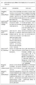
2.1 纳入文献基本情况 纳入文献包括骨关节炎和p38丝裂原活化蛋白激酶信号通路简介类文章20 篇[1-20],p38丝裂原活化蛋白激酶在骨关节炎中的作用的文章10篇[21-30] ,p38丝裂原活化蛋白激酶阻断剂在骨关节炎中的应用的文章16篇[31-46] 。 2.2 纳入文献的研究结果特征 2.2.1 p38丝裂原活化蛋白激酶信号通路简介 丝裂原活化蛋白激酶三级酶联是细胞内重要的信号转导系统之一。目前在哺乳动物体内共鉴定出4个丝裂原活化蛋白激酶亚家族,包括:细胞外信号调节蛋白激酶(extracellular signal-regulated kinase, ERKs)、p38丝裂原活化蛋白激酶、c-Jun氨基末端激酶/应激活化蛋白激酶(c-Jun N-terminal kinase/ stress xctivated protein kinases, JNK/SAPKs)和ERK5/大丝裂素活化蛋白激酶1(big MAP kinase 1, BMK 1)[7]。p38丝裂原活化蛋白激酶属丝氨酸/苏氨酸蛋白激酶,是分布于胞质中具有丝氨酸和酪氨酸双重磷酸化功能的蛋白激酶,可被多种炎症因子和环境应激因子激活,它们被激活后,可将信号传递到转录因子,进而与DNA结合,调节靶基因的表达。p38丝裂原活化蛋白激酶只有在苏氨酸(Thrl 80)和酪氨酸(Tyr 182)双位点同时被磷酸化时才具有全部活性。这两个邻近的位点之间被一氨基酸分开,形成“Thr-Xaa-Tyr”三肽基结构。它是决定激酶活性的关键结构,其长度影响p38丝裂原活化蛋白激酶底物特异性,并在控制其自主磷酸化上起重要作用[8]。 P38是由360个氨基酸构成的,相对分子质量约为38 000的蛋白质[9]。主要有4个亚型,即p38α、p38β、p38γ和p38δ。各同源激酶的氨基酸序列非常相近,但组织分布不同,p38α和p38β分布广泛,几乎可以在所有的组织细胞中得到表达,p38γ主要在骨骼肌,p38δ的表达主要在肺、肾、肠、唾液腺的表皮细胞及卵巢、睾丸、肾上腺和垂体[10]。 P38丝裂原活化蛋白激酶信号转导途径是磷酸化级联反应,需要多级激酶把细胞外信号传递到细胞内,其中需要3个关键激酶:丝裂原活化蛋白激酶、丝裂原活化蛋白激酶激酶、促分裂原活化蛋白激酶激酶[11]。首先,细胞受到刺激后通过中间环节使促分裂原活化蛋白激酶激酶激活,转而激活丝裂原活化蛋白激酶激酶,再由丝裂原活化蛋白激酶激酶对丝裂原活化蛋白激酶双位点磷酸化。P38丝裂原活化蛋白激酶上游激酶是MKK3即MAPKK3、MKK4及MKK6,是P38丝裂原活化蛋白激酶的重要调节因子,作为潜在的治疗目标来调节细胞因子的产生。其中MKK3与MKK6能直接磷酸化酪氨酸、丝氨酸/苏氨酸残基,特异性激活p38丝裂原活化蛋白激酶 [12],MKK3只能对p38α、p38β、p38γ进行磷酸化,而MKK6还可以磷酸化p38β[13]。目前认为MKK4能通过直接磷酸化丝氨酸/苏氨酸残基激活p38丝裂原活化蛋白激酶,但在哺乳动物体内MKK4/p38通路是否具有作用仍需进一步验证[14]。更上游的p38丝裂原活化蛋白激酶激活物可能包括Rho家族的GTP结合蛋白Rac和Cdc42,这些小分子蛋白是通过一组称为p21激活蛋白激酶的丝氨酸/苏氨酸激酶来发挥激活p38的作用。 p38丝裂原活化蛋白激酶被磷酸化激活后,移入核内或转移到其他部位,作用到细胞内相应的靶点并发挥调节功能。p38信号通路调控多种转录因子的基因表达活性,如激活作用转录因子、生长停滞及DNA损伤基因、核转录因子κB、热休克转录因子等,其中,有些转录因子是p38的直接底物,有些是p38的间接底物[15]。p38丝裂原活化蛋白激酶下游底物对于确定p38丝裂原活化蛋白激酶生理功能很重要,活化转录因子2、C/EBP环磷酸腺苷反应元件结合转录因子同源蛋白10和肌细胞增强因子2C是p38丝裂原活化蛋白激酶的主要作用底物[16]。p38丝裂原活化蛋白激酶可以特异性的将活化转录因子2的Thr71磷酸化,通过活化转录因子2磷酸化程度,人们可以测定p38蛋白激酶的活性。激活后的活化转录因子2与核转录因子κB以异二聚体的形式结合于激活蛋白1位点,能正向调节一些细胞因子的基因表达。CHOP10是C/EBP转录因子家族成员,可能参与了应激条件下细胞因子的表达调控。肌细胞增强因子2C磷酸化使转录因子c-Jun转录增加,从而增加其表达。炎症状态下磷酸化的c-Jun可以与激活蛋白1位点结合,从而使炎症相关基因表达增强,在炎症过程中起重要作用[17]。p38丝裂原活化蛋白激酶的作用底物还包括c-myc、MAX、STAT1等。p38丝裂原活化蛋白激酶还参与一些炎症相关事件,例如中性粒细胞的活化,细胞凋亡和一氧化氮合酶的诱导等[18-20]。 2.2.2 p38丝裂原活化蛋白激酶在骨关节炎中的作用 近年来发现,p38丝裂原活化蛋白激酶信号通路可能在骨关节炎的发生发展中发挥重要作用,它与软骨细胞表型的保持和分化、软骨细胞的肥大化和钙化、软骨细胞的凋亡、软骨基质金属蛋白酶的合成、软骨炎性细胞因子的产生等都有密切关系[21]。关节软骨细胞表型主要与其产生的Ⅱ型胶原和蛋白聚糖有关。近年来多数实验认为,多种细胞因子、细胞外基质可以通过细胞表面的整合素,而一氧化氮则是直接进入细胞质内,激活p38来促进关节软骨Ⅱ型胶原和蛋白聚糖的合成,也就是保持透明软骨的表型。而Robbins等[22]发现p38的激活可以阻断COL2AI mRNA的表达,从而阻断Ⅱ型胶原的合成。X型胶原在骨关节病中可作为软骨细胞表型不可逆转的标志,是大量关节软骨细胞发生凋亡,最终导致关节软骨变薄,关节软骨下骨板变硬、增厚的主要原因。转化生长因子β、白细胞介素8和生长相关性癌基因α通过激活p38来诱导X型胶原的表达。软骨细胞凋亡可能是骨性关节炎形成的重要原因,p38信号通路是关节软骨细胞凋亡的上游信号通路之一,一氧化氮、白细胞介素1β均可以通过p38来诱导软骨细胞的凋亡。 软骨在关节负荷和运动中起震动缓冲器的作用,细胞外基质蛋白聚糖和Ⅱ型胶原为这个震动缓冲器功能提供了结构基础。基质金属蛋白酶是一系列破坏细胞外基质的蛋白水解酶,其中基质金属蛋白酶1和基质金属蛋白酶13通过降解蛋白聚糖和Ⅱ型胶原,在软骨不可逆破坏中起关键的作用。骨关节炎的发病机制涉及基质金属蛋白酶,并且已经证实p38丝裂原活化蛋白激酶信号通路在调控软骨细胞分泌基质金属蛋白酶中起作用。骨关节炎发病过程中产生的软骨基质碎片,如纤连蛋白和胶原蛋白会刺激软骨细胞分泌基质金属蛋白酶,这一过程需要p38丝裂原活化蛋白激酶参与。研究表明,p38被炎症因子激活后通过调节激活蛋白1转录因子调节基质金属蛋白酶的表达,从而造成关节软骨损伤[23-25]。 Radons等[26]研究显示,在白细胞介素1β诱导的人软骨细胞分解代谢中P38的激活起到了重要作用,白细胞介素1β通过激活p38导致基质金属蛋白酶13表达增加进而降解Ⅱ型胶原增加。Rasheed等[27]报道在骨关节炎软骨细胞中检测到p38α、p38γ和p38δ的表达,而p38β未见表达;白细胞介素-1β可提高p38α、p38γ的磷酸化,但对p38δ无作用。在多种骨关节炎软骨破坏模型中,p38丝裂原活化蛋白激酶的活性得到验证,例如白细胞介素1β诱导的软骨溶解以及基质降解产物包括纤连蛋白碎片和胶原碎片诱导的软骨损伤[28-29]。 Nadia等[30]证实在骨关节炎软骨中白细胞介素1β可能通过激活JNK、p38丝裂原活化蛋白激酶和核转录因子κB信号通路引起Lipocalin型前列腺素D合成酶的表达上调。实验证实,在骨关节炎软骨细胞中,软骨下骨成骨细胞能诱导激活细胞外信号调节激酶1/2以及灭活p38,导致软骨细胞肥大的改变[31],这为丝裂原活化蛋白激酶信号通路在骨关节炎中的研究提供新的思路。 2.2.3 p38丝裂原活化蛋白激酶阻断剂在骨关节炎中的应用 基于p38丝裂原活化蛋白激酶在骨关节炎中不可忽视的作用,其抑制剂的研究也得到了迅速发展,成为治疗骨关节炎新的靶点。p38丝裂原活化蛋白激酶抑制剂在化学结构上可以分为两类,一类是吡啶咪唑芳基杂环类化合物,为p38丝裂原活化蛋白激酶专一抑制剂,以SB203580的应用最为广泛。第二类化合物包括N,N’-双芳基脲、N,N-双芳基脲、苯甲酮、吡唑酮、吲哚酰胺、二酰胺、喹唑酮、双嘧啶、吡啶氨喹唑啉等九小类。 SB203580是一种常用的特异性p38丝裂原活化蛋白激酶抑制剂,它可以通透细胞,抑制p38丝裂原活化蛋白激酶,并且抑制后续MAPKAP Kinase-2和MAPKAP Kinase-3的激活。通过抑制p38丝裂原活化蛋白激酶,SB203580可以有效抑制一些炎症因子如白细胞介素1β和肿瘤坏死因子α诱导的部分信号转导。但是SB203580并不阻断上游激酶对p38的激活,而是作用于p38三磷酸腺苷结合活性位点Thrl 06,使p38失去了与ATP结合能力,从而使其失去激酶活性[32]。SB203580能大量的抑制白细胞介素1β诱导的基质金属蛋白酶13的表达,但是MEK抑制剂PD98059和JNK抑制剂SP600125则不可以,这也显示出p38的激活对基质金属蛋白酶-13的表达是很重要的[33]。SB203580对p38丝裂原活化蛋白激酶各个亚型的抑制作用是不同的,它能抑制p38α和p38β的活性,但是不能抑制p38γ和p38δ的活性[34]。p38α的抑制剂SB203580能明显抑制白细胞介素1β或纤连蛋白碎片诱导的基质金属蛋白酶13的表达。p38α和p38γ的联合抑制剂BIRB796也能减少基质金属蛋白酶-13的表达和产生,但效果明显不如p38α抑制剂SB203580[35]。 研究显示,在人软骨细胞中,毒胡萝卜素诱导的基质金属蛋白酶13 mRNA的上调作用有49%被SB203580抑制,而衣毒素对它的上调作用有84%被SB203580抑制[36]。Joos等[37]采用基因芯片分析多种p38阻断剂对人骨关节炎软骨细胞炎性相关基因表达的差异,结果发现所有p38阻断剂均可抑制环氧化酶2、膜结合型前列腺素E2合酶1、诱导型一氧化氮合酶、基质金属蛋白酶-13及肿瘤坏死因子受体超家族11B的表达。Lee等[38]发现抑制p38丝裂原活化蛋白激酶活性可以抑制软骨细胞肥大进程,减少Ⅱ型胶原的合成。相关实验证实,p38丝裂原活化蛋白激酶抑制剂能够显著抑制中性粒细胞趋化性和超氧化物产生,减轻炎症反应[39]。 2.2.4 关于p38丝裂原活化蛋白激酶信号转导通路在骨关节炎中作用机制的相关文献分析 见表1。"

| [1]Felson DT. Clinical practice. Osteoarthritis of the knee. N Engl J Med. 2006;354(8):841-848.[2]Abramson SB, Yazici Y. Biologics in development for rheumatoid arthritis: relevance to osteoarthritis. Adv Drug Deliv Rev. 2006;58(2):212-225. [3]Hashimoto S, Setareh M, Ochs RL, et al. Fas/Fas ligand expression and induction of apoptosis in chondrocytes. Arthritis Rheum. 1997;40(10):1749-1755. [4]Kim SJ, Ju JW, Oh CD, et al. ERK-1/2 and p38 kinase oppositely regulate nitric oxide-induced apoptosis in chondrocytes in association with p53, caspase-3 and differentiation status. J Biol Chem. 2002;277(2):1332-1339. [5]Wei L, Sun XJ, Wang Z, et al. CD95-induced osteoarthritis chondrocyte apoptosis and necrosis: dependency on p38 mitogen-activated protein kinase. Arthritis Res Ther.2006; 8(2):R37. [6]Menqshol JA, Vincenti MP, Coon CI, et al. Interleukin -1 induction of collagenase 3 (matrix metalloproteinase 13) gene expression in chondrocytes requires p38, c-Jun N -terminal kinase, and nuclear factor kappaB: differential regulation of collagenase 1 and collagenase3. Arthritis Rheum. 2000;43(4): 801-811.[7]Zhang PJ, Zhu LX, Geng XP. Anhui Yiyao. 2010;14(5): 596-598.张频捷,朱立新,耿小平. p38 MAPK信号传导通路及其抑制剂的研究现状[J]. 安徽医药,2010,14(5):596-598. [8]Papadakis KA, Targan SR. Role of cytokines in the pathogenesis of inflammatory bowel disease. Annu Rev Med. 2000;51:289-98. [9]Morazzani M, de Carvalho DD, Kovacic H, et al. Monolayer versus aggregate balance in survival process for EGF-induced apoptosis in A431 carcinoma cells: Implication of ROS-p38 MAPK-integrin alpha2betal pathway. Int J Cancer. 2004;110(6):788-799. [10]张文军,李荣山. p38丝裂原活化蛋白激酶通路及其研究进展[J]. 医学综述,2007,13(9):663-665.[11]Han J, Lee JD, Bibbs L, et al. A MAP kinase targeted by endotoxin and hyperosmolarity in mammalian cells. Science. 1994;265(5173):808-811 . [12]Han J, Sun P. The pathways to tumor suppression via route p38. Trends Biochem Sci. 2007;32(8):364-371. [13]于波,朱振安. 骨关节炎治疗靶点p38MAPK信号通路研究进展[J]. 国际骨科学杂志,2011,32(6):343-345.[14]Enslen H, Raingeaud J, Davis RJ. Selective activation of p38 mitogen-activated protein (MAP) kinase isoforms by the MAP kinase kinases MKK3 and MKK6. J Biol Chem. 1998;273(3): 1741-1748. [15]Cazillis M, Bringuier AF. Disruption of MKK4 signaling reveals its tumor-suppressor role in embryonic stem cells. Oncogene. 2004;23(27):4735-4744.[16]Obata T, Brown GE, Yaffe MB. MAP kinase pathways activated by stress: the p38MAPK pathway. Crit Care Med. 2000;28(4):67-77. [17]Sugden PH, Clerk A. “Stress-responsive” mitogen-activated protein kinases (c-Jun N-terminal kinases and p38 mitogen-activated protein kinases ) in the myocardium. Circ Res. 1998;83(4):345-352. [18]Suzuki M, Tetsuka T, Yoshida S, et al. The role of p38 mitogen-activated protein kinase in IL-6 and IL-8 production from the TNF-alpha-or IL-1beta-stimulated rheumatoid synovial fibroblasts. FEBS Lett. 2000;465(1):23-27. [19]Juo P, Kuo CJ, Reynolds SE, et al. Fas activation of the p38 mitogen-activated protein kinase signalling pathway requires ICE/CED-3 family proteases. Mol Cell Biol. 1997;17(1):24-35. [20]Badger AM, Cook MN, Lark MW, et al. SB 203580 inhibits p38 mitogen-activated protein kinase, nitric oxide production, and inducible nitric oxide synthase in bovine cartilage-derived chondrocytes. J Immunol. 1998;161(1):467-473. [21]Qin ST, Jiang Q, Huang JH, et al. Zhongguo Guzhi Susong Zazhi. 2008;14(11):776-781.秦泗通,蒋青,黄际河,等. p38 丝裂原活化蛋白激酶抑制剂对大鼠骨性关节炎的影响[J]. 中国骨质疏松杂志,2008,14(11): 776-781.[22]Robbins JR, Thomas B, Tan L, et al. Immortalized human adult articular chondrocytes maintain cartilage-specific phenotype and responses to interleukin-1beta. Arthritis Rheum. 2000;43:2189-2201. [23]Murata M, Yudoh K, Nakamura H, et al. Distinct signaling pathways are involved in hypoxia-and IL-1-induced VEGF expression in human articular chondrocytes. J Orthop Res.2006;24(7):1544-1554. [24]Mengshol JA, Vincenti MP, Coon CI, et al. Interleukin-1 induction of collagenase 3 (matrix metalloproteinase 13) gene expression in chondrocytes requires p38, c-Jun N-terminal kinase, and nuclear factor kappaB :differential regulation of collagenase 1 and collagenase 3. Arthritis Rheum. 2000; 43(4):801-811. [25]Liacini A, Sylvester J, Li WQ, et al. Inhibition of interleukin-1-stimulated MAP kinases, activating protein-1(AP-1) and nuclear factor kappa B ( NF- kappa B ) transcription factors down-regulates matrix metalloproteinase gene expression in articular chondrocytes. Matrix Biology. 2002;21(3):251-262. [26]Radons J, Bosserhoff AK, Grassel S, et al. p38MAPK mediates IL-1- induced down-regulation of aggrecan gene expression in human chondrocytes. Int J Mol Med. 2006; 17(4):661-668. [27]Rasheed Z, Akhtar N, Haqqi TM. Pomegranate extract inhibits th interleukin-1β -induced activation of MKK-3, p38α-MAPK and transcription factor RUNX-2 in human osteoarthritis chondrocytes. Arthritis Res Ther. 2010;12(5):R195. [28]Chowdhury TT, Salter DM, Bader DL, Lee DA. Signal transduction pathways involving p38 MAPK, JNK, NF kappaB and AP-1 influences the response of chondrocytes cultured in agarose constructs to IL-1beta and dynamic compression. Inflamm Res. 2008;57(7):306-313. [29]Ding L, Guo D, Homandberg GA. The cartilage chondrolytic mechanism of fibronectin fragments involves MAP kinases: comparison of three fragments and native fibronectin. Osteoarthritis Cartilage. 2008 Oct;16(10):1253–1262. [30]Zayed N, Li X, Chabane N, et al. Increased expression of lipocalin-type prostaglandin D2 synthase in osteoarthritic cartilage. Arthritis Res Ther. 2008;10(6):R146. [31]Prasadam I, van Gennip S, Friis T, et al. ERK-1/2 and p38 in the regulation of hypertrophic changes of normal articular cartilage chondrocytes induced by osteoarthritic subchondral osteoblasts. Arthritis Rheum. 2010;62(5):1349-1360.[32]Morel C, Ibarz G, Oiry C, et al. Cross-interactions of two p38 mitogen-activated protein (MAP) kinase inhibitors and two cholecystokinin (CCK) receptor antagonists with the CCK1 receptor and p38 MAP kinase. J Biol Chem. 2005;280(22): 21384-21393. [33]Lim H, Kim HP. Matrix metalloproteinase-13 expression in IL-1β-treated chondrocytes by activation of the p38 MAPK/c-Fos/AP-1 and JAK/STAT pathways. Arch Pharm Res. 2011;34(1):109-117. [34]Lee JC, Kumar S, Griswold DE, et al. Inhibition of p38 MAP kinase as a therapeutic strategy.Immunopharmacology.2000;47(2-3):185-201. [35]Long DL, Loeser RF. 38gamma mitogen-activated protein kinase suppresses chondrocyte production of MMP-13 in response to catabolic stimulation. Osteoarthritis Cartilage. 2010;18(9):1203-1210. [36]Hamamura K, Goldring MB, Yokota H. Involvement of p38 MAPK in regulation of MMP-13 mRNA in chondrocytes in response to surviving stress to endoplasmic reticulum. Arch Oral Biol. 2009;54:279-286. [37]Joos H, Albrecht W, Laufer S, et al. Differential effects of p38MAP kinase inhibitors on the expression of inflammation-associated genes in primary, interleukin-1beta- stimulated human chondrocytes. Br J Pharmacol. 2010; 160(5):1252-1262. [38]Stanton LA, Sabari S, Sampaio AV, et al. p38 MAP kinase signalling is required for hypertrophic chondrocyte differentiation. BioChem J. 2004;378(1):53-62. [39]Nick JA, Young SK, Arndt PG, et al. Selective suppression of neutrophil accumulation in ongoing pulmonary inflammation by systemic inhibition of p38 mitogen-activated protein kinase. Jimmunol. 2002;169(9):5260-5269. [40]Brown KK, Heitmeyer SA, Hookfin EB, et al. P38 MAP kinase inhibitors as potential therapeutics for the treatment of joint degeneration and pain associated with osteoarthritis. J Inflamm (Lond). 2008;5:22. [41]Namdari S, Wei L, Moore D, et al. Reduced limb length and worsened osteoarthritis in adult mice after genetic inhibition of p38 MAP kinase activity in cartilage. Arthritis Rheum. 2008; 58(11): 3520-3529. [42]Li TF, Gao L, Sheu TJ, et al. Aberrant hypertrophy in smad3-deficient murine chondrocytes is rescued by restoring transforming growth factor beta-activated kinase 1/activating transcription factor 2 signaling:a potential clinical implication for osteoarthritis. Arthritis Rheum.2010;62(8):2359-2369.[43]Rasheed Z, Akhtar N, Haqqi TM. Pomegranate extract inhibitors the interleukin-1β-induced activation of MKK-3,p38α-MAPK and transcription factor RUNX-2 in human osteoarthritis chondrocytes. Arthritis Research & Therapy.2010;12(5):R195.[44]牛振东,刘恒,曹瑞治,等. 骨关节炎病理进程相关机制的研究进展[J]. 华西医学, 2012,27(11):1741-1744.[45]范素芳,王文雅,张柳. 丝裂原活化蛋白激酶信号通路对骨关节炎软骨的作用[J]. 中国组织工程研究,2012,16(37):7015-7019.[46]杨丰建,俞永林,林伟龙,等. 骨关节炎家兔关节软骨中丝裂原活化蛋白激酶信号通路蛋白表达以及活化状况[J]. 复旦学报:医学版,2011,38(3):221-225. |
| [1] | Huang Dengcheng, Wang Zhike, Cao Xuewei. Comparison of the short-term efficacy of extracorporeal shock wave therapy for middle-aged and elderly knee osteoarthritis: a meta-analysis [J]. Chinese Journal of Tissue Engineering Research, 2021, 25(9): 1471-1476. |
| [2] | Peng Zhihao, Feng Zongquan, Zou Yonggen, Niu Guoqing, Wu Feng. Relationship of lower limb force line and the progression of lateral compartment arthritis after unicompartmental knee arthroplasty with mobile bearing [J]. Chinese Journal of Tissue Engineering Research, 2021, 25(9): 1368-1374. |
| [3] | Liu Xiangxiang, Huang Yunmei, Chen Wenlie, Lin Ruhui, Lu Xiaodong, Li Zuanfang, Xu Yaye, Huang Meiya, Li Xihai. Ultrastructural changes of the white zone cells of the meniscus in a rat model of early osteoarthritis [J]. Chinese Journal of Tissue Engineering Research, 2021, 25(8): 1237-1242. |
| [4] | Liu Xin, Yan Feihua, Hong Kunhao. Delaying cartilage degeneration by regulating the expression of aquaporins in rats with knee osteoarthritis [J]. Chinese Journal of Tissue Engineering Research, 2021, 25(5): 668-673. |
| [5] | Ma Zetao, Zeng Hui, Wang Deli, Weng Jian, Feng Song. MicroRNA-138-5p regulates chondrocyte proliferation and autophagy [J]. Chinese Journal of Tissue Engineering Research, 2021, 25(5): 674-678. |
| [6] | Xie Chongxin, Zhang Lei. Comparison of knee degeneration after anterior cruciate ligament reconstruction with or without remnant preservation [J]. Chinese Journal of Tissue Engineering Research, 2021, 25(5): 735-740. |
| [7] | Cao Xuhan, Bai Zixing, Sun Chengyi, Yang Yanjun, Sun Weidong. Mechanism of “Ruxiang-Moyao” herbal pair in the treatment of knee osteoarthritis based on network pharmacology [J]. Chinese Journal of Tissue Engineering Research, 2021, 25(5): 746-753. |
| [8] | Li Yonghua, Feng Qiang, Tan Renting, Huang Shifu, Qiu Jinlong, Yin Heng. Molecular mechanism of Eucommia ulmoides active ingredients treating synovitis of knee osteoarthritis: an analysis based on network pharmacology [J]. Chinese Journal of Tissue Engineering Research, 2021, 25(5): 765-771. |
| [9] | Song Shan, Hu Fangyuan, Qiao Jun, Wang Jia, Zhang Shengxiao, Li Xiaofeng. An insight into biomarkers of osteoarthritis synovium based on bioinformatics [J]. Chinese Journal of Tissue Engineering Research, 2021, 25(5): 785-790. |
| [10] | Deng Zhenhan, Huang Yong, Xiao Lulu, Chen Yulin, Zhu Weimin, Lu Wei, Wang Daping. Role and application of bone morphogenetic proteins in articular cartilage regeneration [J]. Chinese Journal of Tissue Engineering Research, 2021, 25(5): 798-806. |
| [11] | Zheng Li, Li Dadi, Hu Weifan, Tang Jinlong, Zhao Fengchao. Risk assessment of contralateral knee arthroplasty after unilateral total knee arthroplasty [J]. Chinese Journal of Tissue Engineering Research, 2021, 25(3): 374-379. |
| [12] | Lü Jiaxing, Bai Leipeng, Yang Zhaoxin, Miao Yuesong, Jin Yu, Li Zhehong, Sun Guangpu, Xu Ying, Zhang Qingzhu. Evaluation of internal fixation with proximal femoral nail antirotation in elderly knee osteoarthritis patients with femoral intertrochanteric fractures [J]. Chinese Journal of Tissue Engineering Research, 2021, 25(3): 391-396. |
| [13] | Luo Anyu, Liu Hanlin, Xie Xiaofei, Huang Chen. Effect of antioxidant mixture on structural degeneration of an osteoarthritis rat model [J]. Chinese Journal of Tissue Engineering Research, 2021, 25(23): 3625-3629. |
| [14] | Gao Kun, Chen Dayu, Zhang Yong, Liu Weidong, Sun Shufen, Lai Wenqiang, Ma Dujun, Wu Yihong, Lin Zhanpeng, Jiang Yinglu, Yu Weiji. Achyranthes bidentata alcohol extract inhibits extracellular matrix degradation of the cartilage by regulating synovial fibroblast exosomes [J]. Chinese Journal of Tissue Engineering Research, 2021, 25(23): 3636-3640. |
| [15] | Peng Chao, Liu Yunpeng, Hua Guojun, Yang Jiaji, Wang Xingliang, Wang Xiaolong. Imaging evaluation of the hip-knee-ankle angle and osteoarthritis progression before and after partial meniscectomy for degenerative medial meniscus posterior root tear [J]. Chinese Journal of Tissue Engineering Research, 2021, 25(21): 3368-3373. |
| Viewed | ||||||
|
Full text |
|
|||||
|
Abstract |
|
|||||