Chinese Journal of Tissue Engineering Research ›› 2013, Vol. 17 ›› Issue (22): 4076-4083.doi: 10.3969/j.issn.2095-4344.2013.22.013
Previous Articles Next Articles
Measurement of partial adult proximal femur parameters
Ju Peng1, Qi Xiao-tong1, Liu Xiao-hu2, An Hong1, Jiang Dian-ming1
- 1Department of Orthopedics, the First Affiliated Hospital of Chongqing Medical University, Chongqing 400016, China
2Department of Radiology, the First Affiliated Hospital of Chongqing Medical University, Chongqing 400016, China
-
Online:2013-05-28Published:2013-05-28 -
Contact:Jiang Dian-ming, Professor, Department of Orthopedics, the First Affiliated Hospital of Chongqing Medical University, Chongqing 400016, China jdm571026@vip.163.com -
About author:Ju Peng★, Master, Department of Orthopedics, the First Affiliated Hospital of Chongqing Medical University, Chongqing 400016, China 360101542@qq.com
CLC Number:
Cite this article
Ju Peng, Qi Xiao-tong, Liu Xiao-hu, An Hong, Jiang Dian-ming. Measurement of partial adult proximal femur parameters[J]. Chinese Journal of Tissue Engineering Research, 2013, 17(22): 4076-4083.
share this article
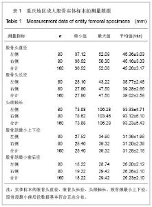
2.1 重庆地区成人股骨实体标本测得的股骨上端参数 游标卡尺对实体标本测量,数据基本符合正态分布。具体测量结果见表1。"
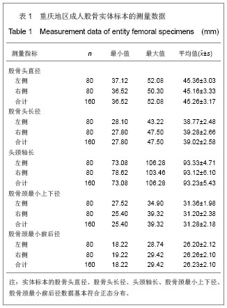
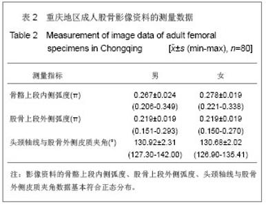
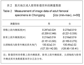
实体测量重庆地区成人股骨标本得出重庆地区成人股骨头直径为(45.26±3.17) mm;股骨头长径为(39.02±2.58) mm;头颈轴长为(93.23±5.43) mm;股骨颈最小上下颈为(31.28±2.18) mm;股骨颈最小前后径为 (26.23±2.10) mm。 2.2 重庆地区成人股骨影像资料测得的股骨上端参数 浩辰CAD2010标准版软件对股骨影像资料测量,数据基本符合正态分布。 具体测量结果为测量重庆地区成人股骨影像学资料得出的重庆地区成年男性股骨上端内侧弧度为(0.267±0.024) π;股骨上端外侧弧度为(0.219± 0.019) π;头颈轴线与股骨外侧皮质夹角为(130.92± 2.31)°;重庆地区成年女性股骨上端内侧弧度为(0.278±0.019) π;股骨上端外侧弧度为(0.219± 0.019) π;头颈轴线与股骨外侧皮质夹角为(130.68± 2.02)°,见表2。"
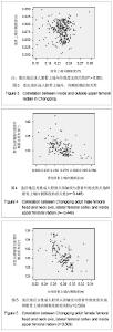
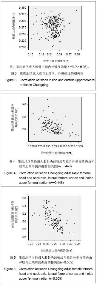
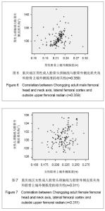
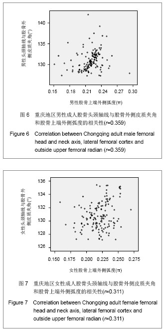
2.3 左右侧及不同性别对测量的重庆地区成人股骨上端参数的影响 在实体测量中,重庆地区成人股骨左右两侧的股骨头直径、股骨头长径、头颈轴长、股骨颈最小上下径、股骨颈最小前后径测量数据差异无显著性意义(P > 0.05);在软件测量中,重庆地区男女之间股骨上端内侧弧度、股骨上端外侧弧度、头颈轴线与股骨外侧皮质夹角也未见差异有显著性意义(P > 0.05)。 2.4 重庆地区成人股骨上端参数的相关性 实体测量的重庆地区成人股骨头直径、股骨头长径、股骨头颈轴长、股骨颈最小前后径和股骨颈最小上下径呈正相关,相关系数为0.246-0.588,提示径线间的变化趋势具有一致性。 软件测量的影像学数据中重庆地区成人股骨上端内、外侧弧度无相关性(P > 0.05);头颈轴线与股骨外侧皮质夹角和股骨上端内侧弧度呈负相关(r男=-0.446, r女=-0.509,P < 0.05),而与股骨上端外侧弧度呈正相关(r男=0.259,r女=0.311,P < 0.05),见图3-7。"
"
| [1]Dai KR. Zhonghua Guke Zazhi. 1997;17(1):3.戴克戎.骨溶解与人工关节后期松动[J].中华骨科杂志,1997, 17(1):3.[2]Chen CF, Liu BL. Nantong Yixueyuan Xuebao. 1984;4(1): 24-27.陈昌富,刘伯亮.成人股骨上端几项测量数值[J].南通医学院学报, 1984,4(1):24-27.[3]Wei B, Li GC, Liang JY, et al. Zhongguo Jiaoxing Waike Zazhi. 2005;3(13):436-438.魏波,李光灿,梁捷予,等.股骨近端解剖学参数分析及对内固定设计的指导意义[J].中国矫形外科杂志,2005,3(13):436-438.[4]Zhao CM, Yao ZX, Zheng YQ, et al. Shanghai Yike Daxue Xuebao. Shanghai Dier Yike Daxue Xuebao. 1987,17:27-30.周朝闵,姚志修,郑乃权,等.X线摄片测量国人股骨上端及其对人工髋关节设计的意义[J].上海第二医科大学学报,1987, 17: 27-30.[5]Zhang CG, Lv HS, Zou DW. Zhonghua Guke Zazhi. 1998; 18(8): 467-470.张纯光,吕厚山,邹德威.国人正常股骨CT测量与假体设计的相关研究[J].中华骨科杂志,1998,18(8):467-470.[6]Lidder S, Lang KJ, Lee HJ, et al. Bilateral hip fractures associated with transient osteoporosis of pregnancy. J R Army Med Corps. 2011;157(2):176-178.[7]Canale ST, Beaty JH. Beijing: People’s Military Medical Press. 2011:2535-2540.Canale ST, Beaty JH.坎贝尔骨科手术学[M].11版.北京:人民军医出版社,2011:2535-2540.[8]Zhang P, Gu ZH, Meng H, et al. Zhongguo Gushang. 1999; 12(6):47-50.张蒲,顾志华,孟和,等.双针固定治疗股骨颈骨折的力学分析[J].中国骨伤, 1999,12(6):47-50. [9]Kawatani Y, Nishida K, Anraku Y, et al. Clinical results of trochanteric fractures treated with the TARGON® proximal femur intramedullary nailing fixation system. Injury. 2011;42 Suppl 4:S22-27.[10]Büttner O, Styger S, Regazzoni P, et al. Stabilization of inter- and subtrochanteric femoral fractures with the PFNΑ®. Oper Orthop Traumatol. 2011;23(5):357-374[11]Rogmark C, Flensburg L, Fredin H. Undisplaced femoral neck fractures--no problems? A consecutive study of 224 patients treated with internal fixation. Injury. 2009;40(3):274-276.[12]Duckworth AD, Bennet SJ, Aderinto J, et al. Fixation of intracapsular fractures of the femoral neck in young patients: risk factors for failure. J Bone Joint Surg Br. 2011;93(6):811- 816.[13]Ma RF, Liu SL, Xu J. Linchuang Guke Zazhi. 2002;5(3):164-166.马若凡,刘尚礼,许杰.人股骨上端测量及其临床意义[J].临床骨科杂志,2002,5(3):164-166. [14]Noble PC, Alexander JW, Lindahl LJ, et al. The anatomic basis of femoral component design. Clin Orthop Relat Res. 1988;(235):148-165. [15]Yuan SM, Shi XP, Wang SC, et al. Jiangsu Daxue Xuebao: Yixue Ban. 2011;21(6):529-531. 袁士明,史小鹏,王素春,等.不同类型股骨转子间骨折内固定选择及疗效分析[J].江苏大学学报:医学版,2011,21(6):529-531.[16]Romero JM, Díaz RA, Oropeza YM. Dynamic hip screw vs. percutaneous compression plate for trochanteric fractures. Acta Ortop Mex. 2008;22(2):115-119.[17]Bali K, Sudesh P, Patel S, et al. Pediatric femoral neck fractures: our 10 years of experience. Clin Orthop Surg. 2011; 3(4):302-308.[18]Khan M, Aleem IS, Poolman RW. Fixation versus primary replacement of displaced femoral neck fractures in the elderly. Indian J Orthop. 2011;45(1):23-26.[19]Cody DD, Hou FJ, Divine GW, et al. Femoral structure and stiffness in patients with femoral neck fracture. J Orthop Res. 2000;18(3):443-448.[20]Chiang CC, Wu HT, Lin CF, et al. Analysis of initial injury radiographs of occult femoral neck fractures in elderly patients: a pilot study. Orthopedics. 2012;35(5):e621-627.[21]Van Embden D, Rhemrev SJ, Genelin F, et al. The reliability of a simplified Garden classification for intracapsular hip fractures. Orthop Traumatol Surg Res. 2012;98(4):405-408.[22]Luo X, He S, Li Z, et al. Systematic review of cemented versus uncemented hemiarthroplasty for displaced femoral neck fractures in older patients. Arch Orthop Trauma Surg. 2012;132(4):455-463.[23]Sergi G, Pintore G, Falci C, et al. Preventive effect of risedronate on bone loss and frailty fractures in elderly women treated with anastrozole for early breast cancer. J Bone Miner Metab. 2012;30(4):461-467.[24]Milovanovic P, Potocnik J, Djonic D, et al. Age-related deterioration in trabecular bone mechanical properties at material level: nanoindentation study of the femoral neck in women by using AFM. Exp Gerontol. 2012;47(2):154-159.[25]Chan AP, Cheung TC, Cheung SY, et al. Disseminated amyloidosis presenting with right proximal femur pathological fracture in a haemodialysis end-stage renal failure patient. Hong Kong Med J. 2011;17(6):495-499.[26]Herman A, Landau Y, Gutman G, et al. Radiological evaluation of intertrochanteric fracture fixation by the proximal femoral nail. Injury. 2012;43(6):856-863.[27]Maravic M, Ostertag A, Cohen-Solal M. Subtrochanteric/ femoral shaft versus hip fractures: incidences and identification of risk factors. J Bone Miner Res. 2012; 27(1): 130-137.[28]Fergus L, Cutfield G, Harris R. Auckland City Hospital's ortho-geriatric service: an audit of patients aged over 65 with fractured neck of femur. N Z Med J. 2011;124(1337):40-54.[29]Glatz U. Joint replacement: how to lower re-surgery rates. Dtsch Med Wochenschr. 2011;136(36):p26.[30]Cuckler JM. Metal-on-metal surface replacement: a triumph of hope over reason: affirms. Orthopedics. 2011;34(9): e439-441.[31]Eberle S, Wutte C, Bauer C, et al. Evaluation of risk for secondary fracture after removal of a new femoral neck plate for intracapsular hip fractures. J Orthop Trauma. 2011;25(12): 721-725.[32]Ruecker AH, Rupprecht M, Gruber M, et al. The treatment of intertrochanteric fractures: results using an intramedullary nail with integrated cephalocervical screws and linear compression. J Orthop Trauma. 2009;23(1):22-30.[33]Zdero R, Keast-Butler O, Schemitsch EH. A biomechanical comparison of two triple-screw methods for femoral neck fracture fixation in a synthetic bone model. J Trauma. 2010; 69(6):1537-1544.[34]Campbell AC, Goyal S, Miller NJ, et al. New technique for revising dynamic hip screw fixations with lag screw in situ. J Orthop Trauma. 2010;24(10):653-655.[35]Roerdink WH, Aalsma AM, Nijenbanning G, et al. Initial promising results of the dynamic locking blade plate, a new implant for the fixation of intracapsular hip fractures: results of a pilot study. Arch Orthop Trauma Surg. 2011;131(4):519-524.[36]Rogmark C, Spetz CL, Garellick G. More intramedullary nails and arthroplasties for treatment of hip fractures in Sweden. Acta Orthop. 2010;81(5):588-592.[37]Eberl R, Singer G, Ferlic P, et al. Post-traumatic coxa vara in children following screw fixation of the femoral neck. Acta Orthop. 2010;81(4):442-445.[38]Bech RD, Lauritsen J, Ovesen O, et al. Local anaesthetic wound infiltration after internal fixation of femoral neck fractures: a randomized, double-blind clinical trial in 33 patients. Hip Int. 2011;21(2):251-259.[39]Henari S, Leonard M, Hamadto M, et al. Review of a single contemporary femoral neck fracture fixation method in young patients. Orthopedics. 2011;34(3):171.[40]Varghese B, Short D, Hangartner T. Development of quantitative computed-tomography-based strength indicators for the identification of low bone-strength individuals in a clinical environment. Bone. 2012;50(1):357-363. |
| [1] | Tian Kechao, Wang Lei, Tao Yong, Yao Tao. Proximal femoral nail antirotation combined with posteromedial wall reconstruction for the treatment of type A2 intertrochanteric fracture in the elderly [J]. Chinese Journal of Tissue Engineering Research, 2021, 25(21): 3337-3342. |
| [2] | Liu Zemin, Lü Xin, Liu Jinyuan, Wang Xiaohu. Epidemiological distribution characteristics of 2 342 cases of hip fracture: a single center analysis [J]. Chinese Journal of Tissue Engineering Research, 2020, 24(32): 5085-5091. |
| [3] | Wei Yong, Li Jun, Zhang Yong, Yu Hao, Xie Jia. Prevalence rate and high-risk factors of preoperative lower-limb deep venous thrombosis in patients with hip fracture [J]. Chinese Journal of Tissue Engineering Research, 2020, 24(27): 4338-4342. |
| [4] | Qin Haikuo, Luo Shixing. Correlation of cortical bone thickness and X-ray gray value in different planes of proximal femur with brittle fracture of female hip [J]. Chinese Journal of Tissue Engineering Research, 2020, 24(18): 2867-2872. |
| [5] | Yang Zhaoxin, Niu Mengye, Lü Jiaxing, Cao Haiying, Kong Lingwei, Zhao Jingxin, Jin Yu. Finite element analysis to verify the rationality of bone cement combined with plate internal fixation for benign proximal femoral lesions [J]. Chinese Journal of Tissue Engineering Research, 2020, 24(15): 2368-2373. |
| [6] | Wang Kexin , Fan Jiang, Li Xue, Yang Shan, Ren Dong, He Chengqi. Long-term intensive family rehabilitation training for postoperative functional recovery in elderly hip fracture patients [J]. Chinese Journal of Tissue Engineering Research, 2020, 24(14): 2158-2163. |
| [7] | Pi Ying, Tian Jing. Relationship of age, pathological grade, and implants with perioperative mortality in patients with hip fracture [J]. Chinese Journal of Tissue Engineering Research, 2020, 24(12): 1835-1840. |
| [8] | Yang Zhaoxin, Niu Mengye, Liu Yuexing, Cao Haiying, Kong Lingwei, Zhao Jingxin, Jin Yu. Stability of the femoral neck and internal fixation with bone cement and steel plate after curettage for benign proximal femoral lesions [J]. Chinese Journal of Tissue Engineering Research, 2020, 24(12): 1841-1846. |
| [9] | Lu Qilin, Cai Xianhua, Shang Ranran, Chen Yanzhao, Xie Wei, Chen Xiongwei, Wu Haiyang. Feasibility of preoperative ratio of C-reactive protein to albumin for assessing the prognosis of older adults with hip fracture undergoing arthroplasty or internal fixation [J]. Chinese Journal of Tissue Engineering Research, 2019, 23(28): 4435-4439. |
| [10] | Wang Wuhua, Liu Xudong, Hu Ling. Prediction of hip fracture in Parkinson’s disease with the combination of geometric structure of proximal femur and bone mineral density [J]. Chinese Journal of Tissue Engineering Research, 2019, 23(24): 3829-3833. |
| [11] | Hao Liang, Zhang Zhonglin, Wang Baodong, Bi Zhenggang. Intertrochanteric fracture of the femur: improvement of internal fixation device, surgical changes and related disputes [J]. Chinese Journal of Tissue Engineering Research, 2019, 23(18): 2927-2935. |
| [12] | Wei Zhihui, Zhang Minghua, Zhang Zhongzu, Jiang Lian. Preoperative skin traction for hip fractures: a systematic review [J]. Chinese Journal of Tissue Engineering Research, 2019, 23(16): 2594-2600. |
| [13] | Zhang Jierong, Xiong Shixi, Tian Xiaolin, Gao Fangmao, Lin Chao, Yang Lixue. Efficacy and safety of proximal femoral anatomical locking compression plate and proximal femoral nail antirotation for long-segment comminuted subtrochanteric fractures of the femur: a non-randomized controlled trial [J]. Chinese Journal of Tissue Engineering Research, 2019, 23(16): 2493-2499. |
| [14] | Guo Jinchao1, Cao Yuan2, Huang Junling1, Ma Jiajia1, Ma Chuang1. Intertrochanteric femoral fractures: an epidemiological analysis of 618 cases [J]. Chinese Journal of Tissue Engineering Research, 2019, 23(12): 1840-1845. |
| [15] | Wan Qian, Yang Xiaohua, Zhang Qingzhu, Feng Zhen, Gu Rui. Curative effects of cemented hemiarthroplasty versus proximal femoral nail anti-rotation for treatment of intertrochanteric fractures of cerebral infarction hemiplegia side in older adults [J]. Chinese Journal of Tissue Engineering Research, 2018, 22(35): 5590-5595. |
| Viewed | ||||||
|
Full text |
|
|||||
|
Abstract |
|
|||||