Chinese Journal of Tissue Engineering Research ›› 2020, Vol. 24 ›› Issue (1): 33-39.doi: 10.3969/j.issn.2095-4344.2006
Previous Articles Next Articles
An increase in proliferation of rat bone mesenchymal stem cells and secretion of vascular endothelial growth factor under negative pressure in vitro
Yang Xiongfeng1, Jiang Huijiao2, Wang Xiaoyi2, Guo Lijiao1, Zhou Qing1, Han Huanhuan1, Li Linlin2, Liao Zhenyu2, Lei Zhen1, Chen Xueling2, Zhang Hongwei1, Wu Xiangwei1
- 1the First Affiliated Hospital of Shihezi University Medical School, Shihezi 832000, Xinjiang Uygur Autonomous Region, China; 2Shihezi University Medical School, Shihezi 832000, Xinjiang Uygur Autonomous Region, China
-
Received:2019-02-16Revised:2019-02-26Accepted:2019-04-15Online:2020-01-08Published:2019-12-11 -
Contact:Wu Xiangwei, MD, Professor, Chief physician, the First Affiliated Hospital of Shihezi University Medical School, Shihezi 832000, Xinjiang Uygur Autonomous Region, China -
About author:Yang Xiongfeng, Master, Physician, the First Affiliated Hospital of Shihezi University Medical School, Shihezi 832000, Xinjiang Uygur Autonomous Region, China -
Supported by:the National Natural Science Foundation of China, No. 81760570; Applied Basic Research Project of Xinjiang Corps, No. 2016AG019; High-Level Talents Start Project of Shihezi University, No. RCZX201538
CLC Number:
Cite this article
Yang Xiongfeng, Jiang Huijiao, Wang Xiaoyi, Guo Lijiao, Zhou Qing, Han Huanhuan, Li Linlin, Liao Zhenyu, Lei Zhen, Chen Xueling, Zhang Hongwei, Wu Xiangwei. An increase in proliferation of rat bone mesenchymal stem cells and secretion of vascular endothelial growth factor under negative pressure in vitro[J]. Chinese Journal of Tissue Engineering Research, 2020, 24(1): 33-39.
share this article
Add to citation manager EndNote|Reference Manager|ProCite|BibTeX|RefWorks

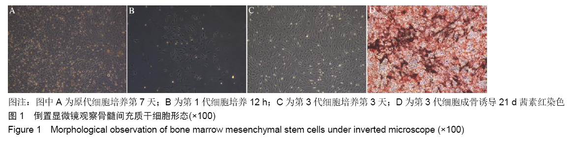
2.1 骨髓间充质干细胞的分离、培养和鉴定结果 原代培养48 h后细胞逐渐贴壁,其他类型的各种杂细胞通过频繁换液逐渐除去,此时贴壁细胞大多呈梭形、多角形,单核细胞状,细胞形态不一,或者为单个或多个细胞的克隆,细胞增殖迅速;培养3-5 d,细胞90%以上融合,融合细胞呈长梭形,细胞集落呈鱼群状生长。传代后4 h可见细胞贴壁呈不规则形并伸出触角;24 h后细胞全部贴壁,可见大部分细胞变为长梭形,具有方向性,部分细胞变为不规则形,杂细胞较少。成骨诱导21 d后茜素红染色可见钙结节红染,见图1。"
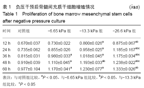
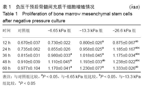
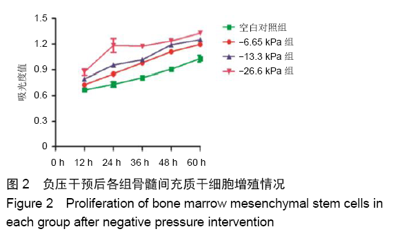
2.2 不同负压条件干预后各组骨髓间充质干细胞的增殖情况 各组细胞给予负压干预后形态均一,生长旺盛,随负压培养时间延长大部分细胞变成不规则的多角形,细胞细长。随着负压压力梯度升高,镜下观察到细胞生长旺盛、密集,并且胞体丰满;与对照组相比,细胞增殖明显增快,细胞集落增大,细胞间连接紧凑。与对照组相比,低负压条件(-6.65 kPa)、中负压条件(-13.3 kPa)、高负压条件(-26.6 kPa)对骨髓间充质干细胞增殖均具有促进作用(P < 0.05),见表1和图2。"

"
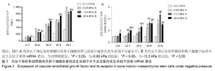
2.3 不同负压条件干预后各组骨髓间充质干细胞培养上清液中血管内皮生长因子水平 负压干预培养后,-6.65 kPa组、-13.3 kPa组、-26.6 kPa组血管内皮生长因子分泌水平在5个时间点均高于对照组,且随着负压培养时间延长呈逐渐上升的趋势。在负压培养12 h时,-13.3 kPa组和 -26.6 kPa组血管内皮生长因子分泌量均高于-6.65 kPa组,然而在负压培养24 h时,-13.3 kPa组和-26.6 kPa组血管内皮生长因子分泌量均低于-6.65 kPa组,且在12 h和24 h时,-13.3 kPa组、-26.6 kPa组血管内皮生长因子分泌量差异无显著性意义。负压干预36 h和48 h时,-26.6 kPa组血管内皮生长因子分泌量高于 -6.65 kPa组和-13.3 kPa组(P < 0.05),然而-6.65 kPa组与-13.3 kPa组比较差异无显著性意义(P > 0.05)。负压干预60 h时,-13.3 kPa组、-26.6 kPa组血管内皮生长因子分泌量均低于-6.65 kPa组(P < 0.05),-26.6 kPa组血管内皮生长因子分泌量显著高于-13.3 kPa组(P < 0.05),见图3A。 2.4 不同负压条件干预后各组骨髓间充质干细胞中血管内皮生长因子受体mRNA表达 负压干预12 h时,-26.6 kPa组血管内皮生长因子受体mRNA表达水平高于-6.65 kPa组、-13.3 kPa组(P < 0.05),-6.65 kPa组、-13.3 kPa组和对照组之间比较差异无显著性意义(P > 0.05)。负压干预 24 h时,-13.3 kPa组、-26.6 kPa组血管内皮生长因子受体mRNA表达水平均高于-6.65 kPa组(P < 0.05),-26.6 kPa组血管内皮生长因子受体mRNA表达水平显著高于 -13.3 kPa组(P < 0.05)。负压干预36 h时,-26.6 kPa组血管内皮生长因子受体mRNA表达水平显著高于-6.65 kPa组、-13.3 kPa组和对照组(P < 0.05),对照组和-6.65 kPa组之间比较差异无显著性意义(P > 0.05)。负压干预48 h时,-13.3 kPa组和-26.6 kPa组血管内皮生长因子受体mRNA表达水平高于-6.65 kPa组和对照组(P < 0.05),-13.3 kPa组和-26.6 kPa组之间比较差异无显著性意义(P > 0.05)。负压干预60 h时,-13.3 kPa组血管内皮生长因子受体mRNA表达水平高于-6.65 kPa组和和-26.6 kPa组,-26.6 kPa组和-6.65 kPa组比较差异无显著性意义(P > 0.05),见图3B。 "


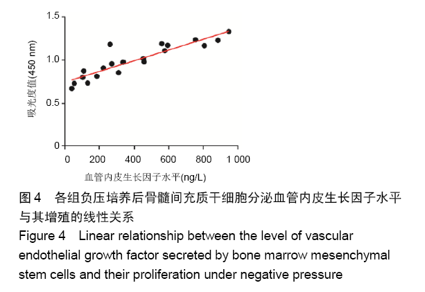
2.5 骨髓间充质干细胞增殖吸光度值与血管内皮生长因子分泌水平线性关系 根据线性关系分析,负压干预后骨髓间充质干细胞增殖指标吸光度值与血管内皮生长因子分泌水平呈正相关,相关系数为0.902,P < 0.05,见图4。"

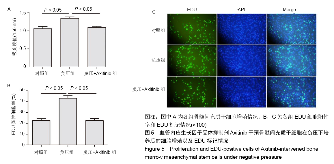
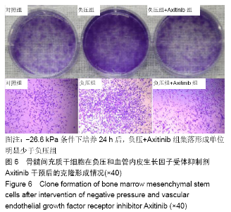
2.6 骨髓间充质干细胞在负压和血管内皮生长因子受体抑制剂Axitinib干预后细胞增殖、克隆形成以及EDU标记情况 -26.6 kPa条件下培养24 h后,血管内皮生长因子受体抑制剂组吸光度值、EDU细胞阳性率及集落形成单位个数明显低于负压组(P < 0.05);与对照组比较,血管内皮生长因子受体抑制剂组吸光度值、EDU细胞阳性率及集落形成单位个数差异无显著性意义(P > 0.05),见图5,6。"
"

| [1] FRIEDENSTEIN AJ, CHAILAKHJAN RK, LALYKINA KS. The development of fibroblast colonies in monolayer cultures of guinea-pig bone marrow and spleen cells. Cell Tissue Kinet. 1970;3(4):393-403. [2] FRIEDENSTEIN AJ, CHAILAKHYAN RK, LATSINIK NV, et al. Stromal cells responsible for transferring the microenvironment of the hemopoietic tissues.Cloning in vitro and retransplantation in vivo. Transplantation.1974;17(4):331-340. [3] ZHOU J, DONG J. Mesenchymal stem cells and mesenchymal- derived endothelial cells. Stem Cells and Cancer Stem Cells. 2012: 277-281. [4] MATHOT F, SHIN AY, VAN WIJNEN AJ. Targeted stimulation of MSCs in peripheral nerve repair. Gene. 2019 Mar 5. doi: 10.1016/j.gene.2019.02.078. [Epub ahead of print] [5] LIU Q, CHEN MX, SUN L, et al. Rational use of mesenchymal stem cells in the treatment of autism spectrum disorders. World J Stem Cells. 2019;11(2):55-72. [6] LIN Y, UMEBAYASHI M, ABDALLAH MN, et al. Combination of polyetherketoneketone scaffold and human mesenchymal stem cells from temporomandibular joint synovial fluid enhances bone regeneration. Sci Rep. 2019;9(1):472. [7] KOMATSU N, KAJIYA M, MOTOIKE S, et al. Type I collagen deposition via osteoinduction ameliorates YAP/TAZ activity in 3D floating culture clumps of mesenchymal stem cell/extracellular matrix complexes. Stem Cell Res Ther. 2018;9(1):342. [8] LING L, FENG X, WEI T, et al. Human amnion-derived mesenchymal stem cell (hAD-MSC) transplantation improves ovarian function in rats with premature ovarian insufficiency (POI) at least partly through a paracrine mechanism. Stem Cell Res Ther. 2019;10(1):46. [9] WANG X, LI C, GONG H. Morphological and functional changes in bone marrow mesenchymal stem cells in rats with heart failure. Exp Ther Med. 2017;13(6):2888-2892. [10] ABDELMONEM M, SHAHIN NN, RASHED LA, et al. Hydrogen sulfide enhances the effectiveness of mesenchymal stem cell therapy in rats with heart failure: In vitro preconditioning versus in vivo co-delivery. Biomed Pharmacother. 2019;112:108584. [11] LIAO S, ZHANG Y, TING S, et al. Potent immunomodulation and angiogenic effects of mesenchymal stem cells versus cardiomyocytes derived from pluripotent stem cells for treatment of heart failure. Stem Cell Res Ther. 2019;10(1):78. [12] MÜLLER-EHMSEN J, WHITTAKER P, KLONER RA, et al. Survival and development of neonatal rat cardiomyocytes transplanted into adult myocardium. J Mol Cell Cardiol. 2002;34(2):107-116. [13] 伞光,宋佳.血管内皮生长因子165转染可促进脂肪间充质干细胞增殖[J].中国组织工程研究, 2015,19(36):5782-5788. [14] 冯文磊,张猛,印双红,等.改良差时贴壁法分离培养鉴定小鼠骨髓间充质干细胞和内皮前体细胞[J].解剖学报, 2015,46(2):282-288. [15] 张猛,张宏伟,冯文磊,等.血管内皮前体细胞通过旁分泌功能促进间充质细胞成骨分化[J].中华医学杂志, 2015,95(16):1253-1257. [16] FLEISCHMANN W, STRECKER W, BOMBELLI M, et al. Vacuum sealing as treatment of soft tissue damage in open fractures. Unfallchirurg. 1993;96(9):488-492. [17] ARGENTA LC, MORYKWAS MJ. Vacuum-assisted closure: a new method for wound control and treatment: clinical experience. Ann Plast Surg. 1997;38(6):563-576 [18] YUAN Y, NIU Y, XIAO W, et al. The Effect and Mechanism of Negative Pressure Wound Therapy on Lymphatic Leakage in Rabbits. J Surg Res. 2019;235:329-339. [19] KARAM RA, REZK NA, ABDEL RAHMAN TM, et al. Effect of negative pressure wound therapy on molecular markers in diabetic foot ulcers. Gene. 2018;667:56-61. [20] SHI J, XI W, YI C, et al. Vacuum sealing drainage promotes experimental pig explosive abdomen wound healing.Xi Bao Yu Fen Zi Mian Yi Xue Za Zhi. 2014;30(3):312-315. [21] BRADLEY BH, CUNNINGHAM M. Biofilms in chronic wounds and the potential role of negative pressure wound therapy: an integrative review. J Wound Ostomy Continence Nurs. 2013;40(2):143-149. [22] ZHANG LJ, WANG C, LUO PF, et al. Debridement combined with vacuum sealing drainage in the treatment of severe infection in abdominal wall due to allogeneic umbilical cord embedded in abdominal wall for immunotherapy. Zhonghua Shao Shang Za Zhi. 2018;34(8):556-558. [23] BORYS S, HOHENDORFF J, FRANKFURTER C, et al. Negative pressure wound therapy use in diabetic foot syndrome-from mechanisms of action to clinical practice. Eur J Clin Invest. 2019; 49(4):e13067. [24] NAM D, SERSHON RA, LEVINE BR, et al. The Use of Closed Incision Negative-Pressure Wound Therapy in Orthopaedic Surgery. J Am Acad Orthop Surg. 2018;26(9):295-302. [25] MU S, HUA Q, JIA Y, et al. Effect of negative-pressure wound therapy on the circulating number of peripheral endothelial progenitor cells in diabetic patients with mild to moderate degrees of ischaemic foot ulcer. Vascular. 2019 Mar 6: doi:10.1177/1708538119836360.[Epub ahead of print] [26] ISHII M, TAKAHASHI M, MURAKAMI J, et al. Vascular endothelial growth factor-C promotes human mesenchymal stem cell migration via an ERK-and FAK-dependent mechanism. Mol Cell Biochem. 2019; 455(1-2):185-193. [27] MENG X, CHEN M, SU W, et al. The differentiation of mesenchymal stem cells to vascular cells regulated by the HMGB1/RAGE axis: its application in cell therapy for transplant arteriosclerosis. Stem Cell Res Ther. 2018;9(1):85. [28] NING J, ZHAO H, CHEN B, et al. Argon Mitigates Impaired Wound Healing Process and Enhances Wound Healing In Vitro and In Vivo. Theranostics. 2019;9(2):477-490. [29] ZHU J, YU A, QI B, et al. Effects of negative pressure wound therapy on mesenchymal stem cells proliferation and osteogenic differentiation in a fibrin matrix. PLoS One. 2014;9(9):e107339. [30] 陶圣祥,余国荣,喻爱喜,等.负压培养对大鼠骨髓基质干细胞增殖及分化的影响[J]. 医学新知, 2007,17(6): 324-326. [31] YANG Z, YAO JF, XU P, et al. Functions and mechanisms of intermittent negative pressure for osteogenesis in human bone marrow mesenchymal stem cells. Mol Med Rep. 2014;9(4):1331-1336. [32] SHOU K, NIU Y, ZHENG X, et al. Enhancement of Bone-Marrow- Derived Mesenchymal Stem Cell Angiogenic Capacity by NPWT for a Combinatorial Therapy to Promote Wound Healing with Large Defect. Biomed Res Int. 2017;2017:7920265. |
| [1] | Liu Cong, Liu Su. Molecular mechanism of miR-17-5p regulation of hypoxia inducible factor-1α mediated adipocyte differentiation and angiogenesis [J]. Chinese Journal of Tissue Engineering Research, 2021, 25(7): 1069-1074. |
| [2] | Li Cai, Zhao Ting, Tan Ge, Zheng Yulin, Zhang Ruonan, Wu Yan, Tang Junming. Platelet-derived growth factor-BB promotes proliferation, differentiation and migration of skeletal muscle myoblast [J]. Chinese Journal of Tissue Engineering Research, 2021, 25(7): 1050-1055. |
| [3] | Ma Zetao, Zeng Hui, Wang Deli, Weng Jian, Feng Song. MicroRNA-138-5p regulates chondrocyte proliferation and autophagy [J]. Chinese Journal of Tissue Engineering Research, 2021, 25(5): 674-678. |
| [4] | Wang Yujiao, Liu Dan, Sun Song, Sun Yong. Biphasic calcium phosphate loaded with advanced platelet rich fibrin can promote the activity of rabbit bone marrow mesenchymal stem cells [J]. Chinese Journal of Tissue Engineering Research, 2021, 25(4): 504-509. |
| [5] | Zhou Jihui, Yao Meng, Wang Yansong, Li Xinzhi, Zhou You, Huang Wei, Chen Wenyao. Influence of novel nanoscaffolds on biological behaviors of neural stem cells and the related gene expression [J]. Chinese Journal of Tissue Engineering Research, 2021, 25(4): 532-536. |
| [6] | Chen Yang, Huang Denggao, Gao Yuanhui, Wang Shunlan, Cao Hui, Zheng Linlin, He Haowei, Luo Siqin, Xiao Jingchuan, Zhang Yingai, Zhang Shufang. Low-intensity pulsed ultrasound promotes the proliferation and adhesion of human adipose-derived mesenchymal stem cells [J]. Chinese Journal of Tissue Engineering Research, 2021, 25(25): 3949-3955. |
| [7] | Zhou Wu, Wang Binping, Wang Yawen, Cheng Yanan, Huang Xieshan. Transforming growth factor beta combined with bone morphogenetic protein-2 induces the proliferation and differentiation of mouse MC3T3-E1 cells [J]. Chinese Journal of Tissue Engineering Research, 2021, 25(23): 3630-3635. |
| [8] | Yu Chengshuai, Du Gang, Pang Shenning, Lao Shan. Chemerin, a pro-inflammatory adipokine, regulates chondrocyte proliferation and metabolism by increasing production of nitric oxide [J]. Chinese Journal of Tissue Engineering Research, 2021, 25(2): 258-263. |
| [9] | Dai Yaling, Chen Lewen, He Xiaojun, Lin Huawei, Jia Weiwei, Chen Lidian, Tao Jing, Liu Weilin. Construction of miR-146b overexpression lentiviral vector and the effect on the proliferation of hippocampal neural stem cells [J]. Chinese Journal of Tissue Engineering Research, 2021, 25(19): 3024-3030. |
| [10] | Ailimaierdan·Ainiwaer, Wang Ling, Gu Li, Dilidaer•Taxifulati, Wang Shan, Yin Hongbin. Effect of transforming growth factor-beta3 on the proliferation and osteogenic capability of osteoblasts [J]. Chinese Journal of Tissue Engineering Research, 2021, 25(17): 2664-2669. |
| [11] | Wang Renxian, Cao Jingjing, Wang Honggang, Wan Ben, Liu Weifeng. Effects of dispersants on aggregation, intracellular distribution and cell proliferation of nano-hydroxyapatite [J]. Chinese Journal of Tissue Engineering Research, 2021, 25(16): 2500-2505. |
| [12] | Wang Huili, Chen Zhijiang, Wu Bingyi. Glia maturation factor-gamma inhibits the proliferation of human colorectal cancer LoVo cells and affects the cytoskeletal motion of human umbilical vein endothelial cells [J]. Chinese Journal of Tissue Engineering Research, 2021, 25(13): 2055-2059. |
| [13] | Jiang Tao, Wu Shuo, Li Zhiqiang, Shou Xi, Mayire·Nuermaimaiti, Ma Chuang, Wei Qin. Platelet-derived growth factor BB promotes the proliferation of bone marrow mesenchymal stem cells of Sprague-Dawley rats [J]. Chinese Journal of Tissue Engineering Research, 2021, 25(13): 1976-1981. |
| [14] | Yan Nan, Si Xiaofeng, Zeng Liang, Tian Wei, Shan Guangdong, Xiong Lishuo, Yang Weijie, Wang Zhengdong. Nerve growth factor interferes with proliferation and alpha-actin expression of skeletal muscle satellite cells in rats [J]. Chinese Journal of Tissue Engineering Research, 2021, 25(13): 2030-2035. |
| [15] | Zhao Chuntao, Qing Mingsong, Yu Langbo, Peng Jiachen . Meta-analysis of total knee arthroplasty guided by kinematic alignment and mechanical alignment [J]. Chinese Journal of Tissue Engineering Research, 2020, 24(9): 1435-1442. |
| Viewed | ||||||
|
Full text |
|
|||||
|
Abstract |
|
|||||