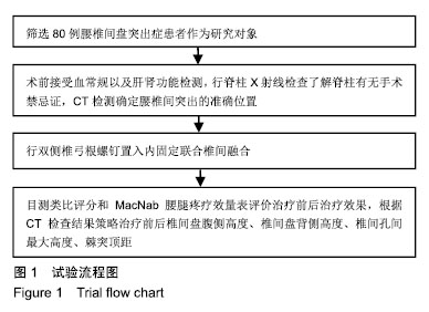
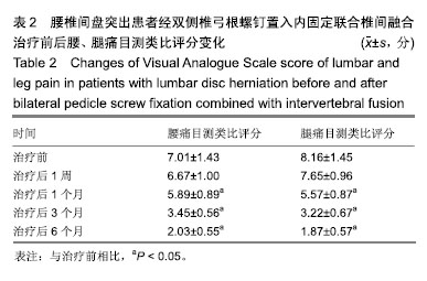
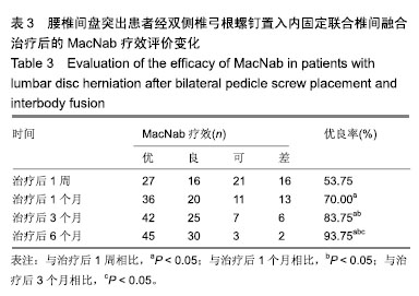
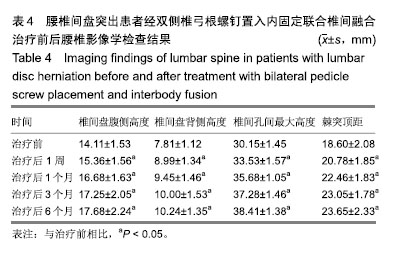
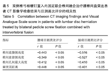
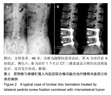
| [1]杨龙彪,高清元,赵东.复发性腰椎间盘突出症应用经皮内镜椎间孔入路微创治疗的疗效观察[J].贵州医药,2017,41(4):394-395.[2]汤培,陈晓君,吴小宝,等.射频消融联合针刀对腰椎间盘突出症疼痛症状的改善效果及1年随访研究[J].中医药临床杂志,2018, 30(8):1531-1534.[3]牛翔科,肖应权,孙凤,等.CT引导下微创治疗腰椎间盘突出症疗效观察[J].中华实用诊断与治疗杂志,2014,28(4):348-349.[4]辛欣,刘新新.腰椎间盘突出症手术治疗的MRI影像学疗效评价[J].中国CT和MRI杂志,2015,13(09):6-8,21.[5]曹兴兵,赵镒汶,黄永辉,等.胸腰椎骨折体位复位结合伤椎椎体内撑开复位椎体内植骨内固定术后矢状面形态的变化[J].中国骨与关节损伤杂志,2015,30(1):31-34. [6]Wang M, Zhou Y, Wang J, et al. A 10-year follow-up study on long-term clinical outcomes of lumbar microendoscopic discectomy. J Neurol Surg A Cent Eur Neurosurg. 2012;73(4): 195-198. [7]Ahn Y. Transforaminal percutaneous endoscopic lumbar discectomy: technical tips to prevent complications. Expert Rev Med Devices. 2012;9(4):361-366. [8]张楠,奚春阳,闫景龙.椎弓根螺钉固定并新型肌骨瓣植骨融合治疗极外侧腰椎间盘突出症临床疗效分析[J].哈尔滨医科大学学报,2016,50(3):223-227.[9]方钧,郑季南,杨培伟.椎弓根钉棒系统联合椎体支柱块治疗胸腰椎骨折的生物力学试验及临床应用[J].颈腰痛杂志,2016,37(3): 176-181. [10]Lee S, Kang JH, Srikantha U, et al. Extraforaminal compression of the L-5 nerve root at the lumbosacral junction: clinical analysis, decompression technique, and outcome. J Neurosurg Spine. 2014;20(4):371-379.[11]Kida K, Tadokoro N, Kumon M, et al. Can cantilever transforaminal lumbar interbody fusion (C-TLIF) maintain segmental lordosis for degenerative spondylolisthesis on a long-term basis? Arch Orthop Trauma Surg. 2014;134(3): 311-315.[12]张人文,莫灼锚,唐树杰.手法治疗腰椎间盘突出症研究进展[J].山东中医药大学学报,2018,42(1):86-89.[13]Lehnert T, Naguib NN, Wutzler S, et al. Analysis of disk volume before and after CT-guided intradiscal and periganglionic ozone-oxygen injection for the treatment of lumbar disk herniation. J Vasc Interv Radiol. 2012;23(11): 1430-1436. [14]Kermani HR, Keykhosravi E, Mirkazemi M, et al. The relationship between morphology of lumbar disc herniation and MRI changes in adjacent vertebral bodies. Arch Bone Jt Surg. 2013;1(2):82-85.[15]黄仕荣,石印玉,詹红生.腰椎间盘突出症临床诊断的思路与程序[J].中国骨伤,2012,25(2):147-151.[16]程俊杰,眭江涛,马原,等.颈椎人工间盘置换与前路减压融合修复单节段颈椎间盘突出症:3年随访[J].中国组织工程研究,2015, 19(53):8529-8536.[17]焦龙兵,吴阳,张兵.经皮椎间孔镜技术在钙化型腰椎间盘突出症的临床应用[J].颈腰痛杂志,2018,39(5):634-636.[18]Lee SH, Daffner SD, Wang JC. Does lumbar disk degeneration increase segmental mobility in vivo? Segmental motion analysis of the whole lumbar spine using kinetic MRI. J Spinal Disord Tech. 2014;27(2):111-116.[19]Fay LY, Wu JC, Tsai TY, et al. Dynamic stabilization for degenerative spondylolisthesis: evaluation of radiographic and clinical outcomes. Clin Neurol Neurosurg. 2013;115(5): 535-541.[20]Wang M, Zhou Y, Wang J, et al. A 10-year follow-up study on long-term clinical outcomes of lumbar microendoscopic discectomy. J Neurol Surg A Cent Eur Neurosurg. 2012;73(4): 195-198.[21]张建洛,杨宏涛,冯宏伟,等.身痛逐瘀汤联合西医治疗腰椎间盘突出症半椎板切除减压术后下肢疼痛及麻木残余症状疗效观察[J].现代中西医结合杂志,2018,27(4):415-418.[22]Gómez FS, Lorza RL, Bobadilla MC, et al. Improving the Process of Adjusting the Parameters of Finite Element Models of Healthy Human Intervertebral Discs by the Multi-Response Surface Method. Materials (Basel). 2017; 10(10):E1116.[23]Wood MJ, McMillen J. The surgical learning curve and accuracy of minimally invasive lumbar pedicle screw placement using CT based computer-assisted navigation plus continuous electromyography monitoring - a retrospective review of 627 screws in 150 patients. Int J Spine Surg. 2014;8: 27. [24]魏社军,王宇,郑标,等.双侧椎弓根螺钉内固定结合椎间融合器植骨融合治疗相邻双节段腰椎间盘突出症合并腰椎管狭窄[J].中医正骨,2015,27(6):46-48[25]徐德利,林浩,陶海鹰.双侧椎弓根螺钉置入内固定修复腰椎间盘突出:椎间高度恢复的影像学评估[J].中国组织工程研究,2015, 19(26):4191-4196.[26]马树伟,吴继功.三种开放手术方式治疗腰4/5椎间盘突出症的疗效对比[J].河北医学,2018,24(7):1155-1161.[27]Li J, Li Y, Kong F, et al. Adjacent segment degeneration after single-level anterior cervical decompression and fusion: disc space distraction and its impact on clinical outcomes. J Clin Neurosci. 2015;22(3):566-569. [28]American Cancer Society. Understanding Radiation Risk from Imaging Tests. https://www.cancer.org/treatment/ understanding-your-diagnosis/tests/understanding-radiation-risk-from-imaging-tests.html. Accessed on 2019-07-16. |