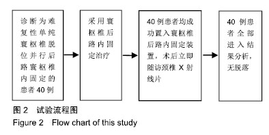
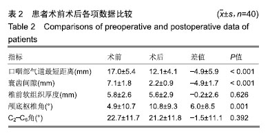
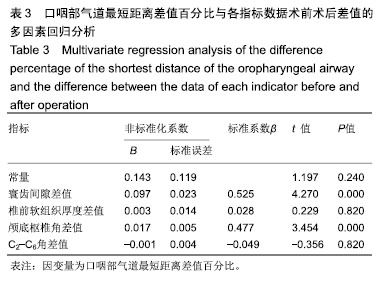
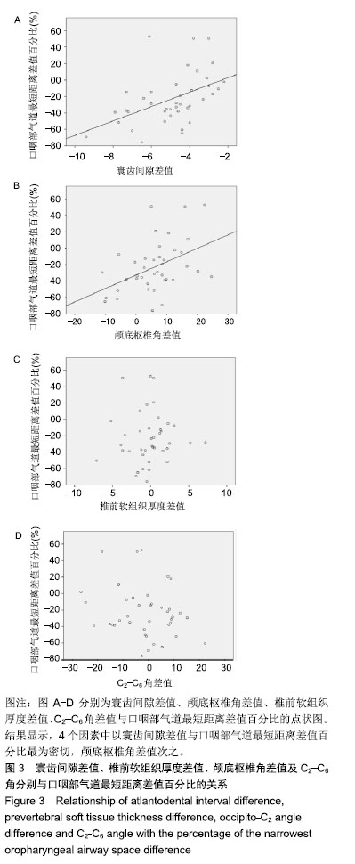
| [1]Xu J, Yin Q, Xia H, et al. New clinical classification system for atlantoaxial dislocation. Orthopedics. 2013; 36(1): e95-e100.[2]Izeki M, Neo M, Ito H, et al. Reduction of atlantoaxial subluxation causes airway stenosis. Spine(Phila Pa 1976).2013;38(9):E513-E520.[3]Battagel JM, Johal A, L'Estrange PR, et al. Changes in airway and hyoid position in response to mandibular protrusion in subjects with obstructive sleep apnoea (OSA). Eur J Orthod. 1999;21(4):363-376.[4]Furuya J, Tamada Y, Suzuki T. Effect of mandibular position on three-dimensional shape of the oropharynx in seated posture. J Oral Rehabil. 2012;39(4): 277-284.[5]Miyata M, Neo M, Fujibayashi S, et al. O-C2 angle as a predictor of dyspnea and/or dysphagia after occipitocervical fusion. Spine (Phila Pa 1976). 2009; 34(2): 184-188.[6]Isono S, Tanaka A, Ishikawa T, et al. Sniffing position improves pharyngeal airway patency in anesthetized patients with obstructive sleep apnea. Anesthesiology. 2005;103(3):489-494.[7]Muto T, Takeda S, Kanazawa M, et al. The effect of head posture on the pharyngeal airway space (PAS). Int J Oral Maxillofac Surg. 2002; 31(6): 579-583.[8]Hasebe D, Kobayashi T, Hasegawa M, et al. Changes in oropharyngeal airway and respiratory function during sleep after orthognathic surgery in patients with mandibular prognathism. Int J Oral Maxillofac Surg. 2011;40(6):584-592.[9]Riley RW, Powell NB, Guilleminault C, et al. Obstructive sleep apnea syndrome following surgery for mandibular prognathism. J Oral Maxillofac Surg. 1987;45(5):450-452.[10]Kawamata A, Fujishita M, Ariji Y, et al. Three-dimensional computed tomographic evaluation of morphologic airway changes after mandibular setback osteotomy for prognathism. Oral Surg Oral Med Oral Pathol Oral Radiol Endod. 2000;89(3):278-287.[11]Izeki M, Neo M, Takemoto M, et al. The O-C2 angle established at occipito-cervical fusion dictates the patient’s destiny in terms of postoperative dyspnea and/or dysphagia. Eur Spine J. 2014;23(2): 328-336.[12]Ota M, Neo M, Aoyama T, et al. Impact of the O-C2 angle on the oropharyngeal space in normal patients. Spine (Phila Pa 1976). 2011; 36(11): E720-E726.[13]Tagawa T, Akeda K, Asanuma Y, et al. Upper airway obstruction associated with flexed cervical position after posterior occipitocervical fusion. J Anesth.2011;25(1):120-122.[14]Hong J, Lim S. Dysphagia after Occipitocervical Fusion. N Engl J Med. 2017;376(22):e46.[15]Izeki M, Neo M, Takemoto M, et al. The O-C2 angle established at occipito-cervical fusion dictates the patient's destiny in terms of postoperative dyspnea and/or dysphagia. Eur Spine J. 2014;23(2): 328-336.[16]Meng Y, Wu T, Liu Z, et al. The impact of the difference in O-C2 angle in the development of dysphagia after occipitocervical fusion: a simulation study in normal volunteers combined with a case-control study. Spine J. 2018.[17]Kaneyama S, Sumi M, Kasahara K, et al. Dysphagia After Occipitothoracic Fusion is Caused by Direct Compression of Oropharyngeal Space Due to Anterior Protrusion of Mid-cervical Spine. Clin Spine Surg. 2017;30(7):314-320.[18]Morizane K, Takemoto M, Neo M, et al. Occipital and external acoustic meatus to axis angle as a predictor of the oropharyngeal space in healthy volunteers: a novel parameter for craniocervical junction alignment. Spine J. 2018;18(5):811-817.[19]Shoda N, Takeshita K, Seichi A, et al. Measurement of occipitocervical angle. Spine (Phila Pa 1976). 2004;29(10):E204-E208.[20]Guo Q, Ni B, Yang J, et al. Relation between alignments of upper and subaxial cervical spine: a radiological study. Arch Orthop Trauma Surg. 2011;131(6): 857-862.[21]Yoshida M, Neo M, Fujibayashi S, et al. Upper-airway obstruction after short posterior occipitocervical fusion in a flexed position. Spine. 2007; 32(8): E267-E270.[22]Redlund-Johnell I. Upper airway obstruction in patients with rheumatoid arthritis and temporomandibular joint destruction. Scand J Rheumatol. 1988;17(4): 273-279. |