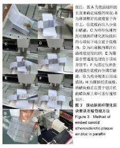
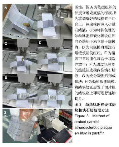
| [1]Sadeghipour ABabaheidarian P. Making formalin-fixed, paraffin embedded blocks. Methods Mol Biol. 2019;1897:253-268. [2]Sorrelle N, Ganguly D, Dominguez ATA, et al. Improved multiplex immunohistochemistry for immune microenvironment evaluation of mouse formalin-fixed, paraffin-embedded tissues. J Immunol. 2019;202(1):292-299. [3]Silakhori M, Naghavi MS, Metselaar HSC, et al. Accelerated thermal cycling test of microencapsulated paraffin wax/polyaniline made by simple preparation method for solar thermal energy storage. Materials (Basel). 2013;6(5):1608-1620. [4]Alcock HE, Stephenson TJ, Royds JA, et al. A simple method for PCR based analyses of immunohistochemically stained, microdissected, formalin fixed, paraffin wax embedded material. Mol Pathol. 1999;52(3):160-163. [5]Hatsukami TS, Ross R, Polissar NL, et al. Visualization of fibrous cap thickness and rupture in human atherosclerotic carotid plaque in vivo with high-resolution magnetic resonance imaging. Circulation. 2000;102(9):959-964.[6]Li F, Yarnykh VL, Hatsukami TS, et al. Scan-rescan reproducibility of carotid atherosclerotic plaque morphology and tissue composition measurements using multicontrast MRI at 3T. J Magn Reson Imaging. 2010;31(1):168-176. [7]Saam T, Ferguson MS, Yarnykh V, et al. Quantitative evaluation of carotid plaque composition by in vivo MRI. Arterioscler Thromb Vasc Biol. 2005;25(1):234-239.[8]Yuan C, Kerwin WS, Yarnykh VL, et al. MRI of atherosclerosis in clinical trials. NMR Biomed. 2006;19(6):636-654.[9]Yoneyama T, Sun J, Hippe DS, et al. In vivo semi-automatic segmentation of multicontrast cardiovascular magnetic resonance for prospective cohort studies on plaque tissue composition: initial experience. Int J Cardiovasc Imaging. 2016;32(1):73-81. [10]Kerwin WS, O'Brien KD, Ferguson MS, et al. Inflammation in carotid atherosclerotic plaque: a dynamic contrast-enhanced MR imaging study. Radiology. 2006;241(2):459-468. [11]Vidal-Perez R, Franco-Gutierrez R, Perez-Perez AJ, et al. Subclinical carotid atherosclerosis predicts all-cause mortality and cardiovascular events in obese patients with negative exercise echocardiography. World J Cardiol. 2019;11(1):24-37.[12]Tarp JB, Sorgaard MH, Christoffersen C, et al. Subclinical atherosclerosis in patients with cyanotic congenital heart disease. Int J Cardiol. 2019;277:97-103. [13]Novo S, Carita P, Lo Voi A, et al. Impact of preclinical carotid atherosclerosis on global cardiovascular risk stratification and events in a 10-year follow-up: comparison between the algorithms of the Framingham Heart Study, the European SCORE and the Italian 'Progetto Cuore'. J Cardiovasc Med (Hagerstown). 2019 ; 20(2):91-96. [14]Bando M, Yamada H, Kusunose K, et al. Comparison of carotid plaque tissue characteristics in patients with acute coronary syndrome or stable angina pectoris: assessment by iPlaque, transcutaneous carotid ultrasonography with integrated backscatter analysis. Cardiovasc Ultrasound. 2015;13:34. [15]Cardellini M, Rovella V, Scimeca M, et al. Chronic kidney disease is linked to carotid nodular calcification, an unstable plaque not correlated to inflammation. Aging Dis. 2019;10(1):71-81. [16]Zhou H, Wang X, Zhu J, et al. Relation of carotid artery plaque to coronary heart disease and stroke in Chinese patients: does hyperglycemia status matter? Exp Clin Endocrinol Diabetes. 2018; 126(3):134-140. [17]Yoon HJ, Kim KH, Park H, et al. Carotid plaque rather than intima-media thickness as a predictor of recurrent vascular events in patients with acute ischemic stroke. Cardiovasc Ultrasound. 2017;15(1):19. [18]Deng F, Hao X, Tang Z, et al. Carotid plaque magnetic resonance imaging and recurrent stroke risk: a protocol for systematic review and meta-analysis. Medicine (Baltimore). 2019;98(18):e15410. [19]Sun R, Wang L, Guan C, et al. Carotid atherosclerotic plaque features in patients with acute ischemic stroke. World Neurosurg. 2018;112:e223-e228. [20]Ota H, Yarnykh VL, Ferguson MS, et al. Carotid intraplaque hemorrhage imaging at 3.0-T MR imaging: comparison of the diagnostic performance of three T1-weighted sequences. Radiology. 2010;254(2):551-563. [21]Wang X, Sun J, Zhao X, et al. Ipsilateral plaques display higher T1 signals than contralateral plaques in recently symptomatic patients with bilateral carotid intraplaque hemorrhage. Atherosclerosis. 2017; 257:78-85.[22]Cai JM, Hatsukami TS, Ferguson MS, et al. Classification of human carotid atherosclerotic lesions with in vivo multicontrast magnetic resonance imaging. Circulation. 2002;106(11):1368-1373. [23]Oikawa M, Ota H, Takaya N, et al. Carotid magnetic resonance imaging. A window to study atherosclerosis and identify high-risk plaques. Circ J. 2009;73(10):1765-1773. [24]Kashiwazaki D, Shiraishi K, Yamamoto S, et al. Efficacy of carotid endarterectomy for mild (<50%) symptomatic carotid stenosis with unstable plaque. World Neurosurg. 2019;121:e60-e69. [25]Lighezan R, Baderca F, Alexa A, et al. The value of the reprocessing method of paraffin-embedded biopsies for transmission electron microscopy. Rom J Morphol Embryol. 2009;50(4):613-617. [26]Nuotio K, Ijas P, Heikkila HM, et al. Morphology and histology of silent and symptom-causing atherosclerotic carotid plaques -Rationale and design of the Helsinki Carotid Endarterectomy Study 2 (the HeCES2). Ann Med. 2018;50(6):501-510. [27]Varrassi M, Sferra R, Gravina GL, et al. Carotid artery plaque characterization with a wide-detector computed tomography using a dedicated post-processing 3D analysis: comparison with histology. Radiol Med. 2019. doi: 10.1007/s11547-019-01026-8.[28]Ota H, Tamura H, Itabashi R, et al. Quantitative characterization of carotid plaque components using MR apparent diffusion coefficients and longitudinal relaxation rates at 3T: a comparison with histology. J Magn Reson Imaging. 2018;48(6):1657-1667. [29]Mondon JA, Howitt J, Tosiano M, et al. A simple osmium post-fixation paraffin-embedment technique to identify lipid accumulation in fish liver using medaka (Oryziaslatipes) eggs and eleutheroembryos as lipid rich models. Mar Pollut Bull. 2011; 63(5-12):86-90. [30]Stockert JC, Lopez-Arias B, Del Castillo P, et al. Replacing xylene with n-heptane for paraffin embedding. Biotech Histochem. 2012; 87(7):464-467. |