Chinese Journal of Tissue Engineering Research ›› 2019, Vol. 23 ›› Issue (28): 4564-4569.doi: 10.3969/j.issn.2095-4344.1331

Previous Articles     Next Articles

Finite element analysis of three-dimensional entity reconstruction of hip joint based on CT image and hip bearing capacity data

Gao Yaodong1, Duan Yuxing1, Guo Pengnian2   

  1. 1College of Mechanical Engineering, Inner Mongolia University of Science and Technology, Baotou 014010, Inner Mongolia Autonomous Region, China; 2Second Department of Orthopedics, First Affiliated Hospital of Baotou Medical College, Inner Mongolia University of Science and Technology, Baotou 014010, Inner Mongolia Autonomous Region, China
  • Online:2019-10-08 Published:2019-10-08
  • Contact: Duan Yuxing, Master, College of Mechanical Engineering, Inner Mongolia University of Science and Technology, Baotou 014010, Inner Mongolia Autonomous Region, China
  • About author:Gao Yaodong, Professor, Master’s supervisor, College of Mechanical Engineering, Inner Mongolia University of Science and Technology, Baotou 014010, Inner Mongolia Autonomous Region, China
  • Supported by:

    the Science Research Foundation of Baotou Medical College, No. BYJJ-QM 2016108 (to GPN)

Abstract:

BACKGROUND: Due to complex anatomical structure and stress of hip, traditional X-ray and CT scanning have some limitations in the diagnosis of hip.
OBJECTIVE: To find an accurate and fast reconstruction method for the entity model of biological skeleton based on CT image, and to study the stress of the human hip bone, and to establish the optimized hip joint model before operation for three-dimensional printing.
METHODS: CT images of hip joint were obtained from one patient (the study was approved by the Ethics Committee of First Affiliated Hospital of Baotou Medical College, and the subject signed the informed consent). The model optical images of human body were developed by medical Mimics software. The reverse design steps of “original CT image-image processing-surface reconstruction-materialization” were adopted. The necessary forward design method needed therein. Firstly, using the core medical Mimics software, the CT image of hip joint was found out from the optical image and noise point, and the vector tomography image was obtained. After carefully separating the image, the multi-layer image was fitted to form three-dimensional image structure. Then in Geomagic software, the surface fitting of the model is completed, and at the same time, Generate surfaces and materialize. In Geomagic Studio software, in order to achieve the precision of the model, it is necessary to make the surface smooth and curve smooth by command, and to meet the requirements of subsequent finite element analysis. Finally, the mesh division, model assignment and finite element analysis of hip joint are completed in Abaqus software. At this point, all the data conversion from the original CT to the entity is completed. At the same time, the Anybody software was used to simulate the human gait simulation, and the data were applied to the finite element analysis of ABAQUS.
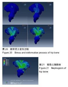
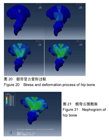
RESULTS AND CONCLUSION: The simplest method was used to complete the reconstruction of CT images, and a good hip model was obtained by Geomagic surface fitting. The important data of hip bearing capacity were obtained by Anybody inverse dynamics analysis. Finite element analysis verifies this result.

Key words: hip arthroplasty, CT images, image processing, surface reconstruction, gait analysis, finite element analysis, three-dimensional entity reconstruction

CLC Number: