Chinese Journal of Tissue Engineering Research ›› 2026, Vol. 30 ›› Issue (18): 4618-4626.doi: 10.12307/2026.681
Previous Articles Next Articles
Secreted modular calcium binding protein regulates autophagy in the acetabular cartilage of rats with developmental dysplasia of the hip
Zheng Wen1, Zhu Dongsheng2, Wang Xiaodong1
- 1Children’s Hospital of Soochow University, Suzhou 215000, Jiangsu Province, China; 2The First People’s Hospital of Lianyungang, Lianyungang 222000, Jiangsu Province, China
-
Received:2025-05-15Accepted:2025-09-08Online:2026-06-28Published:2025-12-04 -
Contact:Wang Xiaodong, PhD, Professor, Children’s Hospital of Soochow University, Suzhou 215000, Jiangsu Province, China Co-corresponding author: Zhu Dongsheng, PhD, Associate chief physician, The First People’s Hospital of Lianyungang, Lianyungang 222000, Jiangsu Province, China -
About author:Zheng Wen, Physician, Children’s Hospital of Soochow University, Suzhou 215000, Jiangsu Province, China -
Supported by:Jiangsu Key Research and Development Program (Social Development), No. BE2022732 (to WXD); Lianyungang Maternal and Child Health Research Project, No. F202319 (to ZDS)
CLC Number:
Cite this article
Zheng Wen, Zhu Dongsheng, Wang Xiaodong. Secreted modular calcium binding protein regulates autophagy in the acetabular cartilage of rats with developmental dysplasia of the hip[J]. Chinese Journal of Tissue Engineering Research, 2026, 30(18): 4618-4626.
share this article
Add to citation manager EndNote|Reference Manager|ProCite|BibTeX|RefWorks
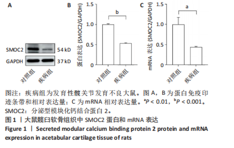
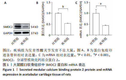
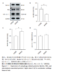
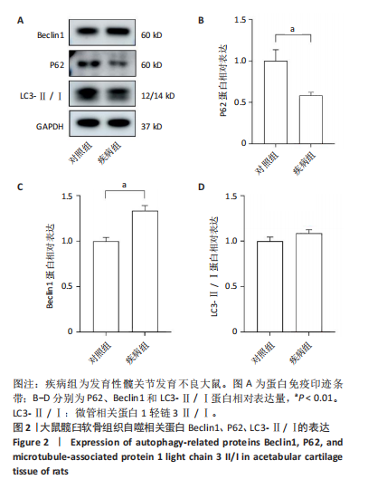
2.1 实验动物数量分析 实验选用新生SD大鼠12只,随机分为2组,每组6只,进入结果分析12只。 2.2 发育性髋关节发育不良大鼠髋臼顶壁软骨组织中SMOC2表达下降 2.2.1 Western Blot检测结果 与对照组相比,疾病组髋臼顶壁软骨组织中SMOC2蛋白的表达下降,见图1A,B。 2.2.2 RT-qPCR检测结果 与对照组相比,疾病组髋臼顶壁软骨组织中Smoc2 mRNA的表达明显下降,见图1C。 2.3 发育性髋关节发育不良大鼠髋臼顶壁软骨组织中自噬相关蛋白表达异常 Western Blot检测结果显示,与对照组相比,疾病组髋臼顶壁软骨组织中P62蛋白表达下降,Beclin1表达升高,LC3-Ⅱ/Ⅰ表达无明显差异,见图2。 2.4 Sh-SMOC2可有效敲低软骨细胞中SMOC2的表达 为探究SMOC2在软骨细胞中的作用,将软骨细"
"
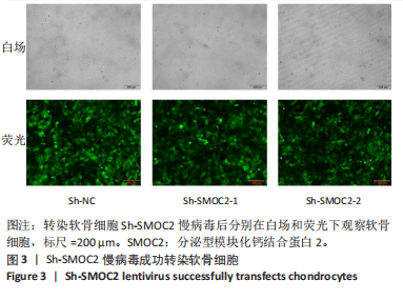
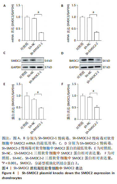
胞分为Sh-NC、Sh-SMOC2-1和Sh-SMOC2-2三组,分别进行质粒转染。转染成功后,细胞发出绿色荧光,见图3。提取3组软骨细胞的RNA和蛋白,进行RT-qPCR检测及Western Blot检测。 2.4.1 RT-qPCR实验结果 与对照组相比,Sh-NC组软骨细胞中Smoc2 mRNA的表达水平未见明显变化(图4A,B);而相较于Sh-NC组,Sh-SMOC2-1和Sh-SMOC2-2组软骨细胞中Smoc2 mRNA的表达均显著下降,见图4A,B。"
2.4.2 Western Blot实验结果 与对照组相比,Sh-NC组软骨细胞中SMOC2蛋白的表达水平未见明显变化;而相较于Sh-NC组,Sh-SMOC2-1和Sh-SMOC2-2组软骨细胞中SMOC2蛋白的表达均显著下降,见图4C-F。由此可见,Sh-SMOC2-1和Sh-SMOC2-2均能有效敲低软骨细胞中SMOC2的表达。"

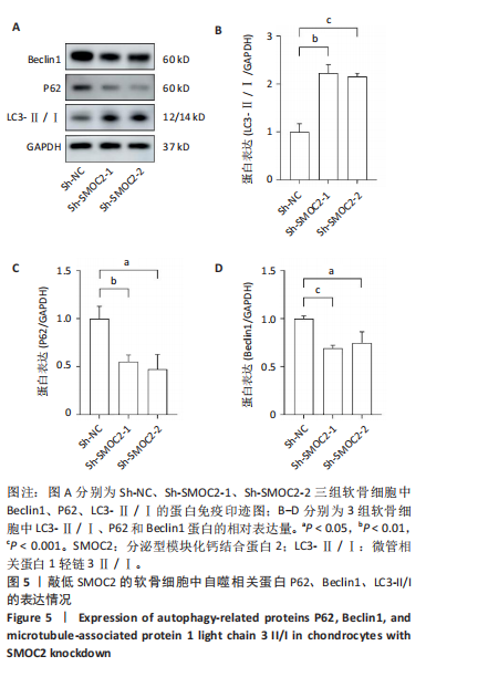
2.5 SMOC2调控软骨细胞自噬 2.5.1 Western Blot检测 提取Sh-NC、Sh-SMOC2-1和Sh-SMOC2-2三组软骨细胞的蛋白,利用Western Blot检测自噬相关分子P62、Beclin-1及LC3-Ⅱ/Ⅰ的表达。结果显示,与Sh-NC组相比,Sh-SMOC2-1和Sh-SMOC2-2组软骨细胞中P62和Beclin-1蛋白的表达下降,LC3-Ⅱ/Ⅰ比值升高,见图5。"
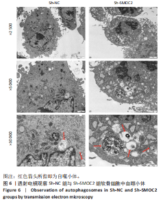
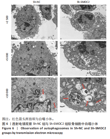
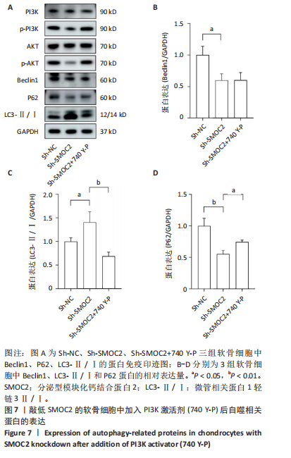
2.5.2 透射电镜观察 收集Sh-NC组和Sh-SMOC2组软骨细胞,置于透射电镜下观察,结果显示,与Sh-NC组相比,Sh-SMOC2组软骨细胞中自噬小体增多,见图6。结果提示,SMOC2是软骨细胞自噬的调控因子。 2.6 SMOC2通过PI3K/AKT信号通路调控软骨细胞自噬相关蛋白表达 为探究SMOC2是否通过PI3K/AKT通路调控软骨细胞自噬相关蛋白,在软骨细胞中加入PI3K激活剂(740 Y-P),软骨细胞分为SH-NC组、 Sh-SMOC2组、Sh-SMOC2+740 Y-P组。提取3组细胞"
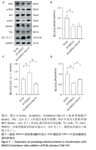
中的蛋白进行Western Blot检测。PI3K、P-PI3K、P-AKT蛋白及软骨细胞自噬相关蛋白表达见图7。结果显示,与SH-NC组相比,Sh-SMOC2组软骨细胞中P62和Beclin1蛋白表达水平下降,LC3-Ⅱ/Ⅰ比值升高。与Sh-SMOC2组相比,Sh-SMOC2+740 Y-P组软骨细胞中P62蛋白表达水平升高,LC3-Ⅱ/Ⅰ比值下降,Beclin1表达无明显差异,见图7。结果提示SMOC2通过PI3K/AKT信号通路调控软骨细胞自噬相关蛋白表达。"
| [1] BAKARMAN K, ALSIDDIKY AM, ZAMZAM M, et al. Developmental Dysplasia of the Hip (DDH): Etiology, Diagnosis, and Management. Cureus. 2023;15(8):e43207. [2] JACOBSEN KK, LABORIE LB, KRISTIANSEN H, et al. Genetics of hip dysplasia - a systematic literature review. BMC Musculoskelet Disord. 2024;25(1):762. [3] SATO T, YAMATE S, UTSUNOMIYA T, et al. Life Course Epidemiology of Hip Osteoarthritis in Japan: A Multicenter, Cross-Sectional Study. J Bone Joint Surg Am. 2024;106(11):966-975. [4] AMAGAMI A, SUGIYAMA H, TONOTSUKA H, et al. Long-term course of developmental dysplasia of the hip: follow-up of the non-operated hips of patients undergoing unilateral rotational acetabular osteotomy for twenty-four years. Arch Orthop Trauma Surg. 2024;144(3):997-1004. [5] LIN NY, BEYER C, GIESSL A, et al. Autophagy regulates TNFα-mediated joint destruction in experimental arthritis. Ann Rheum Dis. 2013;72(5): 761-768. [6] LV X, ZHAO T, DAI Y, et al. New insights into the interplay between autophagy and cartilage degeneration in osteoarthritis. Front Cell Dev Biol. 2022;10:1089668. [7] WANG J, ZHANG Y, CAO J, et al. The role of autophagy in bone metabolism and clinical significance. Autophagy. 2023;19(9):2409-2427. [8] LOTZ MK, CARAMÉS B. Autophagy and cartilage homeostasis mechanisms in joint health, aging and OA. Nat Rev Rheumatol. 2011; 7(10):579-587. [9] PAN Y, YANG Y, FAN M, et al. Progranulin regulation of autophagy contributes to its chondroprotective effect in osteoarthritis. Genes Dis. 2022;10(4):1582-1595. [10] LI M, WEI CB, LI HF, et al. Osteopontin inhibits autophagy via CD44 and avβ3 integrin and promotes cell proliferation in osteoarthritic fibroblast-like synoviocytes. BMC Musculoskelet Disord. 2025;26(1): 274. [11] ANDREI C, MIHAI DP, NITULESCU GM, et al.Modulating Autophagy in Osteoarthritis: Exploring Emerging Therapeutic Drug Targets. Int J Mol Sci. 2024;25(24):13695. [12] LÓPEZ DE FIGUEROA P, LOTZ MK, BLANCO FJ, et al.Autophagy activation and protection from mitochondrial dysfunction in human chondrocytes. Arthritis Rheumatol. 2015;67(4):966-976. [13] HE WG, DENG YX, KE KX, et al. Matricellular Protein SMOC2 Potentiates BMP9-Induced Osteogenic Differentiation in Mesenchymal Stem Cells through the Enhancement of FAK/PI3K/AKT Signaling. Stem Cells Int. 2023;2023:5915988. [14] TAKAHATA Y, HAGINO H, KIMURA A, et al. Smoc1 and Smoc2 regulate bone formation as downstream molecules of Runx2. Commun Biol. 2021;4(1):1199. [15] WANG X, WANG M, ZHOU Z, et al. SMOC2 promoted vascular smooth muscle cell proliferation, migration, and extracellular matrix degradation by activating BMP/TGF-β1 signaling pathway. J Clin Biochem Nutr. 2023;73(2):116-123. [16] ALFAWAZ S, FONG F, PLAGNOL V, et al. Recessive oligodontia linked to a homozygous loss-of-function mutation in the SMOC2 gene. Arch Oral Biol. 2013;58(5):462-466. [17] BLOCH-ZUPAN A, JAMET X, ETARD C, et al. Homozygosity mapping and candidate prioritization identify mutations, missed by whole-exome sequencing, in SMOC2, causing major dental developmental defects. Am J Hum Genet. 2011;89(6):773-781. [18] REN Y, WU Y, HE W, et al. SMOC2 plays a role in heart failure via regulating TGF-β1/Smad3 pathway-mediated autophagy. Open Med (Wars). 2023;18(1):20230752. [19] FENG X, CHEN L, GUO W, et al. Graphene oxide induces p62/SQSTM-dependent apoptosis through the impairment of autophagic flux and lysosomal dysfunction in PC12 cells. Acta Biomater. 2018;81:278-292. [20] NICHOLSON A, DUNNE K, TAAFFE S, et al. Developmental dysplasia of the hip in infants and children. BMJ. 2023;383:e074507. [21] DEZATEUX C, ROSENDAHL K. Developmental dysplasia of the hip. Lancet. 2007;369(9572):1541-1552. [22] RIEDSTRA NS, BOEL F, VAN BUUREN MMA, et al. Acetabular dysplasia and the risk of developing hip osteoarthritis within 4-8 years: An individual participant data meta-analysis of 18,807 hips from the World COACH consortium. Osteoarthritis Cartilage. 2025;33(3):373-382. [23] TANAKA H, CHIBA D, MORI Y, et al. Long-term results of a modified Spitzy shelf operation for developmental dysplasia of the hip in adults and adolescents. Eur J Orthop Surg Traumatol. 2018;28(7):1341-1347. [24] DE SALVO S, SACCO R, MAINARD N, et al. Total hip arthroplasty in patients with common pediatric hip orthopedic pathology. J Child Orthop. 2024;18(2):134-152. [25] GUO YF, SU T, YANG M, et al. The role of autophagy in bone homeostasis. J Cell Physiol. 2021;236(6):4152-4173. [26] LIU S, YAO S, YANG H, et al. Autophagy: Regulator of cell death. Cell Death Dis. 2023;14(10):648. [27] HERNANDEZ PA, WELLS J, USHEVA E, et al. Early-Onset Osteoarthritis originates at the chondrocyte level in Hip Dysplasia. Sci Rep. 2020; 10(1):627. [28] FELSON DT. Osteoarthritis as a disease of mechanics. Osteoarthritis Cartilage. 2013;21(1):10-15. [29] BEECHER BR, MARTIN JA, PEDERSEN DR, et al. Antioxidants block cyclic loading induced chondrocyte death. Iowa Orthop J. 2007;27:1-8. [30] WEN F, GAO J, ZHANG G, et al. ROS-DRP1-mediated excessive mitochondrial fission and autophagic flux inhibition contribute to heat stress-induced apoptosis in goat Sertoli cells. J Anim Sci Biotechnol. 2025;16(1):58. [31] TALEBI M, MOHAMMADI VADOUD SA, HARATIAN A, et al. The interplay between oxidative stress and autophagy: focus on the development of neurological diseases. Behav Brain Funct. 2022;18(1):3. [32] PEI W, HUANG X, NI B, et al. Selective STAT3 Inhibitor Alantolactone Ameliorates Osteoarthritis via Regulating Chondrocyte Autophagy and Cartilage Homeostasis. Front Pharmacol. 2021;12:730312. [33] FLORENCIO-SILVA R, SASSO G, SASSO-CERRI E, et al. Relationship between autophagy and NLRP3 inflammasome during articular cartilage degradation in oestrogen-deficient rats with streptozotocin-induced diabetes. Ann Anat. 2025;257:152318. [34] CHEN X, GONG W, SHAO X, et al.METTL3-mediated m(6)A modification of ATG7 regulates autophagy-GATA4 axis to promote cellular senescence and osteoarthritis progression. Ann Rheum Dis. 2022;81(1): 87-99. [35] MCHUGH J. Aberrant regulation of autophagy linked to OA. Nat Rev Rheumatol. 2022;18(12):672. [36] XU K, HE Y, MOQBEL SAA, et al. SIRT3 ameliorates osteoarthritis via regulating chondrocyte autophagy and apoptosis through the PI3K/Akt/mTOR pathway. Int J Biol Macromol. 2021;175:351-360. [37] VANNAHME C, GÖSLING S, PAULSSON M, et al. Characterization of SMOC-2, a modular extracellular calcium-binding protein. Biochem J. 2003;373(Pt 3):805-814. [38] YAN W, ZHENG L, XU X, et al. Heterozygous LRP1 deficiency causes developmental dysplasia of the hip by impairing triradiate chondrocytes differentiation due to inhibition of autophagy. Proc Natl Acad Sci U S A. 2022;119(37):e2203557119. [39] WANG J, LI X, GUO X, et al. MicroRNA-34a-5p promotes the progression of osteoarthritis secondary to developmental dysplasia of the hip by restraining SESN2-induced autophagy. J Orthop Res. 2024;42(1):66-77. [40] LEE DY, BAHAR ME, KIM CW, et al. Autophagy in Osteoarthritis: A Double-Edged Sword in Cartilage Aging and Mechanical Stress Response: A Systematic Review. J Clin Med. 2024;13(10):3005. [41] WANG W, SUN Y, TANG P, et al. CircTBCK protects against osteoarthritis by regulating extracellular matrix and autophagy. Hum Cell. 2025; 38(2):60. [42] LI Z, LI D, SU H, et al. Autophagy: An important target for natural products in the treatment of bone metabolic diseases. Front Pharmacol. 2022;13:999017. [43] Mayer I A, Arteaga CL. The PI3K/AKT Pathway as a Target for Cancer Treatment. Annu Rev Med. 2016;67:11-28. [44] RAJENDRAN P, SEKAR R, DHAYASANKAR PS, et al.PI3K/AKT Signaling Pathway Mediated Autophagy in Oral Carcinoma - A Comprehensive Review. Int J Med Sci. 2024;21(6):1165-1175. [45] LU M, HUANG L, TANG Y, et al. ARNTL2 knockdown suppressed the invasion and migration of colon carcinoma: decreased SMOC2-EMT expression through inactivation of PI3K/AKT pathway. Am J Transl Res. 2020;12(4):1293-1308. [46] LIU B, DENG X, JIANG Q, et al. Scoparone improves hepatic inflammation and autophagy in mice with nonalcoholic steatohepatitis by regulating the ROS/P38/Nrf2 axis and PI3K/AKT/mTOR pathway in macrophages. Biomed Pharmacother. 2020;125:109895. [47] LI R, ZHENG Y, ZHANG J, et al. Gomisin N attenuated cerebral ischemia-reperfusion injury through inhibition of autophagy by activating the PI3K/AKT/mTOR pathway. Phytomedicine. 2023;110:154644. [48] GAO K, ZHAO Y, SI M, et al.ERS regulates endometrial epithelial cell autophagy through XBP1s-mediated activation of the PI3K/AKT pathway. Sci Rep. 2025;15(1):5943. [49] YE T, XUE F, HU H, et al. Early Emergent and Progressive Aberrant Subchondral Bone Remodeling Coupled with Aggravated Cartilage Degeneration in Developmental Dysplasia of the Hip. Cartilage. 2022; 13(2):19476035221098165. [50] YANG S, ZUSMAN N, LIEBERMAN E, et al. Developmental Dysplasia of the Hip. Pediatrics. 2019;143(1):e20181147. [51] DUAN R, XIE H, LIU ZZ. The Role of Autophagy in Osteoarthritis. Front Cell Dev Biol. 2020;8:608388. [52] XUE JF, SHI ZM, ZOU J, et al. Inhibition of PI3K/AKT/mTOR signaling pathway promotes autophagy of articular chondrocytes and attenuates inflammatory response in rats with osteoarthritis. Biomed Pharmacother. 2017;89:1252-1261. |
| [1] | Zhang Nan, Meng Qinghua, Bao Chunyu. Characteristics and clinical application of ankle joint finite element models [J]. Chinese Journal of Tissue Engineering Research, 2026, 30(9): 2343-2349. |
| [2] | Chen Qiuhan, Yang Long, Yuan Daizhu, Wu Zhanyu, Zou Zihao, Ye Chuan. Peri-knee osteotomy for treatment of knee osteoarthritis: optimization of treatment strategies [J]. Chinese Journal of Tissue Engineering Research, 2026, 30(9): 2303-2312. |
| [3] | Zhang Zizheng, Luo Wang, Liu Changlu. Application value of finite element analysis on unicompartmental knee arthroplasty for medial knee compartmental osteoarthritis [J]. Chinese Journal of Tissue Engineering Research, 2026, 30(9): 2313-2322. |
| [4] | Li Qingbin, Lin Jianhui, Huang Wenjie, Wang Mingshuang, Du Jiankai, Lao Yongqiang. Bone cement filling after enlarged curettage of giant cell tumor around the knee joint: a comparison of subchondral bone grafting and non-grafting [J]. Chinese Journal of Tissue Engineering Research, 2026, 30(8): 1896-1902. |
| [5] | Li Linzhen, Jiao Hongzhuo, Chen Weinan, Zhang Mingzhe, Wang Jianlong, Zhang Juntao. Effect of icariin-containing serum on lipopolysaccharide-induced inflammatory damage in human chondrocytes [J]. Chinese Journal of Tissue Engineering Research, 2026, 30(6): 1368-1374. |
| [6] | Chen Ju, Zheng Jinchang, Liang Zhen, Huang Chengshuo, Lin Hao, Zeng Li. Effect and mechanism of beta-caryophyllene in mice with osteoarthritis [J]. Chinese Journal of Tissue Engineering Research, 2026, 30(6): 1341-1347. |
| [7] | Lyu Guoqing, Aizimaitijiang·Rouzi, Xiong Daohai. Irisin inhibits ferroptosis in human articular chondrocytes: roles and mechanisms [J]. Chinese Journal of Tissue Engineering Research, 2026, 30(6): 1359-1367. |
| [8] | Peng Zhiwei, Chen Lei, Tong Lei. Luteolin promotes wound healing in diabetic mice: roles and mechanisms [J]. Chinese Journal of Tissue Engineering Research, 2026, 30(6): 1398-1406. |
| [9] | Jia Jinwen, Airefate·Ainiwaer, Zhang Juan. Effects of EP300 on autophagy and apoptosis related to allergic rhinitis in rats [J]. Chinese Journal of Tissue Engineering Research, 2026, 30(6): 1439-1449. |
| [10] | Li Hao, Tao Hongcheng, Zeng Ping, Liu Jinfu, Ding Qiang, Niu Chicheng, Huang Kai, Kang Hongyu. Mitogen-activated protein kinase signaling pathway regulates the development of osteoarthritis: guiding targeted therapy with traditional Chinese medicine [J]. Chinese Journal of Tissue Engineering Research, 2026, 30(6): 1476-1485. |
| [11] | You Huijuan, Wu Shuzhen, Rong Rong, Chen Liyuan, Zhao Yuqing, Wang Qinglu, Ou Xiaowei, Yang Fengying. Macrophage autophagy in lung diseases: two-sided effects [J]. Chinese Journal of Tissue Engineering Research, 2026, 30(6): 1516-1526. |
| [12] | Zhang Qian, Huang Dongfeng. Weighted gene co-expression network analysis combined with machine learning to screen and validate biomarkers for osteoarthritis [J]. Chinese Journal of Tissue Engineering Research, 2026, 30(5): 1096-1105. |
| [13] | Liu Kexin, , Hao Kaimin, Zhuang Wenyue, , Li Zhengyi. Autophagy-related gene expression in pulmonary fibrosis models: bioinformatic analysis and experimental validation [J]. Chinese Journal of Tissue Engineering Research, 2026, 30(5): 1129-1138. |
| [14] | Hu Jing, Zhu Ling, Xie Juan, Kong Deying, Liu Doudou. Autophagy regulates early embryonic development in mice via affecting H3K4me3 modification [J]. Chinese Journal of Tissue Engineering Research, 2026, 30(5): 1147-1155. |
| [15] | Bu Yangyang, Ning Xinli, Zhao Chen. Intra-articular injections for the treatment of osteoarthritis of the temporomandibular joint: different drugs with multiple combined treatment options [J]. Chinese Journal of Tissue Engineering Research, 2026, 30(5): 1215-1224. |
| Viewed | ||||||
|
Full text |
|
|||||
|
Abstract |
|
|||||