Chinese Journal of Tissue Engineering Research ›› 2026, Vol. 30 ›› Issue (33): 8838-8844.doi: 10.12307/2026.405
Positive effects of transcranial direct current stimulation in improving muscle and cardiopulmonary endurance
Zhang Yi, Wu Huaduo, Song Yu, Dong Yumo, Zhang Yiguang, Wang Qian, Gao Xin, Jiang Ning
- Institute of Sports and Health, Tianjin Key Laboratory of Physical Fitness Integration and Health Promotion, Tianjin Key Laboratory of Exercise Physiology and Sports Medicine, Tianjin University of Sport, Tianjin 301617, China
-
Received:2025-07-11Revised:2025-11-09Online:2026-11-28Published:2026-06-22 -
Contact:Jiang Ning, PhD, Professor, Institute of Sports and Health, Tianjin Key Laboratory of Physical Fitness Integration and Health Promotion, Tianjin Key Laboratory of Exercise Physiology and Sports Medicine, Tianjin University of Sport, Tianjin 301617, China -
About author:Zhang Yi, MS candidate, Institute of Sports and Health, Tianjin Key Laboratory of Physical Fitness Integration and Health Promotion, Tianjin Key Laboratory of Exercise Physiology and Sports Medicine, Tianjin University of Sport, Tianjin 301617, China -
Supported by:Tianjin Education Commission Scientific Research Project, No. 2021KJ002 (to SY)

CLC Number:
Cite this article
Zhang Yi, Wu Huaduo, Song Yu, Dong Yumo, Zhang Yiguang, Wang Qian, Gao Xin, Jiang Ning. Positive effects of transcranial direct current stimulation in improving muscle and cardiopulmonary endurance[J]. Chinese Journal of Tissue Engineering Research, 2026, 30(33): 8838-8844.
share this article
Add to citation manager EndNote|Reference Manager|ProCite|BibTeX|RefWorks
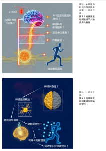
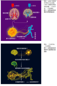
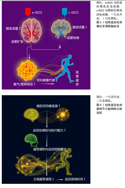
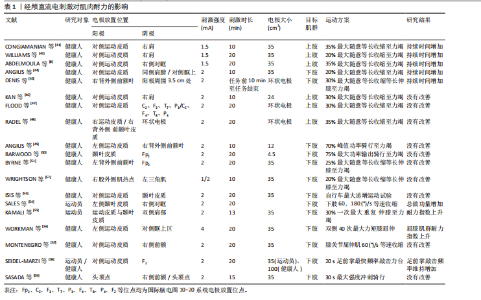
2.1 经颅直流电刺激发展简史 经颅直流电刺激技术有着丰富的发展历程,其起源可追溯到早期对电刺激与神经系统关系的探索。19世纪,科学家们就开始尝试利用电来影响神经活动,为经颅直流电刺激的出现埋下了种子。最初的研究主要集中在对动物模型的电刺激实验,这些研究让人们逐渐认识到电刺激可能对神经功能产生作用。20世纪,随着技术发展和对大脑功能研究的深入,经颅电刺激技术开始有了新的方向,逐渐从理论走向实践,在这个阶段,研究者们开始尝试在人体上进行安全且有效的经颅直流电刺激试验,探索合适的刺激参数和电极放置方法。早期的人体试验主要是为了观察这种刺激对大脑功能的即时影响,比如对简单感知和运动功能的作用。到了21世纪,经颅直流电刺激迎来了快速发展时期。随着神经科学、材料科学等多学科的协同进步,经颅直流电刺激设备得到了极大改进,电极材料更加舒适且能更好地贴合头皮,电源也能更精准地控制电流强度和稳定性。这一时期,经颅直流电刺激在研究领域的应用范围迅速扩大,从基础的大脑功能研究(如对大脑可塑性的探索[10])到对复杂认知功能的研究[11-14]。在临床应用方面,经颅直流电刺激也展现出巨大潜力,它开始应用于神经康复领域,如针对脑卒中患者的运动康复[15-16],通过刺激大脑运动皮质帮助患者改善运动功能;在精神心理疾病治疗上,对抑郁症等疾病的治疗研究也逐渐深入,为这些疾病治疗提供了新的辅助手段。如今,经颅直流电刺激技术仍在不断发展,研究人员一方面致力于进一步优化设备和刺激参数,提高该技术的有效性和安全性;另一方面,经颅直流电刺激技术也在拓展其应用领域,为更多神经系统相关问题的解决提供可能。 2.2 经颅直流电刺激的潜在机制 尽管经颅直流电刺激提升运动表现的精确神经生理学机制尚未完全明确,目前已有若干主流观点对它的作用机制进行了阐释。这些观点认为,经颅直流电刺激的作用主要体现在以下几个方面:调节神经元的静息膜电位,进而改变大脑皮质的兴奋性;增加突触可塑性,调节长时程增强效应;调节神经血管耦合,改善局部脑血流量;调节大脑网络的功能连接,实现脑区的激活与强化。 2.2.1 调节大脑皮质兴奋性 经颅直流电刺激通过调控皮质神经元静息电位,进而改变神经元的兴奋性[17-18]。阳极经颅直流电刺激使神经元去极化,增强皮质兴奋性、提升神经肌肉传导效率,进而提高运动表现(图3);阴极经颅直流电刺激则使神经元超极化,抑制兴奋性[19-20]。 然而,经颅直流电刺激对神经元兴奋性的影响并非仅由电极极性决定,还与神经元的空间排布及电场相互作用有关,例如,阴极经颅直流电刺激刺激小脑可促进运动技能学习,而阳极经颅直流电刺激则相反[21]。这些现象可以通过“轴突调节”机制来解释[22],即经颅直流电刺激电极的极性对轴突兴奋性的影响依赖于轴突相对于电极的方向。 2.2.2 增加突触可塑性 突触可塑性是神经信号传递和功能连接的核心,涉及突触结构的短期和长期调整[23-24]。当突触传递效能增强时会引发长时程增强,而突触传递效能减弱时则导致长时程抑制[25]。长时程增强是由高频刺激引起的持久性增强反应,与细胞膜上的配体和化学离子通道相关,构成了神经环路重塑、学习、记忆等脑功能改善的生理基础[26-27]。阳极经颅直流电刺激通过提高神经元兴奋性促进神经递质释放,激活信号通路,增强突触可塑性。长期来看,经颅直流电刺激通过增强基因转录促进脑源性神经营养因子合成,进一步促进长时程增强和行为学功能改善[26]。研究者推测,经颅直流电刺激可能通过触发长时程增强和长时程抑制实现大脑兴奋性和活动性的持久改变,从而提升运动性能(图4)。 2.2.3 改变局部脑血流 经颅直流电刺激能够调节局部脑血流量,影响大脑皮质和皮质下区域的血流动态[28]。阳极经颅直流电刺激增加局部脑血流量,而阴极经颅直流电刺激则减少局部脑血流量,并且这些变化与皮质兴奋性变化呈正相关[29]。研究表明,经颅直流电刺激的电流密度与任务表现及局部脑血流量相关,并且电流强度与局部脑血流量呈高度相关性[30]。神经血管耦合理论认为,经颅直流电刺激引起的局部脑血流量变化源于神经元与神经血管单元的相互作用,其中阳极经颅直流电刺激促进神经元兴奋,导致血管扩张;而阴极经颅直流电刺激则抑制兴奋,引起血管收缩[31]。通过调节血管收缩或舒张,经颅直流电刺激可影响局部脑血流量,进而提升运动表现(图5)。 2.2.4 调节大脑网络功能连接 脑部构成一个复杂的网络系统,各脑区分工协作,调控包括运动、认知和情绪在内的多项生理功能。功能性磁共振成像研究显示,阳极经颅直流电刺激应用于初级运动皮质或左背外侧前额叶皮质区时可显著增强相关脑区之间的功能连接性,提高受试者的运动和认知能力[32-33],表明经颅直流电刺激可能通过调节大脑网络功能连接发挥作用,激活和加强这些连接,从而改善个体的运动表现(图6)。"

"
2.3 经颅直流电刺激对耐力表现的影响 近年来,经颅直流电刺激作为一种神经生物力学增强技术被引入,用于提高机体能力[34-35],经颅直流电刺激可以增加初级运动皮质的兴奋性、改善平衡性能、加速运动学习[36-37]。此外,经颅直流电刺激增加初级运动皮质兴奋性,从而减少对辅助运动区的依赖,并降低个人在执行任务过程中对努力的感知[38]。一些研究已经探索了经颅直流电刺激与脑成像技术相结合的神经生理学机制,基于功能性近红外光谱的研究发现,经颅直流电刺激提高了双侧感觉运动皮质神经元传输的效率[39],通过增加皮质脊髓兴奋性增强耐力表现[40-42]。 2.3.1 经颅直流电刺激对肌肉耐力的影响 肌肉耐力是指机体受外部阻力作用情况下能坚持的时间或重复次数的能力[43],即对抗疲劳的能力。肌肉耐力在日常生活和体育运动中都具有重要意义。随着科技的发展,非侵入性神经调节技术因可能为提高肌肉耐力提供新的途径逐渐引起了研究人员的关注。该文总结了19篇有关经颅直流电刺激对肌肉耐力影响的研究(表1),总结内容包括研究对象、刺激参数、目标肌群、运动方案以及研究结果。刺激参数影响着经颅直流电刺激的刺激效果,如刺激部位、刺激强度、刺激时间、电极片大小等,在选择刺激参数时,需要综合考虑受试者的个体差异、测试目标以及已有的临床研究证据等。19项研究中最主要的刺激脑区是初级运动皮质,最常用的刺激强度为2 mA,最常用的刺激时间为20 min,最常用的电极片大小为35 cm2。 在经颅直流电刺激干预肌肉耐力的研究中,力竭运动是评价肌肉耐力表现最常用的试验方案,持续运动时间是评价耐力表现最常用的指标,持续时间增加,说明经颅直流电刺激干预对肌肉耐力表现有积极作用。COGIAMANIAN等[41]对24名健康受试者进行1.5 mA、10 min阳极经颅直流电刺激,发现左肘屈肌35%最大随意等长收缩的持续时间增加。WILLIAMS等[42]对18名健康受试者进行肘屈肌20%最大随意收缩至力竭测试,发现阳极经颅直流电刺激干预后运动持续时间增加。ABDELMOULA等[8]对11名健康受试者进行1.5 mA、20 min的经颅直流电刺激,发现右肘屈肌35%最大随意收缩至力竭的持续时间增加。ANGIUS等[44]对9名受试者进行2 mA、10 min的经颅直流电刺激,发现右膝伸肌等长疲劳测试的持续时间增加。DENIS等[45]对20名健康受试者的右背外侧前额叶皮质进行高精度经颅直流电刺激,随后进行30%最大随意收缩等长伸膝至力竭测试,发现阳极刺激组和假刺激组之间没有显著差异,但阳极刺激组耐力持续时间长于假刺激组。然而也有研究指出,经颅直流电刺激干预对持续运动时长没有影响。例如,KAN等[46]、FLOOD等[47]、 RADEL等[48]多篇研究对受试者的上肢进行肌肉耐力试验,发现经颅直流电刺激组和假刺激组之间的持续运动时间无显著差异。ANGIUS等[49]、BARWOOD等[50]、BYRNE等[51]、WRIGHTSON等[52]、ISIS等[53]多篇研究对受试者下肢进行肌肉耐力试验,发现持续运动时间没有显著变化。 此外,总做功量和耐力指数也是评价肌肉耐力水平常用的指标,总做功量增加、耐力指数上升,说明经颅直流电刺激干预对肌肉耐力表现有积极作用。SALES等[54]通过对运动员进行2 mA、20 min的经颅直流电刺激,结果发现等速收缩的总做功量增加。KAMALI等[55]对12名有经验的健美运动员进行2 mA、13 min的经颅直流电刺激,并且目标区域为运动皮质与颞叶皮质同时进行干预,结果发现耐力指数明显上升。WORKMAN等[56]为了确定4 mA经颅直流电刺激的耐受性和对腿部肌肉耐力的影响,对31名健康受试者进行双侧膝关节40次最大力矩屈伸测试,结果发现屈膝肌群的耐力指数上升,并且受试者对4 mA经颅直流电刺激的耐受性良好。但是也有研究发现经颅直流电刺激干预对总做功量没有影响,例如,MONTENEGRO等[57]对13名受试者进行2 mA、20 min的经颅直流电刺激,发现膝关节最大力矩屈伸的疲劳实验中总做功量无显著增加。 还有一项研究通过20 s足前掌最快频率敲击力台测试,探究经颅直流电刺激干预对肌肉耐力表现的影响,结果发现经颅直流电刺激能够减少快速重复运动时的运动疲劳[58]。而另一项对平均功率和峰值功率的研究却未发现显著变化[59] 。 经颅直流电刺激改善肌肉耐力表现的机制可能为:阳极经颅直流电刺激可通过直接调节运动皮质兴奋性、调节运动前区、减轻疲劳相"


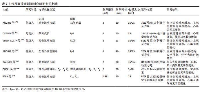
关肌肉疼痛[41];阳极经颅直流电刺激增强皮质兴奋性增强了脊髓机动池的下行驱动,以招募更多的运动单元[42];施加阳极经颅直流电刺激于左侧颞叶皮质是通过调节迷走神经活动 (即副交感神经)和增添愉快感提升肌肉收缩时的总做功量[54]。也有研究表明,在运动皮质和左侧颞叶皮质同时进行经颅直流电刺激也会改善受试者的整体运动表现[55]。 经颅直流电刺激未能显著改善肌肉耐力表现的潜在机制可能为:①刺激参数选择不当:刺激强度、持续时间或电极放置位置等可能不足以诱导或维持皮质兴奋性的有效改变,从而无法优化运动单位募集和下行驱动[44];②个体差异显著:受试者的解剖结构(如颅骨厚度)、基线神经生理状态(如皮质兴奋性水平)、遗传因素以及对刺激的敏感性存在高度异质性[3,5],这些差异导致部分个体对经颅直流电刺激反应微弱或无反应,显著降低了整体干预效果,使得在群体水平上难以观察到耐力提升;③天花板效应:当肌肉功能已经达到最大时,经颅直流电刺激干预不会进一步增强肌肉功能[48]。 2.3.2 经颅直流电刺激对心肺耐力的影响 心肺耐力,也被称为有氧耐力或心肺功能,是指心脏、肺部和血管系统在持续活动中向身体各部位输送氧气和营养的能力。心肺耐力是评估整体健康的关键指标之一,良好的心肺功能可以降低患心血管疾病的风险。增强心肺耐力有助于预防心脏病、高血压、糖尿病、肥胖症等慢性疾病。良好的心肺耐力也能够提高日常活动的效率,减少疲劳感,使个体能够更轻松地完成日常任务和工作。对于运动员和经常参与体育活动的人来说,心肺耐力是提高运动表现和竞技水平的基础。随着对经颅直流电刺激的深入研究,发现经颅直流电刺激可以影响心肺耐力。有研究表明,经颅直流电刺激可以辅助提高运动员的运动能力,包括心肺耐力[60],这表明经颅直流电刺激可能通过影响大脑功能,进而对心肺耐力产生积极作用。该文总结了7篇有关经颅直流电刺激对心肺耐力影响的研究,7项研究中最主要刺激脑区是初级运动皮质,最常用刺激强度为2 mA,最常用刺激时间为20 min,最常用电极片大小为35 cm2。 大脑对心肺功能的调控主要通过自主神经系统实现[61]。有许多脑区在心血管和呼吸功能的调节中扮演关键角色,如颞叶皮质等。OKANO等[62]的研究发现,颞叶皮质上的阳极经颅直流电刺激可以调节自主神经系统活动,通过降低主观感觉疲劳程度和心率来提高受试者的峰值功率输出。在另一项研究中,左颞叶的经颅直流电刺激增加了心率变异性,表明经颅直流电刺激改善了副交感神经对心率的调节[63]。值得注意的是,较高的迷走神经调节增强了自主心功能,其中身体健康归因于运动时心脏迷走神经功能。与非运动员相比,运动员有更高的迷走神经调节,在特定运动任务中他们的心率增加更慢。如果经颅直流电刺激能够改变与自主神经系统相关的大脑区域并增加迷走神经调节,则可以提高受试者在训练中的表现。在运动功能方面,VITOR-COSTA等[64]的研究表明,施加阳极经颅直流电刺激在初级运动皮质上改善了循环性能,增加了疲劳时间,然而,肌肉疲劳和心率等因素未见明显变化。ANGIUS等[40,65]对受试者进行70%峰值功率骑行至力竭测试,分别对运动皮质和左背外侧前额叶皮质进行经颅直流电刺激干预,结果发现左背外侧前额叶的主观感觉疲劳程度和心率都降低,而运动皮质组心率却没有显著变化。BALDARI等[66]对13名受试者进行2 mA、20 min的经颅直流电刺激,结果发现主观感觉疲劳程度和峰值摄氧量等都没有发生显著变化。 评价心肺耐力的核心指标如最大摄氧量、通气阈值等也被应用于经颅直流电刺激的研究中。CODELLA等[67]的研究显示,经过一段时间的经颅直流电刺激干预后,受试者最大摄氧量有明显提高,这可能是由于经颅直流电刺激改善了心肺功能之间的协同作用,使得氧气在肺部的摄取、血液中的运输以及肌肉中的利用更加高效。通气阈值的提高则意味着在运动过程中运动员可以在更高强度下维持有氧代谢,延缓无氧代谢的过早启动,这对于耐力运动表现至关重要。但PARK等[68]的研究表明,2 mA、20 min的阳极经颅直流电刺激干预后受试者通气阈值没有发生显著改变。 经颅直流电刺激对心肺耐力的影响有限,这可能与促进心肺耐力的系统的广泛性有关(即肌肉、神经、心血管、肺和代谢系统)[69]。心肺耐力高度依赖脑干、下丘脑及前额叶皮质对自主神经与代谢系统的动态整合调控,而经颅直流电刺激主要作用于皮质,对皮质下关键中枢的调控深度可能不足,难以有效协调心输出量、肺通气及外周血管阻力的适应性变化[70],只刺激一个皮质可能无法充分激活心肺整合所需的分布式神经网络。因此,当使用总体表现作为结果衡量标准时,仅影响一个系统(即神经系统)可能会产生很小的、难以感知的影响,在心肺任务执行过程中测量经颅直流电刺激后的大脑活动可能是一种更合适的结果测量方法。 经颅直流电刺激对心肺耐力的影响,见表2。"
| [1] MACHADO S, JANSEN P, ALMEIDA V, et al. Is tDCS an Adjunct Ergogenic Resource for Improving Muscular Strength and Endurance Performance? A Systematic Review. Front Psychol. 2019;10:1127. [2] GIORDANO J, BIKSON M, KAPPENMAN ES, et al. Mechanisms and Effects of Transcranial Direct Current Stimulation. Dose-Response. 2017; 15(1):500980030. [3] WIETHOFF S, HAMADA M, ROTHWELL JC. Variability in Response to Transcranial Direct Current Stimulation of the Motor Cortex. Brain Stimul. 2014;7(3):468-475. [4] HASSANZAHRAEE M, NITSCHE MA, ZOGHI M, et al. Determination of anodal tDCS duration threshold for reversal of corticospinal excitability: An investigation for induction of counter-regulatory mechanisms. Brain Stimul. 2020;13(3):832-839. [5] LÓPEZ-ALONSO V, CHEERAN B, RÍO-RODRÍGUEZ D, et al. Inter-individual Variability in Response to Non-invasive Brain Stimulation Paradigms. Brain Stimul. 2014;7(3):372-380. [6] CAULFIELD K, INDAHLASTARI A, NISSIM N, et al. Electric Field Strength From Prefrontal Transcranial Direct Current Stimulation Determines Degree of Working Memory Response: A Potential Application of Reverse‐Calculation Modeling? Neuromodulation. 2022;25(4):578-587. [7] 卞秀玲,王雅娜,王开元,等.经颅直流电刺激技术及其在提升运动表现中的应用[J].体育科学,2018,38(5):66-72. [8] ABDELMOULA A, BAUDRY S, DUCHATEAU J. Anodal transcranial direct current stimulation enhances time to task failure of a submaximal contraction of elbow flexors without changing corticospinal excitability. Neuroscience. 2016;322:94-103. [9] DA SILVA MACHADO DG, BIKSON M, DATTA A, et al. Acute effect of high-definition and conventional tDCS on exercise performance and psychophysiological responses in endurance athletes: a randomized controlled trial. Sci Rep. 2021;11(1):13911. [10] STAGG CJ, NITSCHE MA. Physiological basis of transcranial direct current stimulation. Neuroscientist. 2011;17(1):37-53. [11] TOTH AJ, HARVEY C, GULLANE H, et al. The effect of bipolar bihemispheric tDCS on executive function and working memory abilities. Front Psychol. 2023;14:1275878. [12] TEYMOORI H, AMIRI E, TAHMASEBI W, et al. Effect of tDCS targeting the M1 or left DLPFC on physical performance, psychophysiological responses, and cognitive function in repeated all-out cycling: a randomized controlled trial. J Neuroeng Rehabil. 2023;20(1):97. [13] ETEMADI M, AMIRI E, TADIBI V, et al. Anodal tDCS over the left DLPFC but not M1 increases muscle activity and improves psychophysiological responses, cognitive function, and endurance performance in normobaric hypoxia: a randomized controlled trial. BMC Neurosci. 2023;24(1):25. [14] WELLER S, DERNTL B, PLEWNIA C. Sex matters for the enhancement of cognitive training with transcranial direct current stimulation (tDCS). Biol Sex Differ. 2023;14(1):78. [15] TANG WK, LU H, LEUNG T, et al. Study protocol of a double-blind randomized control trial of transcranial direct current stimulation in post-stroke fatigue. Front Neurol. 2023;14:1297429. [16] SUN X, DONG X, YUAN Q, et al. Effects of transcranial direct current stimulation on patients with post-stroke fatigue: a study protocol for a double-blind randomized controlled trial. Trials. 2022;23(1):200. [17] LABREE B, HOARE DJ, GASCOYNE LE, et al. Determining the Effects of Transcranial Direct Current Stimulation on Tinnitus, Depression, and Anxiety: A Systematic Review. Brain Sci. 2022; 12(4):484. [18] SCHILBERG L, ENGELEN T, TEN OEVER S, et al. Phase of beta-frequency tACS over primary motor cortex modulates corticospinal excitability. Cortex. 2018;103:142-152. [19] FERTONANI A, MINIUSSI C. Transcranial Electrical Stimulation: What We Know and Do Not Know About Mechanisms. Neuroscientist. 2017;23(2):109-123. [20] LATTARI E, ROSA FB, FONSECA JS, et al. Effects on Volume Load and Ratings of Perceived Exertion in Individuals’ Advanced Weight Training After Transcranial Direct Current Stimulation. J Strength Cond Res. 2020;34(1):89-96. [21] BALLARD HK, GOEN J, MALDONADO T, et al. Effects of cerebellar transcranial direct current stimulation on the cognitive stage of sequence learning. J Neurophysiol. 2019;122(2):490-499. [22] LEFAUCHEUR JP, WENDLING F. Mechanisms of action of tDCS: A brief and practical overview. Neurophysiol Clin. 2019;49(4):269-275. [23] BANDEIRA ID, LINS-SILVA DH, BAROUH JL, et al. Neuroplasticity and non-invasive brain stimulation in the developing brain. Prog Brain Res. 2021;264: 57-89. [24] FRASE L, MERTENS L, KRAHL A, et al. Transcranial direct current stimulation induces long-term potentiation-like plasticity in the human visual cortex. Transl Psychiatry. 2021;11(1):17. [25] KRONBERG G, RAHMAN A, SHARMA M, et al. Direct current stimulation boosts hebbian plasticity in vitro. Brain Stimul. 2020;13(2):287-301. [26] CAVALEIRO C, MARTINS J, GONCALVES J, et al. Memory and Cognition-Related Neuroplasticity Enhancement by Transcranial Direct Current Stimulation in Rodents: A Systematic Review. Neural Plast. 2020;2020:4795267. [27] SUDBRACK-OLIVEIRA P, RAZZA LB, BRUNONI AR. Non-invasive cortical stimulation: Transcranial direct current stimulation (tDCS). Int Rev Neurobiol. 2021;159:1-22. [28] WORKMAN CD, FIETSAM AC, PONTO L, et al. Individual Cerebral Blood Flow Responses to Transcranial Direct Current Stimulation at Various Intensities. Brain Sci. 2020;10(11):855. [29] JONES KT, GÖZENMAN F, BERRYHILL ME. The strategy and motivational influences on the beneficial effect of neurostimulation: a tDCS and fNIRS study. Neuroimage. 2015;105:238-247. [30] SHINDE AB, LERUD KD, MUNSCH F, et al. Effects of tDCS dose and electrode montage on regional cerebral blood flow and motor behavior. Neuroimage. 2021;237:118144. [31] BAHR-HOSSEINI M, BIKSON M. Neurovascular-modulation: A review of primary vascular responses to transcranial electrical stimulation as a mechanism of action. Brain Stimul. 2021; 14(4):837-847. [32] YANG K, XI X, WANG T, et al. Effects of transcranial direct current stimulation on brain network connectivity and complexity in motor imagery. Neurosci Lett. 2021;757:135968. [33] ABELLANEDA-PEREZ K, VAQUE-ALCAZAR L, PERELLON-ALFONSO R, et al. Differential tDCS and tACS Effects on Working Memory-Related Neural Activity and Resting-State Connectivity. Front Neurosci. 2019;13:1440. [34] RAZZA LB, LUETHI MS, ZANAO T, et al. Transcranial direct current stimulation versus intermittent theta-burst stimulation for the improvement of working memory performance. Int J Clin Health Psychol. 2023;23(1):100334. [35] XIAO S, SHEN B, ZHANG C, et al. Anodal transcranial direct current stimulation enhances ankle force control and modulates the beta-band activity of the sensorimotor cortex. Cereb Cortex. 2023;33(12):7670-7677. [36] XIAO S, WANG B, YU C, et al. Effects of intervention combining transcranial direct current stimulation and foot core exercise on sensorimotor function in foot and static balance. J Neuroeng Rehabil. 2022;19(1):98. [37] WANG Y, WANG J, ZHANG QF, et al. Neural Mechanism Underlying Task-Specific Enhancement of Motor Learning by Concurrent Transcranial Direct Current Stimulation. Neurosci Bull. 2023; 39(1):69-82. [38] ZENON A, SIDIBE M, OLIVIER E. Disrupting the supplementary motor area makes physical effort appear less effortful. J Neurosci. 2015; 35(23):8737-8744. [39] PATEL R, DAWIDZIUK A, DARZI A, et al. Systematic review of combined functional near-infrared spectroscopy and transcranial direct-current stimulation studies. Neurophotonics. 2020;7(2):20901. [40] ANGIUS L, MAUGER AR, HOPKER J, et al. Bilateral extracephalic transcranial direct current stimulation improves endurance performance in healthy individuals. Brain Stimul. 2018;11(1):108-117. [41] COGIAMANIAN F, MARCEGLIA S, ARDOLINO G, et al. Improved isometric force endurance after transcranial direct current stimulation over the human motor cortical areas. Eur J Neurosci. 2007; 26(1):242-249. [42] WILLIAMS PS, HOFFMAN RL, CLARK BC. Preliminary evidence that anodal transcranial direct current stimulation enhances time to task failure of a sustained submaximal contraction. PLoS One. 2013; 8(12):e81418. [43] 齐玉刚,刘政宇,谭思洁.20-69岁人群肌肉耐力变化规律的试验研究[J].天津体育学院学报, 2016,31(1):69-72. [44] ANGIUS L, PAGEAUX B, HOPKER J, et al. Transcranial direct current stimulation improves isometric time to exhaustion of the knee extensors. Neuroscience. 2016;339:363-375. [45] DENIS G, ZORY R, RADEL R. Testing the role of cognitive inhibition in physical endurance using high-definition transcranial direct current stimulation over the prefrontal cortex. Hum Mov Sci. 2019;67:102507. [46] KAN B, DUNDAS JE, NOSAKA K. Effect of transcranial direct current stimulation on elbow flexor maximal voluntary isometric strength and endurance. Appl Physiol Nutr Metab. 2013; 38(7):734-739. [47] FLOOD A, WADDINGTON G, KEEGAN RJ, et al. The effects of elevated pain inhibition on endurance exercise performance. PeerJ. 2017;5:e3028. [48] RADEL R, TEMPEST G, DENIS G, et al. Extending the limits of force endurance: Stimulation of the motor or the frontal cortex? Cortex. 2017;97:96-108. [49] ANGIUS L, HOPKER JG, MARCORA SM, et al. The effect of transcranial direct current stimulation of the motor cortex on exercise-induced pain. Eur J Appl Physiol. 2015;115(11):2311-2319. [50] BARWOOD MJ, BUTTERWORTH J, GOODALL S, et al. The Effects of Direct Current Stimulation on Exercise Performance, Pacing and Perception in Temperate and Hot Environments. Brain Stimul. 2016;9(6):842-849. [51] BYRNE R, FLOOD A. The influence of transcranial direct current stimulation on pain affect and endurance exercise. Psychol Sport Exerc. 2019;45: 101554-101554. [52] WRIGHTSON JG, TWOMEY R, YEUNG S, et al. No effect of tDCS of the primary motor cortex on isometric exercise performance or perceived fatigue. Eur J Neurosci. 2020; 52(2):2905-2914. [53] ISIS S, ARMELE D, PAULO GL, et al. The effect of tDCS on improving physical performance and attenuating effort perception during maximal dynamic exercise in non-athletes. Neurosci Lett. 2023;794:136991. [54] SALES MM, SOUSA CVD, BROWNE RAV, et al. Transcranial direct current stimulation improves muscle isokinetic performance of young trained individuals. Medicina dello Sport. 2016;69(4):163. [55] KAMALI AM, SAADI ZK, YAHYAVI SS, et al. Transcranial direct current stimulation to enhance athletic performance outcome in experienced bodybuilders. PLoS One. 2019;14(8):e0220363. [56] WORKMAN CD, KAMHOLZ J, RUDROFF T. The Tolerability and Efficacy of 4 mA Transcranial Direct Current Stimulation on Leg Muscle Fatigability. Brain Sci. 2019; 10(1):12. [57] MONTENEGRO R, OKANO A, GURGEL J, et al. Motor cortex tDCS does not improve strength performance in healthy subjects. Motriz. 2015; 21(2):185-193. [58] SEIDEL-MARZI O, RAGERT P. Anodal transcranial direct current stimulation reduces motor slowing in athletes and non-athletes. BMC Neurosci. 2020;21(1):26. [59] SASADA S, ENDOH T, ISHII T, et al. Polarity-dependent improvement of maximal-effort sprint cycling performance by direct current stimulation of the central nervous system. Neurosci Lett. 2017;657:97-101. [60] 鲁盼盼,郭建瑞,欧阳博文,等. 经颅直流电刺激辅助提高运动员运动能力的研究进展[J].中华物理医学与康复杂志,2021,43(4):365-370. [61] HILZ MJ, DEVINSKY O, DOYLE W, et al. Decrease of sympathetic cardiovascular modulation after temporal lobe epilepsy surgery. Brain. 2002;125(Pt 5):985-995. [62] OKANO AH, FONTES EB, MONTENEGRO RA, et al. Brain stimulation modulates the autonomic nervous system, rating of perceived exertion and performance during maximal exercise. Br J Sports Med. 2015;49(18):1213-1218. [63] MONTENEGRO RA, FARINATTI PT, FONTES EB, et al. Transcranial direct current stimulation influences the cardiac autonomic nervous control. Neurosci Lett. 2011;497(1):32-36. [64] VITOR-COSTA M, OKUNO NM, BORTOLOTTI H, et al. Improving Cycling Performance: Transcranial Direct Current Stimulation Increases Time to Exhaustion in Cycling. PLoS One. 2015;10(12): e0144916. [65] ANGIUS L, SANTARNECCHI E, PASCUAL-LEONE A, et al. Transcranial Direct Current Stimulation over the Left Dorsolateral Prefrontal Cortex Improves Inhibitory Control and Endurance Performance in Healthy Individuals. Neuroscience. 2019;419:34-45. [66] BALDARI C, BUZZACHERA CF, VITOR-COSTA M, et al. Effects of Transcranial Direct Current Stimulation on Psychophysiological Responses to Maximal Incremental Exercise Test in Recreational Endurance Runners. Front Psychol. 2018;9:1867. [67] CODELLA R, ALONGI R, FILIPAS L, et al. Ergogenic Effects of Bihemispheric Transcranial Direct Current Stimulation on Fitness: a Randomized Cross-over Trial. Int J Sports Med. 2021;42(1):66-73. [68] PARK SB, SUNG DJ, KIM B, et al. Transcranial Direct Current Stimulation of motor cortex enhances running performance. PLoS One. 2019; 14(2):e0211902. [69] HANSEN D, BONNE K, ALDERS T, et al. Exercise training intensity determination in cardiovascular rehabilitation: Should the guidelines be reconsidered? Eur J Prev Cardiol. 2019;26(18):1921-1928. [70] ROBERTSON CV, MARINO FE. Prefrontal and motor cortex EEG responses and their relationship to ventilatory thresholds during exhaustive incremental exercise. Eur J Appl Physiol. 2015; 115(9):1939-1948. [71] CHEW T, HO KA, LOO CK. Inter- and Intra-individual Variability in Response to Transcranial Direct Current Stimulation (tDCS) at Varying Current Intensities. Brain Stimul. 2015;8(6):1130-1137. [72] JACKSON MP, RAHMAN A, LAFON B, et al. Animal models of transcranial direct current stimulation: Methods and mechanisms. Clin Neurophysiol. 2016;127(11):3425-3454. [73] HORVATH JC, FORTE JD, CARTER O. Evidence that transcranial direct current stimulation (tDCS) generates little-to-no reliable neurophysiologic effect beyond MEP amplitude modulation in healthy human subjects: A systematic review. Neuropsychologia. 2015;66:213-236. |
| [1] | Ma Tian, Luo Yu. Relationship between diaphragmatic ultrasound parameters and core stability tests in young adults [J]. Chinese Journal of Tissue Engineering Research, 2026, 30(33): 8703-8711. |
| [2] | Hao Yilin, Bao Chunyu. Effects of 12-week blood flow restriction training combined with aerobic exercise on cardiorespiratory endurance and cardiovascular function in overweight and obese individuals [J]. Chinese Journal of Tissue Engineering Research, 2026, 30(33): 8778-8784. |
| [3] | Zhang Shuai, Zhu Jiabin. Hydrogels: multifunctional effects from sports injury treatment to sports performance enhancement [J]. Chinese Journal of Tissue Engineering Research, 2026, 30(14): 3687-3693. |
| [4] | Yang Yicheng, Zheng Zhizhen, Liang Shuangxue, Wu Chengliang, Du Yunyun. Influence and implications of basketball shoes' functional parameters on human biomechanics [J]. Chinese Journal of Tissue Engineering Research, 2025, 29(35): 7620-7628. |
| [5] | Guo Hui, Kong Jianda, Tian Chunlan. The role of mitochondrial autophagy-related receptor proteins and signaling pathways in the prevention and treatment of sarcopenia through exercise [J]. Chinese Journal of Tissue Engineering Research, 2024, 28(27): 4397-4404. |
| [6] | Li Yujiao, Su Kunxia. High-intensity endurance exercise influences browning of white adipose tissue in a mouse model of high-fat diet induced obesity [J]. Chinese Journal of Tissue Engineering Research, 2023, 27(5): 707-713. |
| [7] | Lei Senlin, Zhang Minghui, Ma Chunlian, Gao Weifeng, Xia Xiaoyan, Dong Kunwei. Muscle effect, dose-effect relationship, and physiological mechanism of KAATSU-resistance training [J]. Chinese Journal of Tissue Engineering Research, 2023, 27(26): 4254-4264. |
| [8] | Wu Dongzhe, Gao Xiaolin, Shi Yongjin, Li Chuangtao, Wang Hao, Wang Wendi. Correlation analysis between “carbon dioxide tolerance” and cardiorespiratory endurance during exercise [J]. Chinese Journal of Tissue Engineering Research, 2023, 27(23): 3693-3699. |
| [9] | Hao Zhixin, Wu Yixin, Wang Xin, Xia Zhongliang. Effect of anodal transcranial direct current stimulation on muscle strength and endurance: a Meta-analysis [J]. Chinese Journal of Tissue Engineering Research, 2022, 26(20): 3273-3280. |
| [10] | Zhang Shuang, Tan Rui, Wang Chunxiao, Wu Fengyu, Guo Hongyu. MicroRNAs for assessing the motion control of human skeletal muscles [J]. Chinese Journal of Tissue Engineering Research, 2021, 25(17): 2755-2760. |
| [11] | Chen Zegang, Ding Haili, Li Long, Wang Chun. Changes of bone metabolism after different intensity endurance exercises in growing rats [J]. Chinese Journal of Tissue Engineering Research, 2020, 24(35): 5582-5588. |
| [12] | Yang Zhongming1, Jiang Manyi2, Xu Simao3 . Synergistic resistance of moderate-intensity endurance exercise and allicin to rat models of fatty liver induced by high-fat diet [J]. Chinese Journal of Tissue Engineering Research, 2019, 23(31): 5030-5035. |
| Viewed | ||||||
|
Full text |
|
|||||
|
Abstract |
|
|||||