Chinese Journal of Tissue Engineering Research ›› 2026, Vol. 30 ›› Issue (20): 5103-5114.doi: 10.12307/2026.330
Previous Articles Next Articles
Preparation of Cu2+-containing microarc oxidation functional coating on medical magnesium alloy and its anti-tumor and angiogenesis-promoting effects
Lin Kejian1, Chai Yinghong2, Zou Jie1, Huang Ruixin3, Fang Yongchao3, Huang Jing4, Yang Qin1, Luo Xia2, Zhang Hong1
- 1Department of Burn and Plastic Surgery, First Affiliated Hospital of Chengdu Medical College, Chengdu 610599, Sichuan Province, China; 2School of New Energy and Materials, Southwest Petroleum University, Chengdu 610500, Sichuan Province, China; 3School of Pharmacy, Chengdu Medical College, Chengdu 610500, Sichuan Province, China; 4The Fourth People’s Hospital of Zigong, Zigong 643000, Sichuan Province, China
-
Accepted:2025-04-22Online:2026-07-18Published:2025-11-21 -
Contact:Zhang Hong, Associate chief physician, Department of Burn and Plastic Surgery, First Affiliated Hospital of Chengdu Medical College, Chengdu 610599, Sichuan Province, China Luo Xia, Associate professor, Master’s supervisor, School of New Energy and Materials, Southwest Petroleum University, Chengdu 610500, Sichuan Province, China -
About author:Lin Kejian, MS, Department of Burn and Plastic Surgery, First Affiliated Hospital of Chengdu Medical College, Chengdu 610599, Sichuan Province, China -
Supported by:Sichuan Provincial Science and Technology Plan Project, No. 2020YFH0151 (to LX); Sichuan Provincial Key Laboratory of Development and Regeneration Research Fund Project, No. SYS19-09 (to ZH)
CLC Number:
Cite this article
Lin Kejian, Chai Yinghong, Zou Jie, Huang Ruixin, Fang Yongchao, Huang Jing, Yang Qin, Luo Xia, Zhang Hong. Preparation of Cu2+-containing microarc oxidation functional coating on medical magnesium alloy and its anti-tumor and angiogenesis-promoting effects[J]. Chinese Journal of Tissue Engineering Research, 2026, 30(20): 5103-5114.
share this article
Add to citation manager EndNote|Reference Manager|ProCite|BibTeX|RefWorks

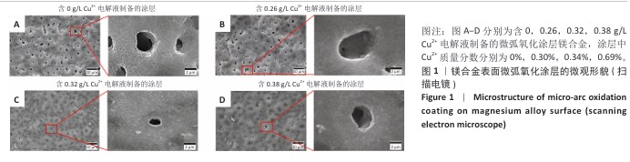
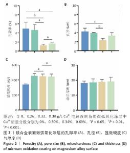
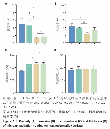
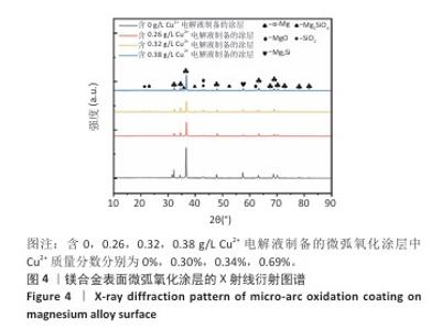
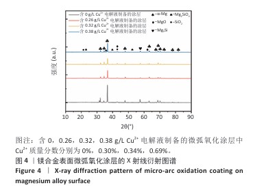
2.1 医用镁合金涂层的形貌表征结果 扫描电镜下可见镁合金微弧氧化膜层表面产生细小的裂纹与孔隙,当微弧氧化电解液中Cu2+质量浓度为0.26 g/L时,镁合金表面微弧氧化涂层较粗糙,表面有较多裂纹和孔隙;当微弧氧化电解液中Cu2+质量浓度为0.32 g/L时,镁合金表面微弧氧化涂层较平整光滑,见图1。随着微弧氧化电解液中Cu2+质量浓度的增加,负载涂层镁合金表面的孔隙率和孔径呈现先减小再增大的趋势,见图2A,B。Cu2+的加入提升了镁合金表面微弧氧化涂层显微硬度,涂层厚度无明显变化,见图2C,D。 能谱分析结果显示,Cu2+在微弧氧化涂层表面分布较为均匀,见图3。等离子体质谱检测显示Cu2+被有效负载于微弧氧化涂层中,随着电解液中Cu2+质量浓度(0,0.26,0.32,0.38 g/L)的增加,微弧氧化涂层中的Cu2+质量分数增加,依次为0%,0.30%,0.34%,0.69%,见图3。X射线衍射仪分析结果显示,在含Cu2+微弧氧化涂层中出现了如SiO2、MgSiO4、Mg2Si等高硬化合物,使得涂层显微硬度得到进一步提升,见图4。 "
"

"
"
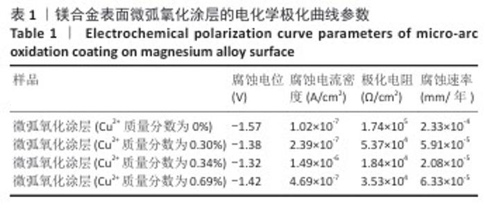
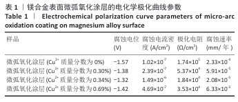
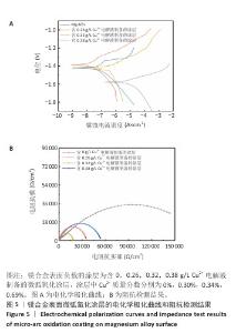
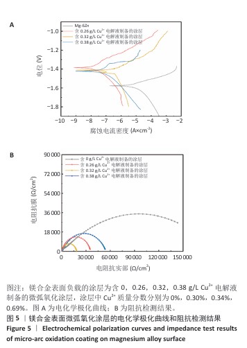
2.2 医用镁合金涂层的耐腐蚀性能检测结果 表1和图5展示了4组微弧氧化涂层的耐腐蚀性能检测结果。与不含Cu2+涂层相比,含Cu2+涂层自腐蚀电位明显正向移动,当涂层中Cu2+质量分数为0.34%时,镁合金表面的腐蚀电位达到最大值,为-1.32 V,电流密度为1.49×10⁻⁷ A/cm2,阻抗值为1.84×104 Ω/cm2,腐蚀速率最小,为2.08×10-5 mm/年。结果表明,Cu2+的添加改善了微弧氧化涂层的缺陷,减小了镁合金表面孔径与孔隙率,有效阻止了腐蚀物质的渗透与扩散,与不含Cu2+微弧氧化涂层相比,添加Cu2+后微弧氧化涂层腐蚀速率降低了1个数量级,呈现出更为优异的耐腐蚀性能。"
"
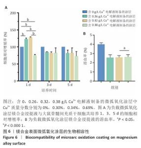
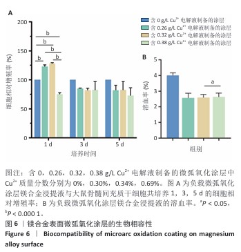
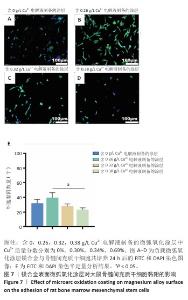
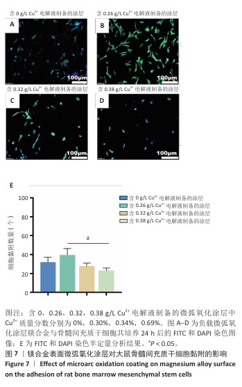
2.3 医用镁合金涂层的生物相容性评估结果 各组大鼠骨髓间充质干细胞的相对增殖率如图6A所示。含Cu2+质量分数0.30%,0.34%微弧氧化涂层组培养1,3,5 d后的大鼠骨髓间充质干细胞相对增殖率均大于80%,符合生物安全材料植入标准;含Cu2+质量分数0.69%微弧氧化涂层组培养1,5 d后的大鼠骨髓间充质干细胞相对增殖率小于80%,不符合生物安全材料植入标准。各组微弧氧化涂层的溶血率均低于5%,见图6B。FITC/DAPI免疫染色结果显示,随着涂层中Cu2+质量分数的增加,镁合金表面黏附的大鼠骨髓间充质干细胞数量呈先增加后减少的趋势,其中含Cu2+质量分数0.30%微弧氧化涂层组黏附细胞数量最多,含Cu2+质量分数0.69%微弧氧化涂层组黏附细胞数量最少,见图7。"
"

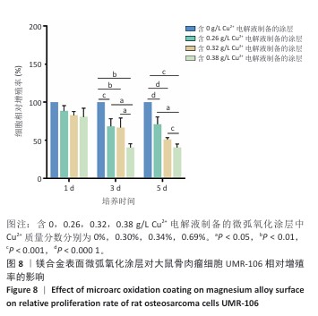
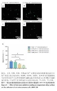
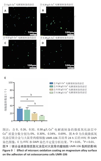
2.4 医用镁合金涂层的体外抗肿瘤性能检测结果 由图8可知,随着培养时间和涂层中Cu2+质量分数的增加,UMR-106细胞相对增殖率呈现减少趋势,培养5 d后,含Cu2+质量分数0.30%,0.34%,0.69%微弧氧化涂层组UMR-106细胞相对细胞增殖率均小于80%。由图9可知,随着涂层中Cu2+质量分数的增加,镁合金表面黏附的UMR-106细胞数量减少。"
"
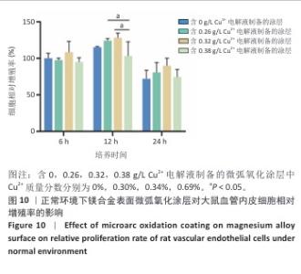
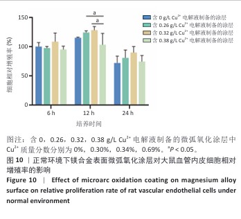
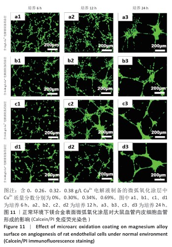
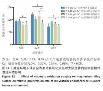
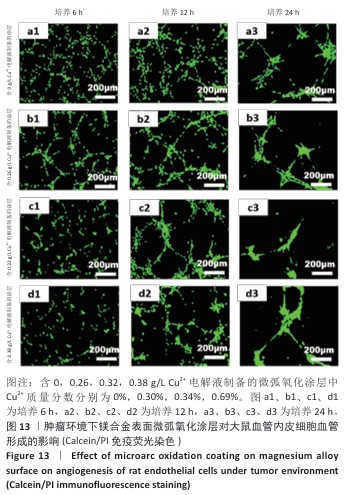
2.5 医用镁合金涂层的体外促血管形成检测结果 正常环境下各组大鼠血管内皮细胞相对增殖率如图10所示。随着培养时间与涂层中Cu2+质量分数的增加,大鼠血管内皮细胞相对增殖率大体呈先升高后降低趋势,其中含Cu2+质量分数0.34%微弧氧化涂层组大鼠血管内皮细胞相对增殖率最高。正常环境下各组大鼠血管内皮细胞Calcein/PI免疫荧光染色结果见图11所示。培养6 h时,各组均未形成成型的网状结构,随着培养时间的延长,各组均出现了血管网状结构;培养12 h时,含Cu2+质量分数0.30%,0.34%微弧氧化涂层组形成的网状结构最为显著,并且具有最多分支节点及最长分支管,此时含Cu2+质量分数0.34%微弧氧化涂层组大鼠血管内皮细胞相对增殖率高达128.24%,表现出优异的成血管能力。 肿瘤环境下各组大鼠血管内皮细胞相对增殖率如图12所示。随着培养时间的增加,大鼠血管内皮细胞相对增殖率呈先升高后降低的趋势,其中培养12 h时大鼠血管内皮细胞相对增殖率最高。肿瘤环境下各组大鼠血管内皮细胞Calcein/PI免疫荧光染色结果见图13所示。培养6 h时,含Cu2+质量分数0%,0.30%微弧氧化涂层组有少量分支节、分支管等血管化结构形成,随着培养时间延长至12 h,各组均出血管网状结构,其中含Cu2+质量分数0.69%微弧氧化涂层组形成的血管网状结构较少,该组大鼠血管内皮细胞相对增殖率为87.97%,明显低于其他3组,血管化程度最弱。"

"
"
"
| [1] FERRARI S, PALMERINI E, STAALS EL, et al. The treatment of nonmetastatic high grade osteosarcoma of the extremity: review of the Itali(1(1an Rizzoli experience. Impact on the future. Cancer Treat Res. 2009;152:275-287. [2] BLÄSIUS F, DELBRÜCK H, HILDEBRAND F, et al. Surgical Treatment of Bone Sarcoma. Cancers (Basel). 2022;14(11):2694. [3] 郭传平,高冬芳,李羡,等.医用镁合金耐腐蚀性能研究进展[J].稀有金属材料与工程,2024,53(8):2390-2404. [4] SARIAN MN, IQBAL N, SOTOUDEHBAGHA P, et al. Potential bioactive coating system for high-performance absorbable magnesium bone implants. Bioact Mater. 2021;12:42-63. [5] LIN X, TAN L, ZHANG Q, et al. The in vitro degradation process and biocompatibility of a ZK60 magnesium alloy with a forsterite-containing micro-arc oxidation coating. Acta Biomater. 2013;9(10):8631-8642. [6] LIANG DY, LIANG PC, YI QQ, et al. Copper coating formed by micro-arc oxidation on pure Mg improved antibacterial activity, osteogenesis, and angiogenesis in vivo and in vitro. Biomed Microdevices. 2021;23(3):39. [7] 石伟.含锶羟基磷灰石涂层对Mg-4Zn合金腐蚀行为及细胞行为的影响[D].长沙:湖南大学,2017. [8] 赵颖,曾利兰,梁涛.可降解镁基金属的生物相容性研究进展[J].金属学报,2017,53(10):1181-1196. [9] NAN M, YANGMEI C, BANGCHENG Y. Magnesium metal--a potential biomaterial with antibone cancer properties. J Biomed Mater Res A. 2014;102(8):2644-2651. [10] ZENG RC, CUI LY, JIANG K, et al. In Vitro Corrosion and Cytocompatibility of a Microarc Oxidation Coating and Poly(L-lactic acid) Composite Coating on Mg-1Li-1Ca Alloy for Orthopedic Implants. ACS Appl Mater Interfaces. 2016;8(15):10014-10028. [11] 纪凌宇.AZ91D镁合金基钙磷/壳聚糖复合抗菌膜层生物相容性研究[D].哈尔滨:哈尔滨商业大学,2016. [12] WU Y, HE G, ZHANG Y, et al. Unique antitumor property of the Mg-Ca-Sr alloys with addition of Zn. Sci Rep. 2016;6:21736. [13] ZHOU M, TIAN M, LI C. Copper-Based Nanomaterials for Cancer Imaging and Therapy. Bioconjug Chem. 2016;27(5):1188-1199. [14] TSVETKOV P, COY S, PETROVA B, et al. Copper induces cell death by targeting lipoylated TCA cycle proteins. Science. 2022;375(6586): 1254-1261. [15] 铙其强.PDA负载金属离子修饰PCL/β-TCP支架表面用于骨组织工程[D].南昌:南昌大学,2021. [16] LIN Z, CAO Y, ZOU J, et al. Improved osteogenesis and angiogenesis of a novel copper ions doped calcium phosphate cement. Mater Sci Eng C Mater Biol Appl. 2020;114:111032. [17] ZHOU Y, HAN S, XIAO L, et al. Accelerated host angiogenesis and immune responses by ion release from mesoporous bioactive glass. J Mater Chem B. 2018;6(20):3274-3284. [18] NOORI A, HOSEINPOUR M, KOLIVAND S, et al. Synergy effects of copper and L-arginine on osteogenic, angiogenic, and antibacterial activities. Tissue Cell. 2022;77:101849. [19] WANG H, ZHAO S, XIAO W, et al. Influence of Cu doping in borosilicate bioactive glass and the properties of its derived scaffolds. Mater Sci Eng C Mater Biol Appl. 2016;58:194-203. [20] HASINOFF BB, WU X, YADAV AA, et al. Cellular mechanisms of the cytotoxicity of the anticancer drug elesclomol and its complex with Cu(II). Biochem Pharmacol. 2015;93(3):266-276. [21] NAGAI M, VO NH, SHIN OGAWA L, et al. The oncology drug elesclomol selectively transports copper to the mitochondria to induce oxidative stress in cancer cells. Free Radic Biol Med. 2012;52(10):2142-2150. [22] LI CY, GAO L, FAN XL, et al. In vitro degradation and cytocompatibility of a low temperature in-situ grown self-healing Mg-Al LDH coating on MAO-coated magnesium alloy AZ31. Bioact Mater. 2020;5(2):364-376. [23] YANG SH, LUO X, LI MY, et al. Effect of MAO Parameters on the Degradation Property of Medical Mg-6Zn Alloy Prepared by Semisolid Powder Molding. Transactions of the Indian Institute of Metals. 2023; 76(8):2273-2283. [24] 杨上挥,罗霞,李铭宇,等.半固态粉末成形温度对Mg-6Zn-xMn显微组织和力学性能的影响[J].粉末冶金材料科学与工程,2022, 27(4):372-381. [25] 秦静.医用镁合金微弧氧化及其在模拟人体环境下的磨损行为[D].西安:西安工业大学,2016. [26] LUO X, LI M, CAI H, et al. Degradation Adaptability Assessment of Semisolid Powder Molded Mg-Zn-Mn Alloys for Orthopedic Applications. ACS Appl Bio Mater. 2023;6(12):5515-5530. [27] GB/T 16886.12-2023,医疗器械生物学评价第12部分:样品制备与参照材料[S]. [28] 侯丽,乔春霞,赵增琳.解读ISO 10993-4:2017《医疗器械生物学评价第4部分:与血液相互作用试验选择》[J].中国医疗设备, 2018,33(11):1-6. [29] GB/T 16886.5-2017,医疗器械生物学评价第5部分:体外细胞毒性试验[S]. [30] GILL J, GORLICK R. Advancing therapy for osteosarcoma. Nat Rev Clin Oncol. 2021;18(10):609-624. [31] ISAKOFF MS, BIELACK SS, MELTZER P, et al. Osteosarcoma: Current Treatment and a Collaborative Pathway to Success. J Clin Oncol. 2015; 33(27):3029-3035. [32] AGARWAL S, CURTIN J, DUFFY B, et al. Biodegradable magnesium alloys for orthopaedic applications: A review on corrosion, biocompatibility and surface modifications. Mater Sci Eng C Mater Biol Appl. 2016;68: 948-963. [33] ZHANG E, YANG L, XU J, et al. Microstructure, mechanical properties and bio-corrosion properties of Mg-Si(-Ca, Zn) alloy for biomedical application. Acta Biomater. 2010;6(5):1756-1762. [34] WONG CC, WONG PC, TSAI PH, et al. Biocompatibility and Osteogenic Capacity of Mg-Zn-Ca Bulk Metallic Glass for Rabbit Tendon-Bone Interference Fixation. Int J Mol Sci. 2019;20(9):2191. [35] XU L, PAN F, YU G, et al. In vitro and in vivo evaluation of the surface bioactivity of a calcium phosphate coated magnesium alloy. Biomaterials. 2009;30(8):1512-1523. [36] ATKINSON RL, ZHANG M, DIAGARADJANE P, et al. Thermal enhancement with optically activated gold nanoshells sensitizes breast cancer stem cells to radiation therapy. Sci Transl Med. 2010;2(55):55ra79. [37] 侯莉,徐斌,宋嘉卓,等.仿生壳聚糖基水凝胶的可控构筑及其对多能干细胞生物学行为影响的分析[J].分析化学,2022,50(10):1520-1532. [38] ZHANG E, YANG L, XU J, et al. Microstructure, mechanical properties and bio-corrosion properties of Mg-Si(-Ca, Zn) alloy for biomedical application. Acta Biomater. 2010;6(5):1756-1762. [39] WU C, ZHOU Y, XU M, et al. Copper-containing mesoporous bioactive glass scaffolds with multifunctional properties of angiogenesis capacity, osteostimulation and antibacterial activity. Biomaterials. 2013;34(2):422-433. [40] ROMERO-SÁNCHEZ LB, MARÍ-BEFFA M, CARRILLO P, et al. Copper-containing mesoporous bioactive glass promotes angiogenesis in an in vivo zebrafish model. Acta Biomater. 2018;68:272-285. [41] AHMED M, QI Y, ZHANG L, et al. Influence of Cu2+ Ions on the Corrosion Resistance of AZ31 Magnesium Alloy with Microarc Oxidation. Materials (Basel). 2020;13(11):2647. [42] FU L, XU JL, WANG FP, et al. Biomimetic deposition of apatite coatings on micro-arc oxidation treated biomedical NiTi alloy. Surf Coat Technol. 2010;204:3294-3299. [43] 刘庆,欧阳雪琼,刘文财,等.流延工艺制备氧化锆燃料电池薄膜的研究进展[J].材料导报,2025,39(10):1-20. [44] 姜炳春,卢立伟,林标智,等.微/纳米颗粒改性镁合金微弧氧化膜层性能的研究进展[J].中国有色金属学报,2024,34(10): 3308-3324. [45] BREWER GJ. Copper toxicity in the general population. Clin Neurophysiol. 2010;121(4):459-460. [46] KIM BE, NEVITT T, THIELE DJ. Mechanisms for copper acquisition, distribution and regulation. Nat Chem Biol. 2008;4(3):176-185. [47] VALKO M, LEIBFRITZ D, MONCOL J, et al. Free radicals and antioxidants in normal physiological functions and human disease. Int J Biochem Cell Biol. 2007;39(1):44-84. [48] XU Y, LIU SY, ZENG L, et al. An Enzyme-Engineered Nonporous Copper(I) Coordination Polymer Nanoplatform for Cuproptosis-Based Synergistic Cancer Therapy. Adv Mater. 2022;34(43):e2204733. [49] ZHONG X, DAI X, WANG Y, et al. Copper-based nanomaterials for cancer theranostics. Wiley Interdiscip Rev Nanomed Nanobiotechnol. 2022;14(4):e1797. [50] 唐思成.CeO2含量对Al2O3-10TiO2复合陶瓷涂层耐腐蚀性能的影响[D].成都:西华大学,2022. [51] HOA BT, VUONG BX. Sol-gel synthesis of nano-bioactive glass 60SiO2-36CaO-4P2O5 (mol.%) without using acid catalysts. Sol-Gel Sci Technol. 2024;110:900-909. [52] SHANBHAG VC, GUDEKAR N, JASMER K, et al. Copper metabolism as a unique vulnerability in cancer. Biochim Biophys Acta Mol Cell Res. 2021;1868(2):118893. [53] WANG X, SCHRÖDER HC, MÜLLER WE. Enzymatically synthesized inorganic polymers as morphogenetically active bone scaffolds: application in regenerative medicine. Int Rev Cell Mol Biol. 2014;313: 27-77. [54] MANSO Y, ADLARD PA, CARRASCO J, et al. Metallothionein and brain inflammation. J Biol Inorg Chem. 2011;16(7):1103-1113. [55] GÉRARD C, BORDELEAU LJ, BARRALET J, et al. The stimulation of angiogenesis and collagen deposition by copper. Biomaterials. 2010; 31(5):824-831. [56] 杨淑,崔文国,魏杰,等.自愈合可注射性透明质酸水凝胶的构建及促进血管生成的研究[J].上海交通大学学报(医学版),2023, 43(12):1480-1492. [57] BARRALET J, GBURECK U, HABIBOVIC P, et al. Angiogenesis in calcium phosphate scaffolds by inorganic copper ion release. Tissue Eng Part A. 2009;15(7):1601-1609. [58] TSVETKOV P, COY S, PETROVA B, et al. Copper induces cell death by targeting lipoylated TCA cycle proteins. Science. 2022;375(6586): 1254-1261. |
| [1] | Wu Yanting, Li Yu, Liao Jinfeng. Magnesium oxide nanoparticles regulate osteogenesis- and angiogenesis-related gene expressions to promote bone defect healing [J]. Chinese Journal of Tissue Engineering Research, 2026, 30(8): 1885-1895. |
| [2] | Liu Dawei, Cui Yingying, Wang Fanghui, Wang Zixuan, Chen Yuhan, Li Yourui, Zhang Ronghe. Epigallocatechin gallate-mediated bidirectional regulation of reactive oxygen species and its application in nanomaterials [J]. Chinese Journal of Tissue Engineering Research, 2026, 30(8): 2101-2112. |
| [3] | Zhou Xiaohui, Wang Siyi, Zhou Qiyun, He Zhao, Jia Yujuan, Wang Yuanbin, Ma Jianwu, Chen Gang, Zheng Feng, Chu Genglei. Nanohydroxyapatite-polyether carbonate urethane electrospinning membrane promotes bone defect repair [J]. Chinese Journal of Tissue Engineering Research, 2026, 30(20): 5134-5142. |
| [4] | Wang Liang, Zhang Xin, He Wei, Wang Jian. Clinical application and prospects of MXene-based materials for the repair of bone defects [J]. Chinese Journal of Tissue Engineering Research, 2026, 30(20): 5282-5294. |
| [5] | Xu Yawei, Meng Shilong, Zhang Xu, Wang Chengjie, Yuan Yifeng, Shi Xiaolin, Wang Jiao, Liu Kang . Repairing bone defects with active ingredients of traditional Chinese medicine combined with hydrogels: successes and challenges [J]. Chinese Journal of Tissue Engineering Research, 2026, 30(20): 5295-5303. |
| [6] | Gong Yukang, Ye Gaoqi, Wang Chenhao, Chen Dejin, Gao Wenshan. Effects and mechanisms of natural polyphenol-based hydrogels in promoting bone repair [J]. Chinese Journal of Tissue Engineering Research, 2026, 30(14): 3675-3686. |
| Viewed | ||||||
|
Full text |
|
|||||
|
Abstract |
|
|||||