Chinese Journal of Tissue Engineering Research ›› 2026, Vol. 30 ›› Issue (21): 5517-5523.doi: 10.12307/2026.314
Previous Articles Next Articles
Anatomical risk factor analysis of posterior cruciate ligament tibial avulsion fracture in adults
Li Gen1, Zhang Xichen1, Sun Yingjin1, He Chenglong1, Gao Xuren1, 2
- 1Xuzhou Medical University, Xuzhou 221004, Jiangsu Province, China; 2Affiliated Hospital of Xuzhou Medical University, Xuzhou 221000, Jiangsu Province, China
-
Accepted:2025-05-06Online:2026-07-28Published:2026-03-05 -
Contact:Gao Xuren, PhD, Chief physician, Associate professor, Xuzhou Medical University, Xuzhou 221004, Jiangsu Province, China; Affiliated Hospital of Xuzhou Medical University, Xuzhou 221000, Jiangsu Province, China -
About author:Li Gen, Master candidate, Physician, Xuzhou Medical University, Xuzhou 221004, Jiangsu Province, China
CLC Number:
Cite this article
Li Gen, Zhang Xichen, Sun Yingjin, He Chenglong, Gao Xuren. Anatomical risk factor analysis of posterior cruciate ligament tibial avulsion fracture in adults[J]. Chinese Journal of Tissue Engineering Research, 2026, 30(21): 5517-5523.
share this article
Add to citation manager EndNote|Reference Manager|ProCite|BibTeX|RefWorks
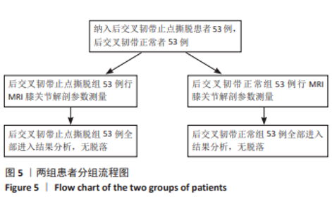
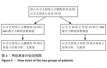
2.1 参与者数量分析 纳入后交叉韧带止点撕脱患者53例,后交叉韧带正常者53例,全部进入结果分析,无脱落。 2.2 试验流程图 见图5。"

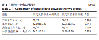
2.3 一般情况比较 两组观察对象性别、年龄、患侧膝、体质量指数比较,差异无显著性意义(P > 0.05),具有可比性,见表1。"
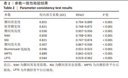
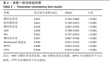
2.4 一致性检验 经ICC分析,测量数据可信度较高(ICC > 0.75,P < 0.001),见表2。"
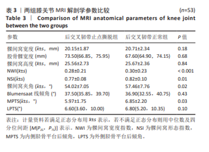
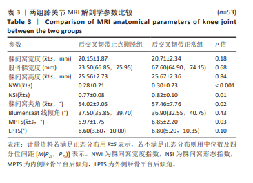
2.5 解剖学参数比较 后交叉韧带止点撕脱组与后交叉韧带正常组解剖学参数比较,两组间髁间窝宽度、髁间窝高度、股骨髁宽度、Blumensaat倾角、LPTS相比差异无显著性意义(P > 0.05)。后交叉韧带止点撕脱组的NWI、NSI、髁间窝夹角、MPTS均小于后交叉韧带正常组,差异有显著性意义(P < 0.05),见表3。"
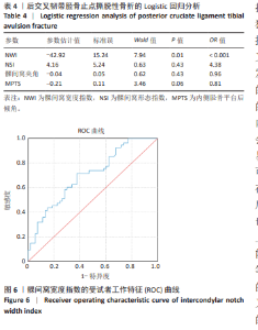
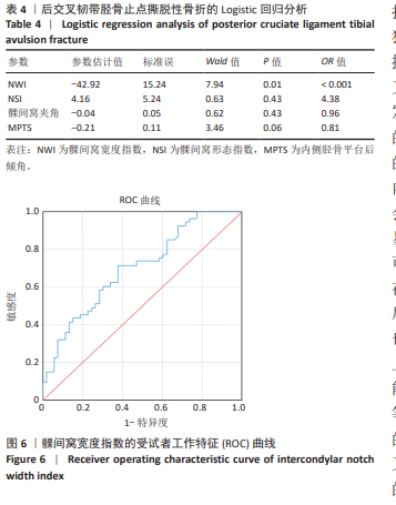
2.6 二元Logistic回归分析 对两组数据进行二元Logistic回归分析,结果显示NSI、髁间窝夹角、MPTS无统计学意义(P > 0.05);NWI的OR值< 0.001,且P < 0.05,故NWI与后交叉韧带胫骨止点撕脱性骨折相关。提示较小的NWI是后交叉韧带胫骨止点撕脱性骨折的独立危险因素,见表4。 2.7 ROC曲线 对独立危险因素NWI进行ROC曲线分析。结果曲线下面积为0.70,最佳截点为0.29,该截点下约登指数为0.340,敏感度71.7%,特异性62.3%。见图6。 2.8 不良事件 此次试验过程中所有患者均未发生任何不良反应。"
| [1] XIN W, GAO Y, ZHENG L, et al. Posterior cruciate ligament tibial attachment sacrifice percentage is higher in cruciate-retaining total knee arthroplasty in patients with discoid lateral meniscus. Arthroplasty. 2024;6(1):19. [2] AKPINAR B, DECLOUETTE B, GONZALEZ-LOMAS G, et al. Posterior Cruciate Ligament Reconstruction Current Concepts Review. Bull Hosp Jt Dis (2013). 2024;82(1):4-9. [3] ZSIDAI B, HORVATH A, WINKLER PW, et al. Different injury patterns exist among patients undergoing operative treatment of isolated PCL, combined PCL/ACL, and isolated ACL injuries: a study from the Swedish National Knee Ligament Registry. Knee Surg Sports Traumatol Arthrosc. 2022;30(10):3451-3460. [4] ZHOU Z, WANG S, XIAO J, et al. The degree of fracture reduction does not compromise the clinical efficacy of arthroscopic reduction and fixation of tibial posterior cruciate ligament avulsion fractures: A retrospective study. Medicine (Baltimore). 2023;102(39):e35356. [5] VAN KUIJK KSR, REIJMAN M, BIERMA-ZEINSTRA SMA, et al. Posterior cruciate ligament injury is influenced by intercondylar shape and size of tibial eminence. Bone Joint J. 2019;101-B(9):1058-1062. [6] NEDAIE S, VIVEKANANTHA P, O’HARA K, et al. Decreased posterior tibial slope is a risk factor for primary posterior cruciate ligament rupture and posterior cruciate ligament reconstruction failure: A systematic review. Knee Surg Sports Traumatol Arthrosc. 2024;32(1):167-180. [7] OWESEN C, SANDVEN-THRANE S, LIND M, et al. Epidemiolgy of surgically treated posterior cruciate ligament injuries in Scandinavia. Knee Surg Sports Traumatol Arthrosc. 2017;25(8):2384-2391. [8] SONIN AH, FITZGERALD SW, HOFF FL, et al. MR imaging of the posterior cruciate ligament: normal, abnormal, and associated injury patterns. Radiographics. 1995;15(3):551-561. [9] LIU F, ZHANG S, XIAO Y, et al. Stenotic intercondylar notch is not a risk factor for posterior cruciate ligament rupture: a morphological analyses using magnetic resonance imaging. Knee Surg Sports Traumatol Arthrosc. 2022;30(5):1711-1717. [10] HODEL S, KABELITZ M, TONDELLI T, et al. Introducing the Lateral Femoral Condyle Index as a Risk Factor for Anterior Cruciate Ligament Injury. Am J Sports Med. 2019;47(10):2420-2426. [11] KIZILGÖZ V, SIVRIOĞLU AK, AYDIN H, et al. The Combined Effect of Body Mass Index and Tibial Slope Angles on Anterior Cruciate Ligament Injury Risk in Male Knees: A Case-Control Study. Clin Med Insights Arthritis Musculoskelet Disord. 2019;12:1179544119867922. [12] BERNHARDSON AS, DEPHILLIPO NN, AMAN ZS, et al. Decreased Posterior Tibial Slope Does Not Affect Postoperative Posterior Knee Laxity After Double-Bundle Posterior Cruciate Ligament Reconstruction. Am J Sports Med. 2019;47(2):318-323. [13] LAPRADE CM, CIVITARESE DM, RASMUSSEN MT, et al. Emerging Updates on the Posterior Cruciate Ligament: A Review of the Current Literature. Am J Sports Med. 2015;43(12):3077-3092. [14] GWINNER C, WEILER A, ROIDER M, et al. Tibial Slope Strongly Influences Knee Stability After Posterior Cruciate Ligament Reconstruction: A Prospective 5- to 15-Year Follow-up. Am J Sports Med. 2017;45(2):355-361. [15] OWESEN C, SANDVEN-THRANE S, LIND M, et al. Epidemiology of surgically treated posterior cruciate ligament injuries in Scandinavia. Knee Surg Sports Traumatol Arthrosc. 2017;25(8):2384-2391. [16] BAYER S, MEREDITH SJ, WILSON KW, et al. Knee Morphological Risk Factors for Anterior Cruciate Ligament Injury: A Systematic Review [published correction appears in J Bone Joint Surg Am. 2020;102(14):e85. doi: 10.2106/JBJS.ER.19.00535]. J Bone Joint Surg Am. 2020;102(8):703-718. [17] JHA V, PANDIT A. Notch Volume Measured on Magnetic Resonance Imaging Is Better Than 2-Dimensional Notch Parameters for Predicting Noncontact Anterior Cruciate Ligament Injury in Males. Arthroscopy. 2021;37(5): 1534-1543.e1. [18] SHEN X, XIAO J, YANG Y, et al. Multivariable analysis of anatomic risk factors for anterior cruciate ligament injury in active individuals. Arch Orthop Trauma Surg. 2019;139(9):1277-1285. [19] HUDEK R, SCHMUTZ S, REGENFELDER F, et al. Novel measurement technique of the tibial slope on conventional MRI. Clin Orthop Relat Res. 2009;467(8):2066-2072. [20] HUANG WT, KANG K, WANG J, et al. Morphological Risk Factors for Posterior Cruciate Ligament Tear and Tibial Avulsion Injuries of the Tibial Plateau and Femoral Condyle. Am J Sports Med. 2023;51(1):129-140. [21] FAN N, ZHENG YC, ZANG L, et al. What is the impact of knee morphology on posterior cruciate ligament avulsion fracture in men and women: a case control study. BMC Musculoskelet Disord. 2021;22(1):100. [22] SOURYAL TO, FREEMAN TR. Intercondylar notch size and anterior cruciate ligament injuries in athletes. A prospective study. Am J Sports Med. 1993; 21(4):535-539. [23] DOMZALSKI M, GRZELAK P, GABOS P. Risk factors for Anterior Cruciate Ligament injury in skeletally immature patients: analysis of intercondylar notch width using Magnetic Resonance Imaging. Int Orthop. 2010;34(5): 703-707. [24] BOURAS T, FENNEMA P, BURKE S, et al. Stenotic intercondylar notch type is correlated with anterior cruciate ligament injury in female patients using magnetic resonance imaging. Knee Surg Sports Traumatol Arthrosc. 2018;26(4):1252-1257. [25] EVERHART JS, FLANIGAN DC, SIMON RA, et al. Association of noncontact anterior cruciate ligament injury with presence and thickness of a bony ridge on the anteromedial aspect of the femoral intercondylar notch. Am J Sports Med. 2010;38(8):1667-1673. [26] FUNG DT, HENDRIX RW, KOH JL, et al. ACL impingement prediction based on MRI scans of individual knees. Clin Orthop Relat Res. 2007;460:210-218. [27] DAVIS TJ, SHELBOURNE KD, KLOOTWYK TE. Correlation of the intercondylar notch width of the femur to the width of the anterior and posterior cruciate ligaments. Knee Surg Sports Traumatol Arthrosc. 1999;7(4):209-214. [28] DIENST M, SCHNEIDER G, ALTMEYER K, et al. Correlation of intercondylar notch cross sections to the ACL size: a high resolution MR tomographic in vivo analysis. Arch Orthop Trauma Surg. 2007;127(4):253-260. [29] ALENTORN-GELI E, PELFORT X, MINGO F, et al. An Evaluation of the Association Between Radiographic Intercondylar Notch Narrowing and Anterior Cruciate Ligament Injury in Men: The Notch Angle Is a Better Parameter Than Notch Width. Arthroscopy. 2015;31(10):2004-2013. [30] AL-SAEED O, BROWN M, ATHYAL R, et al. Association of femoral intercondylar notch morphology, width index and the risk of anterior cruciate ligament injury. Knee Surg Sports Traumatol Arthrosc. 2013;21(3):678-682. [31] MA S, TIAN XD, TAN YT, et al. [Clinical study of intercondylar fossa formation to prevent intercondylar fossa impingement after high tibia osteotomy]. Zhongguo Gu Shang. 2023;36(10):943-948. [32] KOH JRD, LOH SYJ. All-inside posterior cruciate ligament reconstruction-A systematic review of current practice. J Orthop. 2024;55:1-10. [33] BERNHARDSON AS, DEPHILLIPO NN, DANEY BT, et al. Posterior Tibial Slope and Risk of Posterior Cruciate Ligament Injury. Am J Sports Med. 2019;47(2):312-317. [34] YIN B, ZHAO P, CHEN J, et al. Decreased lateral posterior tibial slope and medial tibial depth are underlying anatomic risk factors for posterior cruciate ligament injury: a case-control study. BMC Musculoskelet Disord. 2022;23(1):689. [35] YOON KH, LEE JH, KIM SG, et al. Effect of Posterior Tibial Slopes on Graft Survival Rates at 10 Years After Primary Single-Bundle Posterior Cruciate Ligament Reconstruction. Am J Sports Med. 2023;51(5):1194-1201. [36] SHELBURNE KB, KIM HJ, STERETT WI, et al. Effect of posterior tibial slope on knee biomechanics during functional activity. J Orthop Res. 2011;29(2): 223-231. [37] BERNHARDSON AS, AMAN ZS, DEPHILLIPO NN, et al. Tibial Slope and Its Effect on Graft Force in Posterior Cruciate Ligament Reconstructions. Am J Sports Med. 2019;47(5):1168-1174. [38] GIFFIN JR, STABILE KJ, ZANTOP T, et al. Importance of tibial slope for stability of the posterior cruciate ligament deficient knee. Am J Sports Med. 2007;35(9):1443-1449. [39] AGNESKIRCHNER JD, HURSCHLER C, STUKENBORG-COLSMAN C, et al. Effect of high tibial flexion osteotomy on cartilage pressure and joint kinematics: a biomechanical study in human cadaveric knees. Winner of the AGA-DonJoy Award 2004. Arch Orthop Trauma Surg. 2004;124(9):575-584. [40] WINKLER PW, WAGALA NN, CARROZZI S, et al. Low posterior tibial slope is associated with increased risk of PCL graft failure. Knee Surg Sports Traumatol Arthrosc. 2022;30(10):3277-3286. [41] STURNICK DR, VACEK PM, DESARNO MJ, et al. Combined anatomic factors predicting risk of anterior cruciate ligament injury for males and females. Am J Sports Med. 2015;43(4):839-847. [42] HUANG YL, JUNG J, MULLIGAN CMS, et al. A Majority of Anterior Cruciate Ligament Injuries Can Be Prevented by Injury Prevention Programs: A Systematic Review of Randomized Controlled Trials and Cluster-Randomized Controlled Trials With Meta-analysis. Am J Sports Med. 2020;48(6): 1505-1515. |
| [1] | Shi Yaozhou, Jia Fanglin, Zhang Heling, Song Hanlin, Gao Haoran, Gao Xiao, Sun Wei, Feng Hu. Establishment and validation of a prediction model for axial symptoms after laminectomy with lateral mass screw fixation [J]. Chinese Journal of Tissue Engineering Research, 2026, 30(9): 2269-2277. |
| [2] | Zhang Zizheng, Luo Wang, Liu Changlu. Application value of finite element analysis on unicompartmental knee arthroplasty for medial knee compartmental osteoarthritis [J]. Chinese Journal of Tissue Engineering Research, 2026, 30(9): 2313-2322. |
| [3] | Zhao Feifan, Cao Yujing. Risk factors and coping strategies of internal fixation failure in treatment of intertrochanteric fracture with proximal femoral nail antirotation [J]. Chinese Journal of Tissue Engineering Research, 2026, 30(9): 2323-2333. |
| [4] | Li Sa, Sun Ning, Sun Zhaozhong, Feng Zhimeng, Li Xuedong. Evaluation parameters and specific region of C6 nerve oppression by uncinate process degeneration [J]. Chinese Journal of Tissue Engineering Research, 2026, 30(9): 2294-2302. |
| [5] | Li Qingbin, Lin Jianhui, Huang Wenjie, Wang Mingshuang, Du Jiankai, Lao Yongqiang. Bone cement filling after enlarged curettage of giant cell tumor around the knee joint: a comparison of subchondral bone grafting and non-grafting [J]. Chinese Journal of Tissue Engineering Research, 2026, 30(8): 1896-1902. |
| [6] | Abuduwupuer·Haibier, Shang Qisong Song Xinghua. Analysis of factors for recurrent fractures of vertebral and adjacent vertebrae after osteoporotic compression fracture in the elderly patients with underlying diseases [J]. Chinese Journal of Tissue Engineering Research, 2026, 30(3): 642-651. |
| [7] | Yang Peng, Xu Chenghan, Zhou Yingjie, Chai Xubin, Zhuo Hanjie, Li Lin, Shi Jinyu. A meta-analysis of risk factors for residual back pain after vertebral augmentation for osteoporotic vertebral compression fractures [J]. Chinese Journal of Tissue Engineering Research, 2026, 30(3): 731-739. |
| [8] | Wang Ting, Yang Yang, Li Yuping, Yang Lin. Association between environmental exposure to endocrine disrupting chemicals and the risk of type 1 diabetes [J]. Chinese Journal of Tissue Engineering Research, 2026, 30(23): 5964-5971. |
| [9] | Huang Zhijian, Zhang Cheng, He Haijun, Dong Yawei, Sun Yifei, Gao Rui, Du Pengcheng. Correlation analysis between acetabular coverage and the onset of idiopathic osteonecrosis of the femoral head [J]. Chinese Journal of Tissue Engineering Research, 2026, 30(21): 5485-5493. |
| [10] | Peng Yujian, Xie Yu, Wang Qianliang, Jiang Fengxian. Perioperative hidden blood loss and risk factors in transforaminal lumbar interbody fusion calculated by a new method [J]. Chinese Journal of Tissue Engineering Research, 2026, 30(21): 5549-5555. |
| [11] | Zhang Junwei, Chen Lingling, Ma Zhenyuan, Nie Weizhi, Li Chaohui, Wang Haitao, Duan Laibao, Hou Jinyong, Bi Hongzheng. Three-dimensional displacement and risk factors of midshaft clavicle fractures treated with titanium elastic intramedullary nailing [J]. Chinese Journal of Tissue Engineering Research, 2026, 30(2): 269-277. |
| [12] | Xu Feng, Gu Dongyang, Zhu Zihao, Li Qiujie, Wan Xianglin. Relationship between spatio-temporal gait characteristics and fall risk in stroke patients [J]. Chinese Journal of Tissue Engineering Research, 2026, 30(16): 4038-4044. |
| [13] | La Rui, Wu Qian, Zhang Zhongtai, Xu Wu, Ding Qingfeng, Zhang Zhigang, Jiang Dinghua, Huang Lixin, Wang Shenghao. Analysis of risk factors for secondary fractures after hip fracture surgery in the elderly [J]. Chinese Journal of Tissue Engineering Research, 2026, 30(15): 3920-3928. |
| [14] | Ma Le, Song Yuke, Zhong Xianxing, Zhang Wensheng. Risk factors for axial symptoms following posterior cervical laminoplasty: a systematic review and meta-analysis [J]. Chinese Journal of Tissue Engineering Research, 2026, 30(15): 3983-3992. |
| [15] | Yan Wenjian, Li Yinghui, Zhang Yong. Daily diet and structural damage of the knee joint: a large-scale genetic analysis based on UK and FinnGen databases [J]. Chinese Journal of Tissue Engineering Research, 2026, 30(11): 2877-2885. |
| Viewed | ||||||
|
Full text |
|
|||||
|
Abstract |
|
|||||