Chinese Journal of Tissue Engineering Research ›› 2025, Vol. 29 ›› Issue (14): 2951-2957.doi: 10.12307/2025.611
Previous Articles Next Articles
Aerobic exercise mitigates liver fibrosis in db/db diabetes mice by regulating transforming growth factor beta/Smad pathway
Huang Chaolu1, Huang Yi2, Wu Changyan1, 3, Li Fangfei1, Li Haiyan4
- 1Department of Clinical Medicine, Qiandongnan Vocational and Technical College for Nationalities, Kaili 556000, Guizhou Province, China; 2School of Basic Medicine, Guizhou Medical University, Guiyang 550000, Guizhou Province, China; 3School of Basic Medicine, Guizhou University of Traditional Chinese Medicine, Guiyang 550000, Guizhou Province, China; 4Department of Rehabilitation, The First Affiliated Hospital of Wenzhou Medical University, Wenzhou 325000, Zhejiang Province, China
-
Received:2024-05-09Accepted:2024-06-26Online:2025-05-18Published:2024-09-28 -
Contact:Li Haiyan, PhD, Chief physician, Master’s supervisor, Department of Rehabilitation, The First Affiliated Hospital of Wenzhou Medical University, Wenzhou 325000, Zhejiang Province, China -
About author:Huang Chaolu, Assistant teacher, Department of Clinical Medicine, Qiandongnan Vocational and Technical College for Nationalities, Kaili 556000, Guizhou Province, China -
Supported by:National Natural Science Foundation of China, No. 81501954 (to LHY); Funded Project of Wenzhou Science and Technology Bureau, No. Y2020053 (to LHY); Funded Project of Qiandongnan Vocational and Technical College for Nationalities, No. 21zyyjyb05 (to HCL)
CLC Number:
Cite this article
Huang Chaolu, Huang Yi, Wu Changyan, Li Fangfei, Li Haiyan. Aerobic exercise mitigates liver fibrosis in db/db diabetes mice by regulating transforming growth factor beta/Smad pathway[J]. Chinese Journal of Tissue Engineering Research, 2025, 29(14): 2951-2957.
share this article
Add to citation manager EndNote|Reference Manager|ProCite|BibTeX|RefWorks
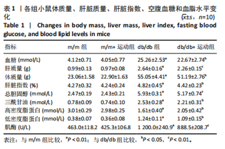
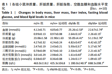
2.1 实验动物数量分析 实验选用db/db小鼠20只,m/m小鼠20只。中途无脱失,全部进入结果分析。 2.2 有氧运动对小鼠体质量、肝脏质量、肝脏指数、空腹血糖和血脂水平的影响 运动12周后,与m/m组和m/m+运动组相比,db/db组小鼠体质量、空腹血糖、肝脏质量和肝脏指数均显著增加(P < 0.01);与db/db组相比,db/db+运动组小鼠体质量、空腹血糖、肝脏质量和肝脏指数均明显下降(P < 0.05,P < 0.05,P < 0.01,P < 0.05)。此外,生化检测结果显示,db/db组小鼠三酰甘油、总胆固醇、低密度脂蛋白、肌酶水平均显著高于m/m组和m/m+运动组(P < 0.01),高密度脂蛋白水平显著低于m/m组和m/m+运动组(P < 0.01);与db/db组相比,db/db+运动组三酰甘油、总胆固醇、低密度脂蛋白、肌酶水平明显降低(P < 0.05,P < 0.01,P < 0.05,P < 0.01),高密度脂蛋白水平明显升高(P < 0.05),见表1。"
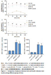
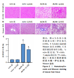
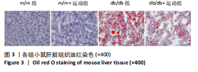
2.3 有氧运动对葡萄糖耐量和胰岛素耐量的影响 与m/m组和m/m+运动组相比,db/db组小鼠出现严重的葡萄糖耐量和胰岛素耐量受损,葡萄糖耐量测试和胰岛素耐量测试的曲线下面积显著增加(P < 0.01)。12周有氧运动干预后,db/db+运动组葡萄糖耐量和胰岛素不耐受均得到改善,葡萄糖耐量测试和胰岛素耐量测试的曲线下面积显著减少(P < 0.01,P < 0.05),见图1。 2.4 各组小鼠肝脏组织苏木精-伊红染色和Masson染色结果 苏木精-伊红染色结果显示,m/m组小鼠的肝脏结构没有明显变化。db/db组小鼠表现出肝细胞膨胀和排列紊乱,边界不清晰,大量炎症细胞浸润,许多肝细胞呈气球状,且存在一些圆形脂质液泡。在12周运动干预后,这些病理变化得到了明显改善,见图2A。除了脂质积累外,肝纤维化是糖尿病诱导肝损伤的另一个主要并发症。应用Masson染色以评估有氧运动对肝纤维化的影响。与m/m组和m/m+运动组相比,db/db小鼠可见明显的胶原纤维沉积,胶原容积分数显著增加(P < 0.01);与db/db组相比,db/db+运动组小鼠肝脏纤维化症状减轻,胶原容积分数明显减少(P < 0.01),见图2B,C。 2.5 各组小鼠肝脏组织油红O染色结果 m/m组和m/m+运动组小鼠肝组织中未见明显红色脂滴,db/db组小鼠肝组织中存在明显的红色脂滴并积累成团,在12周运动干预后,红色脂滴明显减少,见图3。"

"


"

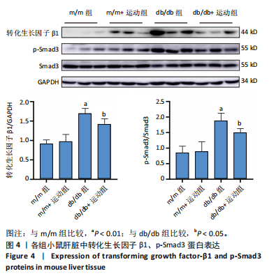
2.6 有氧运动对转化生长因子β1、p-Smad3蛋白表达的影响 转化生长因子β/Smad信号通路是参与肝纤维化的重要细胞内信号转导途径[16]。为了验证有氧运动对糖尿病小鼠肝脏纤维化的改善作用,检测了转化生长因子β1、p-Smad3蛋白表达。结果显示,与m/m组和m/m+运动组相比,db/db组小鼠肝脏中转化生长因子β1、p-Smad3蛋白表达明显增加(P < 0.01);与db/db组相比,db/db+运动组转化生长因子β1、p-Smad3蛋白表达明显下降(P < 0.05),见图4。"

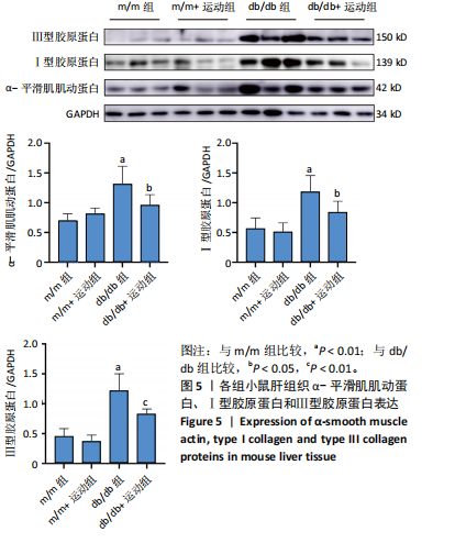
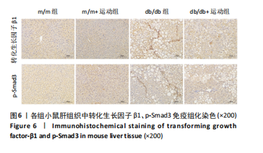
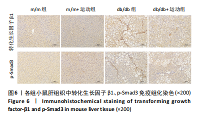
2.7 有氧运动对α-平滑肌肌动蛋白、Ⅰ型胶原蛋白和Ⅲ型胶原蛋白表达的影响 与m/m组和m/m+运动组相比,db/db组小鼠肝脏中α-平滑肌肌动蛋白、Ⅰ型胶原蛋白和Ⅲ型胶原蛋白表达明显增加(P < 0.01);与db/db组相比,db/db+运动组肝脏中α-平滑肌肌动蛋白、Ⅰ型胶原蛋白和Ⅲ型胶原蛋白表达显著下降(P < 0.05,P < 0.05,P < 0.01),见图5。 2.8 各组小鼠肝脏组织中转化生长因子β1、p-Smad3免疫组化结果 db/db组小鼠肝脏中转化生长因子β1、p-Smad3表达明显高于m/m组和m/m+运动组;与db/db组相比,db/db+运动组肝脏中转化生长因子β1、p-Smad3表达明显减少,见图6。"
"
| [1] SUN H, SAEEDI P, KARURANGA S, et al. IDF Diabetes Atlas: Global, regional and country-level diabetes prevalence estimates for 2021 and projections for 2045. Diabetes Res Clin Pract. 2022;183:109119. [2] JIANG Y, SUI D, LI M, et al. Ginsenoside Re Improves Inflammation and Fibrosis in Hepatic Tissue by Upregulating PPARγ Expression and Inhibiting Oxidative Stress in db/db Mice. Evid Based Complement Alternat Med. 2021;2021:9003603. [3] FALCK-YTTER Y, YOUNOSSI ZM, MARCHESINI G, et al. Clinical features and natural history of nonalcoholic steatosis syndromes. Semin Liver Dis. 2001;21(1):17-26. [4] LEE CH, LUI DT, LAM KS. Non-alcoholic fatty liver disease and type 2 diabetes: An update. J Diabetes Investig. 2022;13(6):930-940. [5] GHARBIA S, NAZARIE SR, DINESCU S, et al. Adipose-Derived Stem Cells (ADSCs) Supplemented with Hepatocyte Growth Factor (HGF) Attenuate Hepatic Stellate Cell Activation and Liver Fibrosis by Inhibiting the TGF-β/Smad Signaling Pathway in Chemical-Induced Liver Fibrosis Associated with Diabetes. Cells. 2022;11(21):3338. [6] KIM HY, SAKANE S, EGUILEOR A, et al. The Origin and Fate of Liver Myofibroblasts. Cell Mol Gastroenterol Hepatol. 2024;17(1):93-106. [7] FABREGAT I, CABALLERO-DÍAZ D. Transforming Growth Factor-β-Induced Cell Plasticity in Liver Fibrosis and Hepatocarcinogenesis. Front Oncol. 2018;8:357. [8] DEWIDAR B, MEYER C, DOOLEY S, et al. TGF-β in Hepatic Stellate Cell Activation and Liver Fibrogenesis-Updated 2019. Cells. 2019;8(11): 1419. [9] QIU D, SONG S, CHEN N, et al. NQO1 alleviates renal fibrosis by inhibiting the TLR4/NF-κB and TGF-β/Smad signaling pathways in diabetic nephropathy. Cell Signal. 2023;108:110712. [10] ZHANG Z, LI X, ZHANG J, et al. Chrono-Aerobic Exercise Optimizes Metabolic State in DB/DB Mice through CLOCK-Mitophagy-Apoptosis. Int J Mol Sci. 2022;23(16):9308. [11] MA Y, KUANG Y, BO W, et al. Exercise Training Alleviates Cardiac Fibrosis through Increasing Fibroblast Growth Factor 21 and Regulating TGF-β1-Smad2/3-MMP2/9 Signaling in Mice with Myocardial Infarction. Int J Mol Sci. 2021;22(22):12341. [12] WANG J, POLAKI V, CHEN S, et al. Exercise Improves Endothelial Function Associated with Alleviated Inflammation and Oxidative Stress of Perivascular Adipose Tissue in Type 2 Diabetic Mice. Oxid Med Cell Longev. 2020;2020:8830537. [13] HOFMANN SM, DONG HJ, LI Z, et al. Improved insulin sensitivity is associated with restricted intake of dietary glycoxidation products in the db/db mouse. Diabetes. 2002;51(7):2082-2089. [14] YAO J, ZHAO Y. Lp-PLA2 silencing ameliorates inflammation and autophagy in nonalcoholic steatohepatitis through inhibiting the JAK2/STAT3 pathway. PeerJ. 2023;11:e15639. [15] ZHANG Y, LIU Y, LIU X, et al. Exercise and Metformin Intervention Prevents Lipotoxicity-Induced Hepatocyte Apoptosis by Alleviating Oxidative and ER Stress and Activating the AMPK/Nrf2/HO-1 Signaling Pathway in db/db Mice. Oxid Med Cell Longev. 2022;2022:2297268. [16] ZHANG J, WANG W, CUI X, et al. Ganoderma lucidum ethanol extracts ameliorate hepatic fibrosis and promote the communication between metabolites and gut microbiota g_Ruminococcus through the NF-κB and TGF-β1/Smads pathways. J Ethnopharmacol. 2024;322:117656. [17] WANG Q, JIANG L, WANG J, et al. Abrogation of hepatic ATP-citrate lyase protects against fatty liver and ameliorates hyperglycemia in leptin receptor-deficient mice. Hepatology. 2009;49(4):1166-1175. [18] KANALEY JA, COLBERG SR, CORCORAN MH, et al. Exercise/Physical Activity in Individuals with Type 2 Diabetes: A Consensus Statement from the American College of Sports Medicine. Med Sci Sports Exerc. 2022;54(2):353-368. [19] GENG L, LIAO B, JIN L, et al. Exercise Alleviates Obesity-Induced Metabolic Dysfunction via Enhancing FGF21 Sensitivity in Adipose Tissues. Cell Rep. 2019;26(10):2738-2752.e4. [20] 中华医学会糖尿病学分会.中国2型糖尿病防治指南(2020年版)[J].中华糖尿病杂志,2021,13(4):315-409. [21] ZHOU Z, WANG M, HUANG C, et al. Treadmill exercise training alleviates diabetes-induced depressive-like behavior and cognitive impairment by improving hippocampal CA1 neurons injury in db/db mice. Brain Res Bull. 2022;190:84-96. [22] WANG Y, GUO Y, XU Y, et al. HIIT Ameliorates Inflammation and Lipid Metabolism by Regulating Macrophage Polarization and Mitochondrial Dynamics in the Liver of Type 2 Diabetes Mellitus Mice. Metabolites. 2022;13(1):14. [23] LI X, JIAO Y, XING Y, et al. Diabetes Mellitus and Risk of Hepatic Fibrosis/Cirrhosis. Biomed Res Int. 2019;2019:5308308. [24] 吴玉婷,徐利芬,杨宇石,等. lncRNA-MALAT1通过调节SIRT1介导的肝星状细胞活化调控2型糖尿病并发肝纤维化的机制研究[J].中国免疫学杂志,2022,38(17):2054-2056,2063. [25] MULLUGETA Y, CHAWLA R, KEBEDE T, et al. Dyslipidemia associated with poor glycemic control in type 2 diabetes mellitus and the protective effect of metformin supplementation. Indian J Clin Biochem. 2012;27(4):363-369. [26] XIONG Y, PENG Q, CAO C, et al. Effect of Different Exercise Methods on Non-Alcoholic Fatty Liver Disease: A Meta-Analysis and Meta-Regression. Int J Environ Res Public Health. 2021;18(6):3242. [27] BATALLER R, BRENNER DA. Liver fibrosis. J Clin Invest. 2005;115(2):209-218. [28] LEE UE, FRIEDMAN SL. Mechanisms of hepatic fibrogenesis. Best Pract Res Clin Gastroenterol. 2011;25(2):195-206. [29] ZHANG R, LI W, JIANG X, et al. Ferulic Acid Combined With Bone Marrow Mesenchymal Stem Cells Attenuates the Activation of Hepatic Stellate Cells and Alleviates Liver Fibrosis. Front Pharmacol. 2022;13:863797. [30] HU HH, CHEN DQ, WANG YN, et al. New insights into TGF-β/Smad signaling in tissue fibrosis. Chem Biol Interact. 2018;292:76-83. [31] HANG N, GUO F, SONG Y. HOXC8/TGF-β1 positive feedback loop promotes liver fibrosis and hepatic stellate cell activation via activating Smad2/Smad3 signaling. Biochem Biophys Res Commun. 2023;662:39-46. [32] COLBERG SR, SIGAL RJ, FERNHALL B, et al. Exercise and type 2 diabetes: the American College of Sports Medicine and the American Diabetes Association: joint position statement executive summary. Diabetes Care. 2010;33(12):2692-2696. [33] GUO X, QU FX, ZHANG JD, et al. Amygdalin and exercise training exert a synergistic effect in improving cardiac performance and ameliorating cardiac inflammation and fibrosis in a rat model of myocardial infarction. Appl Physiol Nutr Metab. 2024;49(3):360-374. [34] 扈盛,袁海燕,胡萌,等.中等强度跑台运动干预2型糖尿病模型大鼠肝脏α-SMA和Ⅳ型胶原的变化[J].中国组织工程研究,2021, 25(11):1723-1727. [35] HE H, ZHONG Y, WANG H, et al. Smad3 Mediates Diabetic Dyslipidemia and Fatty Liver in db/db Mice by Targeting PPARδ. Int J Mol Sci. 2023; 24(14):11396. |
| [1] | Yin Lu, Jiang Chuanfeng, Chen Junjie, Yi Ming, Wang Zihe, Shi Houyin, Wang Guoyou, Shen Huarui. Effect of Complanatoside A on the apoptosis of articular chondrocytes [J]. Chinese Journal of Tissue Engineering Research, 2025, 29(8): 1541-1547. |
| [2] | Aikepaer · Aierken, Chen Xiaotao, Wufanbieke · Baheti. Osteogenesis-induced exosomes derived from human periodontal ligament stem cells promote osteogenic differentiation of human periodontal ligament stem cells in an inflammatory microenvironment [J]. Chinese Journal of Tissue Engineering Research, 2025, 29(7): 1388-1394. |
| [3] | Zhang Haojun, Li Hongyi, Zhang Hui, Chen Haoran, Zhang Lizhong, Geng Jie, Hou Chuandong, Yu Qi, He Peifeng, Jia Jinpeng, Lu Xuechun. Identification and drug sensitivity analysis of key molecular markers in mesenchymal cell-derived osteosarcoma [J]. Chinese Journal of Tissue Engineering Research, 2025, 29(7): 1448-1456. |
| [4] | Sun Yuting, Wu Jiayuan, Zhang Jian. Physical factors and action mechanisms affecting osteogenic/odontogenic differentiation of dental pulp stem cells [J]. Chinese Journal of Tissue Engineering Research, 2025, 29(7): 1531-1540. |
| [5] | Yu Ting, Lyu Dongmei, Deng Hao, Sun Tao, Cheng Qian. Icariin pretreatment enhances effect of human periodontal stem cells on M1-type macrophages [J]. Chinese Journal of Tissue Engineering Research, 2025, 29(7): 1328-1335. |
| [6] | Zhao Ruihua, Chen Sixian, Guo Yang, Shi Lei, Wu Chengjie, Wu Mao, Yang Guanglu, Zhang Haoheng, Ma Yong. Wen-Shen-Tong-Du Decoction promoting spinal cord injury repair in mice [J]. Chinese Journal of Tissue Engineering Research, 2025, 29(6): 1118-1126. |
| [7] | Zheng Lin, Jin Wenjun, Luo Shanshan, Huang Rui, Wang Jie, Cheng Yuting, An Zheqing, Xiong Yue, Gong Zipeng, Liao Jian. Eucommia ulmoides promotes alveolar bone formation in ovariectomized rats [J]. Chinese Journal of Tissue Engineering Research, 2025, 29(6): 1159-1167. |
| [8] | Zhang Debao, Wang Peng, Li Kun, Zhang Shaojie, Li Zhijun, Li Shuwen, Wu Yimin. Epidural fibrous scar formation in rabbits following autologous ligamentum flavum intervention [J]. Chinese Journal of Tissue Engineering Research, 2025, 29(6): 1168-1175. |
| [9] | Ji Huihui, Jiang Xu, Zhang Zhimin, Xing Yunhong, Wang Liangliang, Li Na, Song Yuting, Luo Xuguang, Cui Huilin, Cao Ximei. SR9009 combined with indolepropionic acid alleviates inflammation in C2C12 myoblasts through the nuclear factor-kappa B signaling pathway [J]. Chinese Journal of Tissue Engineering Research, 2025, 29(6): 1220-1229. |
| [10] | He Bo, Chen Wen, Ma Suilu, He Zhijun, Song Yuan, Li Jinpeng, Liu Tao, Wei Xiaotao, Wang Weiwei, Xie Jing . Pathogenesis and treatment progress of flap ischemia-reperfusion injury [J]. Chinese Journal of Tissue Engineering Research, 2025, 29(6): 1230-1238. |
| [11] | Zhang Wenhua, Li Xun, Zhang Weichao, Li Xinying, Ma Guoao, Wang Xiaoqiang . Promoting myogenesis based on the SphK1/S1P/S1PR2 signaling pathway: a new perspective on improving skeletal muscle health through exercise [J]. Chinese Journal of Tissue Engineering Research, 2025, 29(6): 1265-1275. |
| [12] | Lan Shuangli, Xiang Feifan, Deng Guanghui, Xiao Yukun, Yang Yunkang, Liang Jie. Naringin inhibits iron deposition and cell apoptosis in bone tissue of osteoporotic rats [J]. Chinese Journal of Tissue Engineering Research, 2025, 29(5): 888-898. |
| [13] | Lang Mecuo, Zhang Yilin, Wang Li. MiR-338-3p affects proliferation and apoptosis of alveolar bone osteoblasts by targeting receptor activator of nuclear factor-kappaB ligand [J]. Chinese Journal of Tissue Engineering Research, 2025, 29(5): 899-907. |
| [14] | Zhi Fang, Zhu Manhua, Xiong Wei, Lin Xingzhen. Analgesic effect of acupuncture in a rat model of lumbar disc herniation [J]. Chinese Journal of Tissue Engineering Research, 2025, 29(5): 936-941. |
| [15] | Xu Tianjie, Fan Jiaxin, Guo Xiaoling, Jia Xiang, Zhao Xingwang, Liu kainan, Wang Qian. Metformin exerts a protective effect on articular cartilage in osteoarthritis rats by inhibiting the PI3K/AKT/mTOR pathway [J]. Chinese Journal of Tissue Engineering Research, 2025, 29(5): 1003-1012. |
| Viewed | ||||||
|
Full text |
|
|||||
|
Abstract |
|
|||||