Chinese Journal of Tissue Engineering Research ›› 2023, Vol. 27 ›› Issue (22): 3573-3579.doi: 10.12307/2023.353
Previous Articles Next Articles
CT study on the normal development and variation of the atlas
Lan Zuozhen1, 2, Qian Yanan3, Chi Jincheng3, Huang Bingxu2, Duan Shaoyin1, 3
- 1The Third Clinical Medical College, Fujian Medical University, Fuzhou 350004, Fujian Province, China; 2Department of Radiology, Xiamen Children’s Hospital, Xiamen 361006, Fujian Province, China; 3Department of Medical Imaging, Zhongshan Hospital of Xiamen University, Xiamen 361004, Fujian Province, China
-
Received:2022-03-09Accepted:2022-05-21Online:2023-08-08Published:2022-11-02 -
Contact:Duan Shaoyin, Professor, Chief physician, MD, The Third Clinical Medical College, Fujian Medical University, Fuzhou 350004, Fujian Province, China; Department of Medical Imaging, Zhongshan Hospital of Xiamen University, Xiamen 361004, Fujian Province, China -
About author:Lan Zuozhen, Physician, The Third Clinical Medical College, Fujian Medical University, Fuzhou 350004, Fujian Province, China; Department of Radiology, Xiamen Children’s Hospital, Xiamen 361006, Fujian Province, China
CLC Number:
Cite this article
Lan Zuozhen, Qian Yanan, Chi Jincheng, Huang Bingxu, Duan Shaoyin. CT study on the normal development and variation of the atlas[J]. Chinese Journal of Tissue Engineering Research, 2023, 27(22): 3573-3579.
share this article
Add to citation manager EndNote|Reference Manager|ProCite|BibTeX|RefWorks
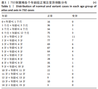
2.1 寰椎CT数据 符合入选标准的寰椎样本732例,其中正常发育679例(92.76%),发育畸形或变异53例,占7.24%,见表1。"
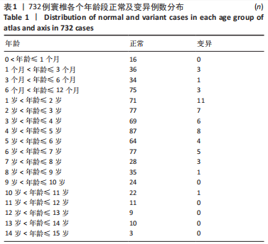
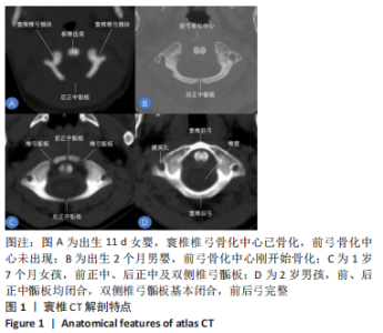
2.2 寰椎骨化中心、骺板影像解剖与特征 寰椎骨化中心共3个,包括椎弓侧块2个和前弓1个;骺板共4个,包括2个椎弓、1个前正中、1个后正中骺板,见图1。"
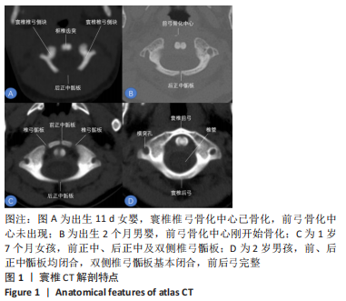
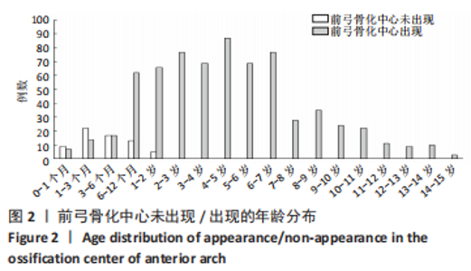
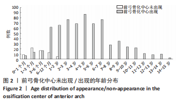
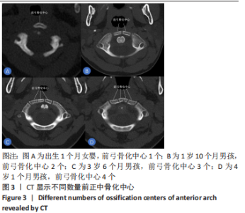
2.3 寰椎骨化中心的出现 双侧椎弓骨化中心出生时均已骨化,出现于胎儿期;前弓骨化中心出现于生后2 d-24个月,年龄中位数为6个月,骨化中心出现的年龄四分位距为4-11个月,见图2。732例数据中前弓骨化中心数量不等,见图3,无前弓有10例,占1.37%;1个骨化中心有458例,占62.57%;2个骨化中心256例,占34.97%;多个骨化中心8例,占1.09%。"
"

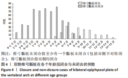
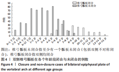
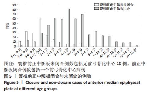
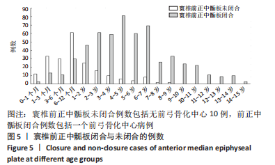
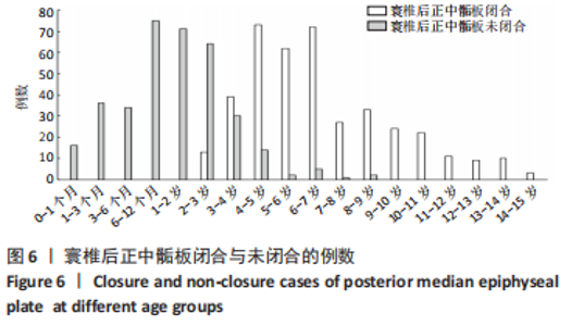
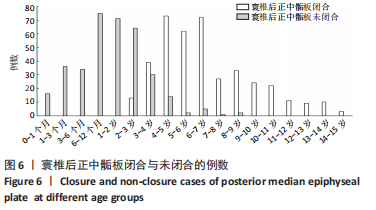
2.4 寰椎骺板闭合 寰椎4个骺板,即椎弓、前正中及后正中骺板。双侧椎弓骺板闭合中位年龄是6.2岁,年龄四分位距为4.5-7岁,见图4,椎弓骺板未闭合的最大年龄是8岁9个月,最小闭合年龄为4岁2个月。前正中骺板:无前弓、2个及以上骨化中心构成前正中骺板,此次试验有264例,前正中骺板出现率为37.43%;骺板闭合年龄中位数为4.8岁,年龄四分位距为2.8-6.0岁,未闭合最大年龄是8岁5个月,闭合的最小年龄为3 d,见图5。后正中骺板闭合:闭合年龄中位数为2.8岁,年龄四分位距为2-3.8岁,后正中骺板未闭合的最大年龄是8岁11个月,闭合的最小年龄是2岁2个月,见图6。"
"
"
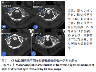
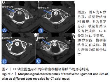
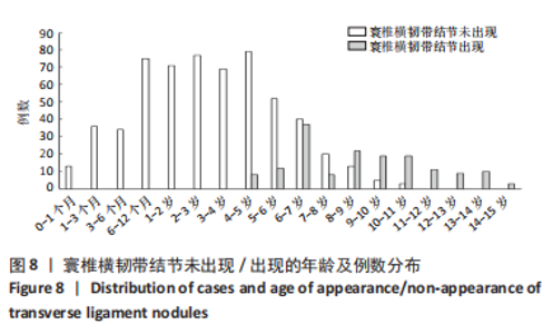
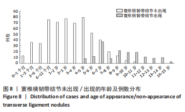
2.5 横韧带结节 横韧带结节在青少年寰椎CT轴位上清晰显示,见图7。横韧带结节出现的中位年龄为9岁,年龄四分位距为5.5-10岁,最小出现年龄为4岁8个月,最大未出现年龄是10岁7个月,见图8。"
"

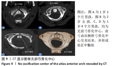
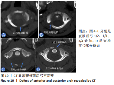
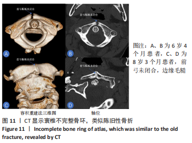
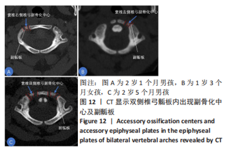
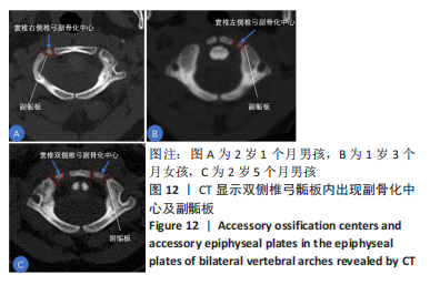
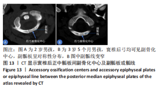
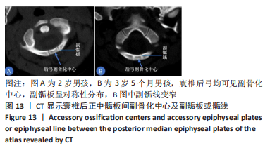
2.6 寰椎变异或畸形 寰椎变异或畸形共53例,包括寰椎骨环不完整27例,内含后正中骺板不闭合即脊柱隐性裂11例、寰椎无前弓(图9)10例、前后弓缺失(图10)4例、寰椎不对称骨环类陈旧性不愈合骨折(图11)4例(含并发2例无前弓病例);副骨化中心及副骺板23例(图12,13);寰枕关节畸形3例(其中1例为Klippel-Feil综合征)。"

"

"
"
"
| [1] BAILEY DK. The normal cervical spine in infants and children. Radiology, 1952;59(5):712-719. [2] MCALLISTER AS, NAGARAJ U, RADHAKRISHNAN R. Emergent Imaging of Pediatric Cervical Spine Trauma. Radiographics. 2019;39(4):1126-1142. [3] CALVY TM, SEGALL HD, GILLES FH, et al. CT anatomy of the craniovertebral junction in infants and children. AJNR Am J Neuroradiol. 1987;8(3):489-494. [4] JIN M, ASADOORIAN M, HILLER LP, et al. Hypertrophy of the anterior arch of the atlas associated with congenital nonunion of the posterior arch: a retrospective case-control study. Spine J. 2014;14(7): 1155-1158. [5] 王华伟.寰椎枕化寰椎侧块形态解剖学研究及有关致病机制的生物力学分析[D].北京:中国人民解放军医学院,2019. [6] 冯会梅,王星,张少杰,等.有限元法分析0-6岁儿童枕寰枢复合体发育及其生物力学的变化特征[J]. 中国组织工程研究,2018, 22(23):3710-3715. [7] 徐雪彬. 数字儿童颈部薄层断面与影像解剖对比及其三维可视化应用研究[D].呼和浩特:内蒙古医科大学,2021. [8] 吴春立,尤壮志,史君,等.学龄前期儿童寰枢椎椎弓根形态特征发育的临床解剖学研究[J].局解手术学杂志,2017,26(8):547-551. [9] AVELLINO AM, MANN FA, GRADY MS, et al. The misdiagnosis of acute cervical spine injuries and fractures in infants and children: the 12-year experience of a level I pediatric and adult trauma center. Childs Nerv Syst. 2005;21(2):122-127. [10] 田忠祥,赵永峰,王丹,等.多层螺旋CT图像后处理技术诊断寰枢椎骨折与脱位价值探讨[J].医学影像学杂志,2018,28(6):1039-1040. [11] DONG L, GE C, XU Z, et al. Kinematic MRI Analysis of Reducible Atlantoaxial Dislocation for Decompression. Biomed Res Int. 2020; 2020:5395071. [12] GIOVANNINI I, ZABOTTI A, CICCIO C, et al. Axial Psoriatic Disease: Clinical and Imaging Assessment of an Underdiagnosed Condition. J Clin Med. 2021;10(13):2845. [13] CHAUDHARY K, DHAWALE A, SHAH A, et al. The technique of using three-dimensional and multiplanar reformatted computed tomography for preoperative planning in pediatric craniovertebral anomalies. N Am Spine Soc J. 2021;7:100073. [14] BOOTH TN. Cervical spine evaluation in pediatric trauma. AJR Am J Roentgenol. 2012;198(5):W417-W425. [15] ELLIOTT S. The odontoid process in children--is it hypoplastic? Clin Radiol. 1988;39(4):391-393. [16] CALVY TM, SEGALL HD, GILLES FH, et al. CT anatomy of the craniovertebral junction in infants and children. AJNR Am J Neuroradiol. 1987;8(3):489-494. [17] 胡贤铧,胡云地,曹志刚,等.齿突游离小骨MSCT和MRI表现特点和临床诊断(附15例报道)[J].医学影像学杂志,2017,27(12): 2276-2279. [18] ROPPER AE. From Anatomic to Genetic Understanding of Developmental Craniovertebral Junction Abnormalities. Neurospine. 2020;17(4):859-861. [19] GROVER PJ, HARRIS LS, THOMPSON D. Craniovertebral junction fixation in children less than 5 years. Eur Spine J. 2020;29(5):961-969. [20] KIM HJ. The Mandible: An Atlas of Osteological and Radiological Anatomy. Anat Cell Biol. 2022;55(1):1-2. [21] NAOUM S, VASILIADIS AV, KOUTSERIMPAS C, et al. Finite Element Method for the Evaluation of the Human Spine: A Literature Overview. J Funct Biomater. 2021;12(3):43. [22] 丁延,宋瑞鹏,蔡一鸣,等.儿童寰椎椎弓根螺钉最佳进钉点及进钉方法[J].中华实验外科杂志,2020,37(11):2114-2116. [23] 于永涛,张少杰,刘颖,等.学龄期儿童寰枢椎数字化三维形态测量研究[J].中国脊柱脊髓杂志,2017,27(1):69-74. [24] LIU C, KAMARA A, YAN Y. Biomechanical study of C1 posterior arch crossing screw and C2 lamina screw fixations for atlantoaxial joint instability. J Orthop Surg Res. 2020;15(1):156. [25] VIOLA A, KOZMA I, SUVEGH D. Surgery for craniovertebral junction pathologies: minimally invasive anterior submandibular retropharyngeal key-hole approach. BMC Surg. 2021;21(1):199. [26] ABUAMARA S, DACHER JN, LECHEVALLIER J. Posterior arch bifocal fracture of the atlas vertebra: a variant of Jefferson fracture. J Pediatr Orthop B. 2001;10(3):201-204. [27] SELB J, OGDEN TM, DUBB J, et al. Comparison of a layered slab and an atlas head model for Monte Carlo fitting of time-domain near-infrared spectroscopy data of the adult head. J Biomed Opt. 2014;19(1):16010. [28] WANG JC, NUCCION SL, FEIGHAN JE, et al. Growth and development of the pediatric cervical spine documented radiographically. J Bone Joint Surg Am. 2001;83(8):1212-1218. [29] SASSON DC. Atlas of Anatomy, Fourth Edition. Plast Reconstr Surg. 2021;147(6):1486. [30] PALANCAR CA, TORRES-TAMAYO N, GARCÍA-MARTÍNEZ D, et al. Comparative anatomy and 3D geometric morphometrics of the El Sidrón atlases (C1). J Hum Evol. 2020;149:102897. [31] LEHMAN VT, BLACK DF, DELONE DR, et al. Current concepts of cross-sectional and functional anatomy of the cerebellum: a pictorial review and atlas. Br J Radiol. 2020;93(1106):20190467. [32] JOAQUIM AF, GHIZONI E, TEDESCHI H, et al. Upper cervical injuries - a rational approach to guide surgical management. J Spinal Cord Med. 2014;37(2):139-151. [33] CHAUHAN AK, CHANDRA PS, GOYAL N, et al. Weak Ligaments and Sloping Joints: A New Hypothesis for Development of Congenital Atlantoaxial Dislocation and Basilar Invagination. Neurospine. 2020; 17(4):843-856. [34] 栾钦花.不同年龄人群寰椎横韧带结节厚度变化规律的CT研究[D].济南:山东大学,2014. [35] DONG L, GE C, XU Z, et al. Kinematic MRI Analysis of Reducible Atlantoaxial Dislocation for Decompression. Biomed Res Int. 2020; 2020:5395071. [36] 李贵林,宋跃明,何兴民,等.寰枢关节旋转运动CT扫描的临床意义[J].临床骨科杂志,2006,9(4):292-294. [37] XU G, WANG D, CHEN J, et al. Spiral CT measurement for atlantoaxial pedicle screw trajectory and its clinical application. Am J Transl Res. 2021;13(4):2555-2562. eCollection 2021. [38] SINGH AK, SHEIKH AI, PANDEY TK, et al. Congenital Mobile Atlantoaxial Dislocation with Cervicomedullary Astrocytoma in Pediatric Patient. Neurol India. 2021;69(1):194-197. [39] MA F, HE H, LIAO Y, et al. Classification of the facets of lateral atlantoaxial joints in patients with congenital atlantoaxial dislocation. Eur Spine J. 2020;29(11):2769-2777. [40] SENOGLU M, SAFAVI-ABBASI S, THEODORE N, et al. The frequency and clinical significance of congenital defects of the posterior and anterior arch of the atlas. J Neurosurg Spine. 2007;7(4):399-402. [41] MCALLISTER AS, NAGARAJ U, RADHAKRISHNAN R. Emergent Imaging of Pediatric Cervical Spine Trauma. Radiographics. 2019;39(4):1126-1142. [42] HA BJ, WON YD, RYU JI, et al. Relationship between the atlantodental interval and T1 slope after atlantoaxial fusion in patients with rheumatoid arthritis. BMC Surg. 2020;20(1):269. [43] LABUDA R, NWOTCHOUANG B, IBRAHIMY A, et al. A new hypothesis for the pathophysiology of symptomatic adult Chiari malformation Type I. Med Hypotheses. 2022;158:110740. [44] SHKARUBO AN, NIKOLENKO VN, CHERNOV IV, et al. [Anatomy of anterior craniovertebral junction in endoscopic transnasal approach]. Zh Vopr Neirokhir Im N N Burdenko. 2020;84(4):46-53. [45] SPINNATO P, ZARANTONELLO P, GUERRI S, et al. Atlantoaxial rotatory subluxation/fixation and Grisel’s syndrome in children: clinical and radiological prognostic factors. Eur J Pediatr. 2021;180(2):441-447. [46] PINI N, CECCOLI M, BERGONZINI P, et al. Grisel’s Syndrome in Children: Two Case Reports and Systematic Review of the Literature. Case Rep Pediatr. 2020;2020:8819758. [47] WENGER KJ, HATTINGEN E, PORTO L. Magnetic Resonance Imaging as the Primary Imaging Modality in Children Presenting with Inflammatory Nontraumatic Atlantoaxial Rotatory Subluxation. Children (Basel). 2021; 8(5):329. [48] 刘利君,彭明惺,唐学阳,等.儿童寰枢椎旋转畸形发病机制的解剖学研究[J].中华小儿外科杂志,2003,24(5):51-53. |
| [1] | Bao Kai, Song Wenhui, Liu Changwen, Liang Kaiheng, Wang Jiajia. Posterior single-segment pedicle screw fixation for unstable atlas fractures [J]. Chinese Journal of Tissue Engineering Research, 2023, 27(4): 594-599. |
| [2] | Pan Baoshun, Fang Zhen, Gao Mingjie, Fang Guiming, Chen Jinshui. Design for posterior atlantoaxial internal fixation system with fusion cage based on imaging data [J]. Chinese Journal of Tissue Engineering Research, 2022, 26(9): 1372-1376. |
| [3] | Li Jian, Guan Tianmin, Zhu Ye. Mechanics analysis of sacral lumbarization based on finite element method [J]. Chinese Journal of Tissue Engineering Research, 2022, 26(33): 5249-5253. |
| [4] | Ren Dong, Zhu Ye, Lei Lei, Wang Yuren. Orthopedic force applied to the rib influences the displacement and rotation angle of thoracic vertebrae: a finite element analysis [J]. Chinese Journal of Tissue Engineering Research, 2022, 26(18): 2812-2816. |
| [5] | Xiong Feng, Li Kun, Zhou Shuyu, Wang Peng, Dang Yexing, Li Zhijun, Zhang Shaojie. Feasibility of atlantoaxial pedicle screw or lateral mass screw fixation in preschool children [J]. Chinese Journal of Tissue Engineering Research, 2021, 25(36): 5804-5809. |
| [6] | Xu Guanghua, Liu Hongyu, Zhang Lifu, Xie Shaoming, Deng Kun, Zhao Changyi. Influence of taekwondo competition on cortical thickness in the calcaneus and stress distribution in the trabecular bone [J]. Chinese Journal of Tissue Engineering Research, 2021, 25(35): 5582-5587. |
| [7] | Li Qiujiang, Fang Xiaomin, Wang Yinbin, Hu Xuehua, Cai Lijun. Current states and tendency of stem cells in the treatment of intervertebral disc degeneration: bibliometric analysis [J]. Chinese Journal of Tissue Engineering Research, 2021, 25(31): 5000-5011. |
| [8] | Cui Qingda, Wang Haige, Zhao Haijun, Liu Wei, Bi Zhenggang. Removal of transepiphyseal plate after fixation for a certain period of time: growth inhibition of epiphyseal plate after a period of observation [J]. Chinese Journal of Tissue Engineering Research, 2020, 24(3): 372-377. |
| [9] | Zhang Bin, Li Zhijun, Ning Pengfei, Liu Ying, Cao Li, Zhang Fengying, Li Xiaohe. Finite element analysis of transoral anterior atlantoaxial plate fixation in a teenager [J]. Chinese Journal of Tissue Engineering Research, 2019, 23(28): 4535-4540. |
| [10] | Gao Yaodong, Duan Yuxing, Guo Pengnian. Finite element analysis of three-dimensional entity reconstruction of hip joint based on CT image and hip bearing capacity data [J]. Chinese Journal of Tissue Engineering Research, 2019, 23(28): 4564-4569. |
| [11] | Wu Chun-li, Zhang Pei. Digital measurement of atlas and axis pedicles with different ages [J]. Chinese Journal of Tissue Engineering Research, 2013, 17(26): 4896-4903. |
| Viewed | ||||||
|
Full text |
|
|||||
|
Abstract |
|
|||||