Chinese Journal of Tissue Engineering Research ›› 2022, Vol. 26 ›› Issue (29): 4651-4657.doi: 10.12307/2022.850
Previous Articles Next Articles
Effects of metformin on cognitive dysfunction in preeclampsia rats
Zhao Junguo1, Chen Xin1, Jiang Ruohan1, Guo Ying1, Gao Bo1, 2
- 1Department of Radiology, The Affiliated Hospital of Guizhou Medical University, Guiyang 550004, Guizhou Province, China; 2Key Laboratory of Brain Imaging, Guizhou Medical University, Guiyang 550001, Guizhou Province, China
-
Received:2021-08-14Accepted:2021-10-12Online:2022-10-18Published:2022-03-28 -
Contact:Gao Bo, MD, Professor, Department of Radiology, The Affiliated Hospital of Guizhou Medical University, Guiyang 550004, Guizhou Province, China; Key Laboratory of Brain Imaging, Guizhou Medical University, Guiyang 550001, Guizhou Province, China -
About author:Zhao Junguo, Master candidate, Department of Radiology, The Affiliated Hospital of Guizhou Medical University, Guiyang 550004, Guizhou Province, China -
Supported by:the National Natural Science Foundation of China (General Program), No. 81871333 (to GB); the Seventh-Batch Thousand Innovation and Enterprising Talent Project of Guizhou Province, No. GZQ202007086 (to GB); Guizhou Provincial Science and Technology Project, No. Qiankehe Support [2020]4Y159 (to GB); the Key Laboratory Construction Project of Guizhou Medical University (to GB)
CLC Number:
Cite this article
Zhao Junguo, Chen Xin, Jiang Ruohan, Guo Ying, Gao Bo. Effects of metformin on cognitive dysfunction in preeclampsia rats[J]. Chinese Journal of Tissue Engineering Research, 2022, 26(29): 4651-4657.
share this article
Add to citation manager EndNote|Reference Manager|ProCite|BibTeX|RefWorks
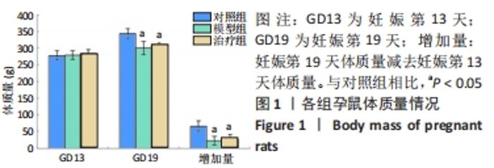
2.1 实验动物数量分析 最终纳入实验为每组7只大鼠,排除6只(其中3只为假孕,在妊娠后期才能发现假孕;3只死亡,其中2只为灌胃时灌胃针误入气管引起窒息死亡,1只为分娩时虚弱无力分娩失败死亡),已重新补入。 2.2 孕鼠体质量情况 妊娠第13天,3组大鼠体质量相比差异无显著性意义(F=0.360 8,P=0.702 1);妊娠第19天,3组间对比差异有显著性意义(F=16.04,P=0.000 1);将妊娠第19天体质量减去妊娠第13天体质量后得到妊娠第13-19天体质量增加量,3组间相比差异有显著性意义(F=20.37,P < 0.000 1);模型组和治疗组大鼠妊娠第19天的体质量均明显低于对照组;模型组和治疗组大鼠妊娠第13-19天体质量增加量显著低于对照组,见图1。"

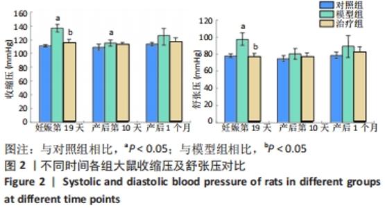
2.3 各组大鼠收缩压和舒张压对比 将妊娠第19天3组大鼠的收缩压和舒张压分别进行单因素方差分析,结果显示收缩压和舒张压在组间对比中差异均有显著性意义(收缩压对比,F=72.32,P < 0.000 1;舒张压对比,F=41.02,P < 0.000 1)。产后第10天3组大鼠对比分析,结果分别为:收缩压F=4.025,P=0.035 9;舒张压F=1.939,P=0.172 8;产后1个月3组间对比结果为:收缩压F=3.764,P=0.043 1;舒张压F=2.562,P=0.104 9。妊娠第19天模型组大鼠的收缩压及舒张压较对照组显著增高,治疗组大鼠收缩压及舒张压较模型组显著降低;产后第10天模型组大鼠收缩压仍较对照组显著增高,见图2。"

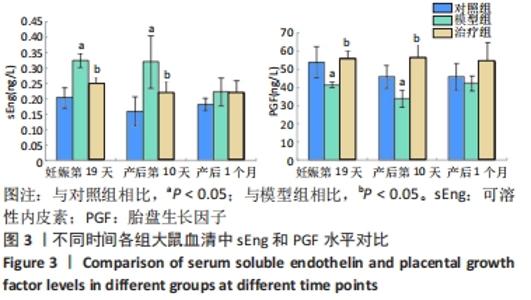
2.4 各组大鼠血清中sEng和PGF水平对比 3组大鼠妊娠第19天和产后第10天分别进行单因素方差分析,大鼠血清中sEng水平差异均有显著性意义(妊娠第19天:F=18.68,P=0.000 3;产后第10天:F=13.02,P=0.000 3);产后1个月3组间进行对比,差异无显著性意义(F=2.249,P=0.134 3)。3组大鼠在妊娠在第19天和产后第10天、产后1个月3个时间点分别进行单因素方差分析,大鼠血清中PGF水平差异均有显著性意义(妊娠第19天:F=6.075,P=0.021 4;产后第10天:F=16.64,P=0.000 3;产后1个月:F=4.037,P=0.036 8)。妊娠第19天和产后第10天模型组大鼠血清中sEng水平显著高于对照组,治疗组大鼠血清中sEng水平显著低于模型组。妊娠第19天和产后第10天模型组大鼠血清中PGF水平显著低于对照组,治疗组大鼠血清中PGF水平显著高于模型组,见图3。"
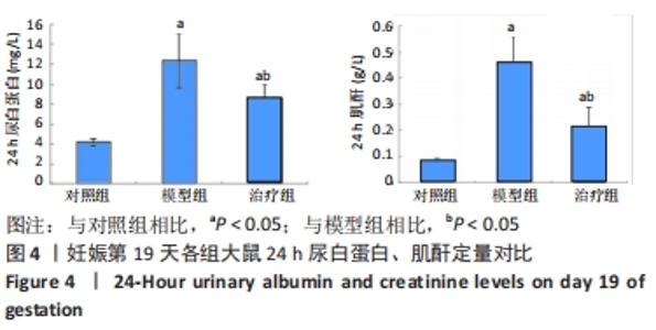
2.5 妊娠第19天24 h尿白蛋白、肌酐定量对比 妊娠第19天将3组的24 h尿白蛋白及肌酐水平分别进行单因素方差分析,3组比较差异均有显著性意义(白蛋白:F=36.000,P < 0.000 1;肌酐:F=41.480,P < 0.000 1)。模型组大鼠24 h尿白蛋白及肌酐均较对照组显著增高;二甲双胍治疗后大鼠尿白蛋白及肌酐较模型组均显著减低,但仍明显高于对照组,见图4。"

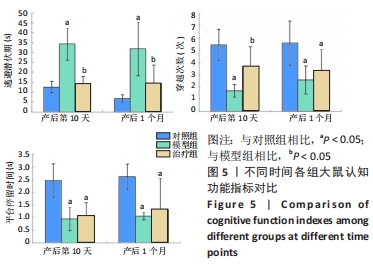
2.6 各组大鼠逃避潜伏期、穿越平台次数及平台逗留时间对比 产后第10天及产后1个月3组潜伏期、穿越平台次数及平台逗留时间对比差异有显著性意义。产后第10天对应的统计量为:潜伏期F=32.570,P < 0.000 1;穿越平台次数F=15.000,P=0.000 2;平台逗留时间F=15.930,P=0.000 1。产后1个月对应的统计量为:潜伏期F=12.480,P=0.000 4;穿越平台次数F=6.604,P=0.007 1;平台逗留时间F=7.527,P=0.005 0。模型组大鼠产后第10天、产后1个月的逃避潜伏期较对照组显著延长;治疗组大鼠产后第10天、产后1个月的逃避潜伏期较模型组显著缩短。模型组大鼠产后第10天及产后1个月的穿越平台次数明显低于对照组,产后第10天治疗组穿越平台次数较模型组显著增多。模型组和治疗组大鼠产后第10天及产后1个月的平台逗留时间较对照组显著缩短,见图5。"
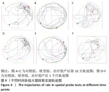
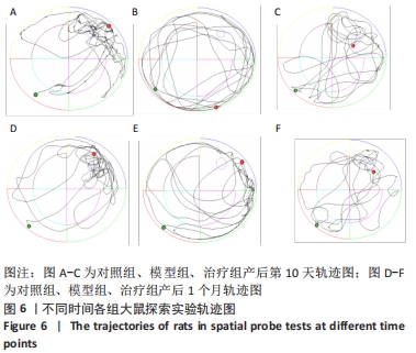
分别从产后第10天及产后1个月各组中各选取1只大鼠(共6只)的探索实验轨迹图以代表每只大鼠所在组别,可以直观地观察到大鼠的运动轨迹,对照组大鼠在平台所在象限运动路程最长,模型组大鼠趋于在池子边缘运动,治疗组大鼠在平台所在象限的运动轨迹较模型组明显增多,见图6。"
| [1] MOL BWJ, ROBERTS CT, THANGARATINAM S, et al. Pre-eclampsia. Lancet. 2016;387(10022):999-1011. [2] WEBSTER K, FISHBURN S, MARESH M, et al. Diagnosis and management of hypertension in pregnancy: summary of updated NICE guidance. BMJ. 2019;366:l5119. [3] CHAIWORAPONGSA T, CHAEMSAITHONG P, YEO L, et al. Pre-eclampsia part 1: current understanding of its pathophysiology. Nat Rev Nephrol. 2014;10(8):466-480. [4] KUMASAWA K. Animal Models in Preeclampsia. Preeclampsia. 2018: 141-155. [5] KOLOVETSIOU-KREINER V, MOERTL MG, PAPOUSEK I, et al. Maternal cardiovascular and endothelial function from first trimester to postpartum. PLoS One. 2018;13(5):e0197748. [6] BAIJNATH S, SOOBRYAN N, MACKRAJ I, et al. The optimization of a chronic nitric oxide synthase (NOS) inhibition model of pre-eclampsia by evaluating physiological changes. Eur J Obstet Gynecol Reprod Biol. 2014;182:71-75. [7] ZHU H, ZHU W, HU R, et al. The effect of pre-eclampsia-like syndrome induced by L-NAME on learning and memory and hippocampal glucocorticoid receptor expression: A rat model. Hypertens Pregnancy. 2017;36(1):36-43. [8] LIU W, QIAO F, LIU H, et al. Low molecular weight heparin improves proteinuria in rats with L-NAME induced preeclampsia by decreasing the expression of nephrin, but not podocin. Hypertens Pregnancy. 2015;34(1):24-35. [9] IJOMONE OK, SHALLIE PD, NAICKER T. Nꭃ-nitro-l-arginine methyl model of pre-eclampsia elicits differential IBA1 and EAAT1 expressions in brain. J Chem Neuroanat. 2019;100:101660. [10] ALONSO-VENTURA V, LI Y, PASUPULETI V, et al. Effects of preeclampsia and eclampsia on maternal metabolic and biochemical outcomes in later life: a systematic review and meta-analysis. Metabolism. 2020; 102:154012. [11] SIA WW, PERTMAN SM, YAN RM, et al. Are Preeclampsia and Adverse Obstetrical Outcomes Predictors of Cardiovascular Disease? A Case-Control Study of Women With Heart Disease. J Obstet Gynaecol Can. 2019;41(12):1760-1767. [12] HEIDEMA WM, SCHOLTEN RR, VAN DRONGELEN J, et al. Metabolic Syndrome After Preeclamptic Pregnancy: A Longitudinal Cohort Study. J Womens Health (Larchmt). 2019;28(3):357-362. [13] WIEGMAN MJ, ZEEMAN GG, AUKES AM, et al. Regional distribution of cerebral white matter lesions years after preeclampsia and eclampsia. Obstet Gynecol. 2014;123(4):790-795. [14] 吴牧熙,沈桂权,高波.先兆子痫对脑结构和功能影响的研究进展[J].磁共振成像,2019,10(12):924-927. [15] FIELDS JA, GAROVIC VD, MIELKE MM, et al. Preeclampsia and cognitive impairment later in life. Am J Obstet Gynecol. 2017;217(1):74.e1-74.e11. [16] IJOMONE OK, SHALLIE P, NAICKER T. Changes in the structure and function of the brain years after Pre-eclampsia. Ageing Res Rev. 2018; 47:49-54. [17] BASIT S, WOHLFAHRT J, BOYD HA. Pre-eclampsia and risk of dementia later in life: nationwide cohort study. BMJ. 2018;363:k4109. [18] 张阳,杨慧,杨柳,等.二甲双胍在妊娠期高血压疾病中的研究现状和前景展望[J].中华妇产科杂志,2020,55(8):567-571. [19] IJOMONE OK, SHALLIE PD, NAICKER T. Oligodendrocytes Death Induced Sensorimotor and Cognitive Deficit in N-nitro-L-arginine methyl Rat Model of Pre-eclampsia. Neurochem Res. 2020;45(4):902-914. [20] HU J, ZHANG J, ZHU B. Protective effect of metformin on a rat model of lipopolysaccharide-induced preeclampsia. Fundam Clin Pharmacol. 2019;33(6):649-658. [21] ZHANG X, LI Q, JIANG W, et al. LAMA5 promotes human umbilical vein endothelial cells migration, proliferation, and angiogenesis and is decreased in preeclampsia. J Matern Fetal Neonatal Med. 2020;33(7): 1114-1124. [22] MARTINEZ-FIERRO ML, HERNADEZ-DELGADILLO GP, FLORES-MENDOZA JF, et al. Fibroblast Growth Factor Type 2 (FGF2) Administration Attenuated the Clinical Manifestations of Preeclampsia in a Murine Model Induced by L-NAME. Front Pharmacol. 2021;12:663044. [23] POSSOMATO-VIEIRA JS, KHALIL RA. Mechanisms of Endothelial Dysfunction in Hypertensive Pregnancy and Preeclampsia. Adv Pharmacol. 2016;77:361-431. [24] NEMA J , SUNDRANI D , JOSHI S . Prenatal vitamin D supplementation reduces blood pressure and improves placental angiogenesis in an animal model of preeclampsia. Food Funct. 2020;11(12):10413-10422. [25] NAVARATNAM K, ABREU P, CLARKE H, et al. Evaluation of agreement of placental growth factor (PlGF) tests and the soluble FMS-like tyrosine kinase 1 (sFlt-1)/PlGF ratio, comparison of predictive accuracy for pre-eclampsia, and relation to uterine artery Doppler and response to aspirin. J Matern Fetal Neonatal Med. 2019;32(2):179-187. [26] BENSCHOP L, SCHALEKAMP-TIMMERMANS S, BROERE-BROWN ZA, et al. Placental Growth Factor as an Indicator of Maternal Cardiovascular Risk After Pregnancy. Circulation. 2019;139(14):1698-1709. [27] POON LC, GALINDO A, SURBEK D, et al. From first-trimester screening to risk stratification of evolving pre-eclampsia in second and third trimesters of pregnancy: comprehensive approach. Ultrasound Obstet Gynecol. 2020;55(1):5-12. [28] CHEN J, KHALIL RA. Matrix Metalloproteinases in Normal Pregnancy and Preeclampsia. Prog Mol Biol Transl Sci. 2017;148:87-165. [29] MARKOVIC S, ROUSSEL T, NEEMAN M, et al. Deuterium Magnetic Resonance Imaging and the Discrimination of Fetoplacental Metabolism in Normal and L-NAME-Induced Preeclamptic Mice. Metabolites. 2021;11(6):376. [30] COATES BJ, BRODERICK TL, BATIA LM, et al. MgSO4 prevents left ventricular dysfunction in an animal model of preeclampsia. Am J Obstet Gynecol. 2006;195(5):1398-1403. [31] ROBERTS TJ, CHAPMAN AC, CIPOLLA MJ. PPAR-gamma agonist rosiglitazone reverses increased cerebral venous hydraulic conductivity during hypertension. Am J Physiol Heart Circ Physiol. 2009;297(4): H1347-1353. [32] YANG X, GUO L, SUN X, et al. Protective effects of hydrogen-rich saline in preeclampsia rat model. Placenta. 2011;32(9):681-686. [33] LI Y, YANG N, WANG B, et al. Effect and mechanism of prophylactic use of tadalafil during pregnancy on l-NAME-induced preeclampsia-like rats. Placenta. 2020;99:35-44. [34] ZHANG Y, LIU X, YANG L, et al. Current Researches, Rationale, Plausibility, and Evidence Gaps on Metformin for the Management of Hypertensive Disorders of Pregnancy. Front Pharmacol. 2020;11: 596145. [35] SONG WY, DING H, DUNN T, et al. Low-dose metformin treatment in the subacute phase improves the locomotor function of a mouse model of spinal cord injury. Neural Regen Res. 2021;16(11):2234-2242. [36] PONIEDZIAŁEK-CZAJKOWSKA E, MIERZYŃSKI R, DŁUSKI D, et al. Prevention of Hypertensive Disorders of Pregnancy-Is There a Place for Metformin? J Clin Med. 2021;10(13):2805. [37] LIU T, HONG L, YANG Y, et al. Metformin reduces proteinuria in spontaneously hypertensive rats by activating the HIF-2α-VEGF-A pathway. Eur J Pharmacol. 2021;891:173731. [38] DENG-BRYANT Y, LEUNG LY, CAUDLE K, et al. Cognitive Evaluation Using Morris Water Maze in Neurotrauma. Methods Mol Biol. 2016; 1462:539-551. [39] WHITING MD, KOKIKO-COCHRAN ON. Assessment of Cognitive Function in the Water Maze Task: Maximizing Data Collection and Analysis in Animal Models of Brain Injury. Methods Mol Biol. 2016; 1462:553-571. [40] WANG HL, LIU FL, LI RQ, et al. Electroacupuncture improves learning and memory functions in a rat cerebral ischemia/reperfusion injury model through PI3K/Akt signaling pathway activation. Neural Regen Res. 2021;16(6):1011-1016. [41] LIU X, ZHAO W, LIU H, et al. Developmental and Functional Brain Impairment in Offspring from Preeclampsia-Like Rats. Mol Neurobiol. 2016;53(2):1009-1019. [42] KUCUK M, UGUR YILMAZ C, ORHAN N, et al. The Effects of Lipopolysaccharide on the Disrupted Blood-Brain Barrier in a Rat Model of Preeclampsia. J Stroke Cerebrovasc Dis. 2018;27(12):3411-3418. [43] YANG X, DING Y, YANG M, et al. Nestin Improves Preeclampsia-Like Symptoms by Inhibiting Activity of Cyclin-Dependent Kinase 5. Kidney Blood Press Res. 2018;43(2):616-627. |
| [1] | Wang Jianping, Zhang Xiaohui, Yu Jinwei, Wei Shaoliang, Zhang Xinmin, Xu Xingxin, Qu Haijun. Application of knee joint motion analysis in machanism based on three-dimensional image registration and coordinate transformation [J]. Chinese Journal of Tissue Engineering Research, 2022, 26(在线): 1-5. |
| [2] | FAN Yaru, LI Ruixin , LI Fengji, LUO Rui, LIU Hao, YAN Yingbin. Characterization and photothermal effect of indocyanine green encapsulated poly lactic acid-co-glycolic acid microspheres [J]. Chinese Journal of Tissue Engineering Research, 2022, 26(在线): 1-6. |
| [3] | Li Huo, Wang Peng, Gao Jianming, Jiang Haoran, Lu Xiaobo, Peng Jiang. Relationship between revascularization and internal microstructure changes in osteonecrosis of the femoral head [J]. Chinese Journal of Tissue Engineering Research, 2022, 26(9): 1323-1328. |
| [4] | Bao Xianguo, Gao Zengxin, Wu Zhanpo, Chen Youmin, Cheng Qinghua, Lu Haitao, Guo Changzheng, Xu Shuai. Correlation between lumbar posterior muscle and local kyphosis in patients with degenerative thoracolumbar kyphosis [J]. Chinese Journal of Tissue Engineering Research, 2022, 26(9): 1418-1423. |
| [5] | Zhang Haobo, Zhao Yunan, Yang Xuejun. Role and therapeutic implications of pyroptosis in intervertebral disc degeneration [J]. Chinese Journal of Tissue Engineering Research, 2022, 26(9): 1445-1451. |
| [6] | Jing Jinpeng, Zhang Yue, Liu Xiaomin, Liu Yi. Traditional Chinese medicine injection for promoting blood circulation in prevention of deep vein thrombosis after orthopedic surgery: network meta-analysis [J]. Chinese Journal of Tissue Engineering Research, 2022, 26(9): 1467-1476. |
| [7] | Gu Zhengqiu, Xu Fei, Wei Jia, Zou Yongdi, Wang Xiaolu, Li Yongming. Exploratory study on talk test as a measure of intensity in blood flow restriction training [J]. Chinese Journal of Tissue Engineering Research, 2022, 26(8): 1154-1159. |
| [8] | Kong Yamin, Yan Juntao, Ma Bingxiang, Li Huawei. Massage vibration intervenes with MyoD expression and proliferation and differentiation of muscle satellite cells in rats with sciatic nerve injury [J]. Chinese Journal of Tissue Engineering Research, 2022, 26(8): 1160-1166. |
| [9] | Wu Cong, Jia Quanzhong, Liu Lun. Relationship between transforming growth factor beta1 expression and chondrocyte migration in adult articular cartilage after fragmentation [J]. Chinese Journal of Tissue Engineering Research, 2022, 26(8): 1167-1172. |
| [10] | Li Zhiyi, He Pengcheng, Bian Tianyue, Xiao Yuxia, Gao Lu, Liu Huasheng. Bibliometric and visualized analysis of ferroptosis mechanism research [J]. Chinese Journal of Tissue Engineering Research, 2022, 26(8): 1202-1209. |
| [11] | Xiang Xinjian, Liu Fang, Wu Liangliang, Jia Daping, Tao Yue, Zhao Zhengnan, Zhao Yu. High-dose vitamin C promotes the survival of autologous fat transplantation in rats [J]. Chinese Journal of Tissue Engineering Research, 2022, 26(8): 1242-1246. |
| [12] | Zhu Chan, Han Xuke, Yao Chengjiao, Zhang Qiang, Liu Jing, Shao Ming. Acupuncture for Parkinson’s disease: an insight into the action mechanism in animal experiments [J]. Chinese Journal of Tissue Engineering Research, 2022, 26(8): 1272-1277. |
| [13] | Tian Chuan, Zhu Xiangqing, Yang Zailing, Yan Donghai, Li Ye, Wang Yanying, Yang Yukun, He Jie, Lü Guanke, Cai Xuemin, Shu Liping, He Zhixu, Pan Xinghua. Bone marrow mesenchymal stem cells regulate ovarian aging in macaques [J]. Chinese Journal of Tissue Engineering Research, 2022, 26(7): 985-991. |
| [14] | Wu Weiyue, Guo Xiaodong, Bao Chongyun. Application of engineered exosomes in bone repair and regeneration [J]. Chinese Journal of Tissue Engineering Research, 2022, 26(7): 1102-1106. |
| [15] | Zhou Hongqin, Wu Dandan, Yang Kun, Liu Qi. Exosomes that deliver specific miRNAs can regulate osteogenesis and promote angiogenesis [J]. Chinese Journal of Tissue Engineering Research, 2022, 26(7): 1107-1112. |
| Viewed | ||||||
|
Full text |
|
|||||
|
Abstract |
|
|||||