Chinese Journal of Tissue Engineering Research ›› 2022, Vol. 26 ›› Issue (15): 2361-2366.doi: 10.12307/2022.591
Previous Articles Next Articles
Imaging analysis of risk factors for knee anterior cruciate ligament injury
Zhang Wei1, 2, Liu Yunpeng1, 2, Wang Xingliang2, Peng Chao1, Hua Guojun2
- 1Wuxi Clinical College of Anhui Medical University, Wuxi 214000, Jiangsu Province, China; 2the 904 Hospital of the Joint Logistics Support Force of Chinese PLA, Wuxi 214000, Jiangsu Province, China
-
Received:2021-07-27Revised:2021-07-29Accepted:2021-09-11Online:2022-05-28Published:2022-01-06 -
Contact:Liu Yunpeng, Chief physician, the 904 Hospital of the Joint Logistics Support Force of Chinese PLA, Wuxi 214000, Jiangsu Province, China -
About author:Zhang Wei, Master candidate, Wuxi Clinical College of Anhui Medical University, Wuxi 214000, Jiangsu Province, China -
Supported by:the Science and Technology Development Fund of Wuxi City, No. CSE31N1618 (to LYP); the Scientific Research Project of Wuxi Municipal Health and Family Planning Commission, No. Q201772 (to WXL)
CLC Number:
Cite this article
Zhang Wei, Liu Yunpeng, Wang Xingliang, Peng Chao, Hua Guojun. Imaging analysis of risk factors for knee anterior cruciate ligament injury[J]. Chinese Journal of Tissue Engineering Research, 2022, 26(15): 2361-2366.
share this article
Add to citation manager EndNote|Reference Manager|ProCite|BibTeX|RefWorks

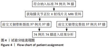
2.1 参与者数量分析 74例患者的影像资料检测结果全部进入结果分析。 2.2 试验分组流程图 见图4。"
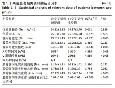
2.3 两组患者相关资料比较 见表1。"

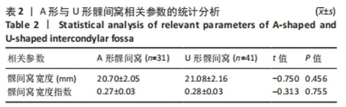
两组患者间体质量指数、年龄、股骨双髁宽度、胫骨髁间隆突内侧高度、胫骨髁间隆突外侧高度相比差异无显著性意义(P > 0.05),前交叉韧带断裂组患者间髁间窝宽度、髁间窝宽度指数、胫骨髁间隆突宽度、胫骨髁间隆突宽度与髁间窝宽度之比低于前交叉韧带完整组(P < 0.05);前交叉韧带断裂组A形髁间窝更常见,前交叉韧带完整组U形髁间窝更常见,两组间髁间窝形态比较差异有显著性意义(P < 0.05)。W形髁间窝由于数量稀少未进行统计学比较。 2.4 不同形态髁间窝的相关参数比较 将A形髁间窝与U形髁间窝的相关参数进行比较,二者的髁间窝宽度、髁间窝宽度指数比较差异无显著性意义(P > 0.05),W形髁间窝由于数量稀少未进行统计学比较,详见表2。"

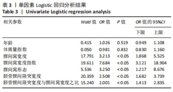
2.5 单因素Logistic回归分析结果 将年龄、体质量指数、髁间窝宽度、髁间窝宽度指数、髁间窝形态、胫骨髁间隆突宽度、胫骨髁间隆突宽度与髁间窝宽度之比纳入单因素Logistic回归分析模型,结果显示髁间窝宽度、髁间窝宽度指数、髁间窝形态、胫骨髁间隆突参数、胫骨髁间隆突宽度与髁间窝宽度之比均与前交叉韧带断裂之间存在相关性(P < 0.05),年龄、体质量指数和前交叉韧带断裂之间不存在相关性(P > 0.05),见表3。"

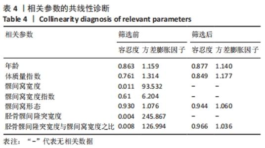
2.6 相关参数共线性诊断结果 见表4。"
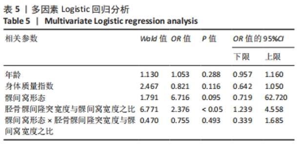
对上述参数进行共线性诊断,发现多个参数之间存在明显的多重共线性,考虑到胫骨髁间隆突宽度与髁间窝宽度之比为其中2个参数的比值,髁间窝宽度指数与髁间窝宽度高度相关,且研究主要探讨胫骨髁间隆突与股骨髁间窝的匹配性对前交叉韧带断裂危险程度的影响,因此这4个参数只保留胫骨髁间隆突宽度与髁间窝宽度之比。筛选之后的相关参数容忍度远大于0.1,方差膨胀因子小于3,可以认为年龄、体质量指数、髁间窝形态和胫骨髁间隆突宽度与髁间窝宽度之比之间不存在多重共线性。 2.7 多因素Logistic回归分析结果 见表5。"
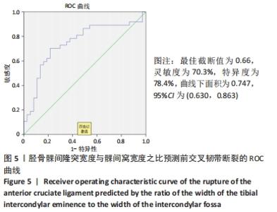
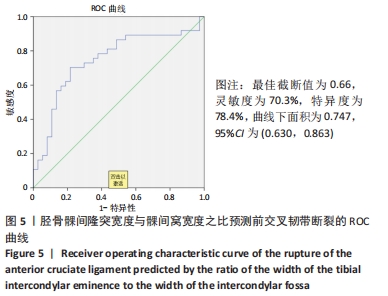
排除多重共线性后,将髁间窝形态、胫骨髁间隆突宽度与髁间窝宽度之比纳入多因素Logistic回归分析模型,年龄和体质量指数虽然在单因素Logistic回归分析中显示与前交叉韧带断裂没有相关性,考虑到既往相关研究依然将其纳入多因素Logistic回归分析模型中。结果显示,胫骨髁间隆突宽度与髁间窝宽度之比是前交叉韧带断裂的危险因素(P < 0.05),引入亚变量髁间窝形态×胫骨髁间隆突宽度与髁间窝宽度之比进行交互作用分析后,结果显示髁间窝形态与胫骨髁间隆突宽度与髁间窝宽度之比之间不存在交互作用(P > 0.05),这表明髁间窝形态不影响胫骨髁间隆突与髁间窝宽度之比与前交叉韧带断裂之间的相关性。 2.8 ROC曲线 利用ROC曲线分析胫骨髁间隆突宽度与髁间窝宽度之比对前交叉韧带断裂的预测价值,结果显示最佳截断值为0.66,灵敏度为70.3%,特异度为78.4%,曲线下面积为0.747,95%CI(0.630,0.863),见图5。"
| [1] KENNEDY JC, WEINBERG HW, WILSON AS. The anatomy and function of the anterior cruciate ligament. As determined by clinical and morphological studies. J Bone Joint Surg Am. 1974;56(2):223-235. [2] SANDERS TL, KREMERS HM, BRYAN AJ, et al. Incidence of Anterior Cruciate Ligament Tears and Reconstruction: A 21-Year Population-Based Study. Am J Sports Med. 2016;44(6):1502-1507. [3] GN B, CENA E, AN B, et al. Anterior cruciate ligament injury risk factors in football: a narrative review. J Sports Med Phys Fitness. 2019;59(10): 1724-1738. [4] KAEDING CC, LÉGER-ST-JEAN B, MAGNUSSEN RA. Epidemiology and Diagnosis of Anterior Cruciate Ligament Injuries. Clin Sports Med. 2017; 36(1):1-8. [5] SOURYAL TO, MOORE HA, EVANS JP. Bilaterality in anterior cruciate ligament injuries: associated intercondylar notch stenosis. Am J Sports Med. 1988;16(5):449. [6] GORMELI CA, GORMELI G, BURAK Y, et al. The effect of the intercondylar notch width index on anterior cruciate ligament injuries: A study on groups with unilateral and bilateral ACL injury. Acta Orthop Belg. 2015;81(2):240-244. [7] SONNERY-COTTET B, ARCHBOLD P, CUCURULO T, et al. The influence of the tibial slope and the size of the intercondylar notch on rupture of the anterior cruciate ligament. J Bone Joint Surg Br. 2011;93(11):1475. [8] LI R, YUAN X, FANG Z, et al. A decreased ratio of height of lateral femoral condyle to anteroposterior diameter is a risk factor for anterior cruciate ligament rupture. BMC Musculoskelet Disord. 2020;21(1): 402-407. [9] BASUKALA B, JOSHI A, PRADHAN I. The Effect of the Intercondylar Notch Shape and Notch Width Index on Anterior Cruciate Ligament Injuries. J Nepal Health Res Counc. 2019;17(45):532-536. [10] 王海蛟,黄竞敏,吴疆,等.股骨髁间窝形态、髁间窝宽度指数与前交叉韧带损伤间关系的相关性研究[J].天津医科大学学报, 2016,22(3):218-221. [11] IRIUCHISHIMA T, GOTO B, FU FH. The occurrence of ACL injury influenced by the variance in width between the tibial spine and the femoral intercondylar notch. Knee Surg Sports Traumatol Arthrosc. 2020;28(11):3625-3630. [12] STURNICK DR, ARGENTIERI EC, VACEK PM, et al. A decreased volume of the medial tibial spine is associated with an increased risk of suffering an anterior cruciate ligament injury for males but not females. J Orthop Res. 2015;32(11):1451-1457. [13] 星月.前交叉韧带撕裂模式及膝关节稳定性的MR定量评估[D].上海:上海交通大学,2020. [14] SOURYAL TO, FREEMAN TR. Intercondylar notch size and anterior cruciate ligament injuries in athletes. A prospective study.Am J Sports Med. 1993;21(4):535-539. [15] ECK C, MARTINS C, VYAS SM, et al. Femoral intercondylar notch shape and dimensions in ACL-injured patients. Knee Surg Sports Traumatol Arthrosc. 2010;18(9):1257-1262. [16] 薛海滨,敖英芳,于长隆,等.前交叉韧带断裂和重建对膝关节软骨退变影响的实验研究[J]. 中华外科杂志,2002,40(4):304-307. [17] BOURAS T, FENNEMA P, BURKE S, et al. Stenotic intercondylar notch type is correlated with anterior cruciate ligament injury in female patients using magnetic resonance imaging. Knee Surg Sports Traumatol Arthrosc. 2018;26(4): 1252-1257. [18] JHA V, PANDIT A. Notch volume measured on MRI is better than two-dimensional notch parameters for predicting non-contact Anterior cruciate ligament injury in males. Arthroscopy. 2021;37(5):1534-1543.e1. [19] ZHANG C, XIE G, FANG Z, et al. Assessment of relationship between three dimensional femoral notch volume and anterior cruciate ligament injury in Chinese Han adults: a retrospective MRI study. Int Orthop. 2019;43(5):1231-1237. [20] WILSON R, BARHORST AA. Intercondylar Notch Impingement of the Anterior Cruciate Ligament: A Cadaveric In Vitro Study Using Robots. J Healthc Eng. 2018;2018:1-27. [21] BEYNNON BD, STURNICK DR, ARGENTIERI EC, et al. A Sex-Stratified Multivariate Risk Factor Model for Anterior Cruciate Ligament Injury. J Athl Train. 2015;50(10):1094-1096. [22] DOMZALSKI M, GRZELAK P, GABOS P. Risk factors for Anterior Cruciate Ligament injury in skeletally immature patients: analysis of intercondylar notch width using Magnetic Resonance Imaging. Int Orthop. 2010;34(5):703-707. [23] 刁硕.非接触性前交叉韧带损伤的解剖学危险因素研究[D].长春:吉林大学,2020. [24] LAPRADE RF, BURNETT QM. Femoral intercondylar notch stenosis and correlation to anterior cruciate ligament injuries. A prospective study. Am J Sports Med. 1994;22(2):198-202. [25] FAHIM SM, DHAWAN T, JAGADEESH N, et al. The relationship of anterior cruciate ligament injuries with MRI based calculation of femoral notch width, notch width index, notch shape - A randomized control study. J Clin Orthop Trauma. 2021;17(172):5-10. [26] GRASSI A, SIGNORELLI C, URRIZOLA F, et al. Patients With Failed Anterior Cruciate Ligament Reconstruction Have an Increased Posterior Lateral Tibial Plateau Slope: A Case-Controlled Study. Arthroscopy. 2019;35(4):1172-1182. [27] CANTIVALLI A, ROSSO F, BONASIA DE, et al. High Tibial Osteotomy and Anterior Cruciate Ligament Reconstruction/Revision. Clin Sports Med. 2019;38(3):417-433. |
| [1] | Pan Baoshun, Fang Zhen, Gao Mingjie, Fang Guiming, Chen Jinshui. Design for posterior atlantoaxial internal fixation system with fusion cage based on imaging data [J]. Chinese Journal of Tissue Engineering Research, 2022, 26(9): 1372-1376. |
| [2] | Yao Xiaoling, Peng Jiancheng, Xu Yuerong, Yang Zhidong, Zhang Shuncong. Variable-angle zero-notch anterior interbody fusion system in the treatment of cervical spondylotic myelopathy: 30-month follow-up [J]. Chinese Journal of Tissue Engineering Research, 2022, 26(9): 1377-1382. |
| [3] | Wu Bingshuang, Wang Zhi, Tang Yi, Tang Xiaoyu, Li Qi. Anterior cruciate ligament reconstruction: from enthesis to tendon-to-bone healing [J]. Chinese Journal of Tissue Engineering Research, 2022, 26(8): 1293-1298. |
| [4] | Yang Kuangyang, Wang Changbing. MRI evaluation of graft maturity and knee function after anterior cruciate ligament reconstruction with autogenous bone-patellar tendon-bone and quadriceps tendon [J]. Chinese Journal of Tissue Engineering Research, 2022, 26(6): 963-968. |
| [5] | Li Jie, Zhang Haitao, Chen Jinlun, Ye Pengcheng, Zhang Hua, Zhou Bengen, Zhao Changqing, Sun Youqiang, Chen Jianfa, Xiang Xiaobing, Zeng Yirong. Anterior cruciate ligament rupture and patellofemoral joint stability before sagittal and axial measurement using MRI [J]. Chinese Journal of Tissue Engineering Research, 2022, 26(6): 969-972. |
| [6] | Xu Kuishuai, Zhang Liang, Chen Jinli, Ren Zhongkai, Zhao Xia, Li Tianyu, Yu Tengbo. Effect of force line changes on lower limb joints after medial open wedge high tibial osteotomy [J]. Chinese Journal of Tissue Engineering Research, 2022, 26(6): 821-826. |
| [7] | Zhang Jian, Lin Jianping, Zhou Gang, Fang Yehan, Wang Benchao, Wu Yongchang. Semi-quantitative MRI evaluation of cartilage degeneration in early knee osteoarthritis [J]. Chinese Journal of Tissue Engineering Research, 2022, 26(3): 425-429. |
| [8] | Liu Yubo, Zhang Huizeng, Zhang Tongrun, Sui Gengyi, Ma Nan, Cheng Xu, Gao Xupeng, Xu Jing, Wang Chaoliang. Correlation analysis between the morphological changes of ankle acupoints and the ankle function after ankle fracture surgery [J]. Chinese Journal of Tissue Engineering Research, 2022, 26(3): 440-445. |
| [9] | Li Lu, Tang Wen. Screw placement in posterior route pedicle on subaxial cervical spine: how to improve the therapeutic effect with the development of artificial intelligence [J]. Chinese Journal of Tissue Engineering Research, 2022, 26(3): 474-479. |
| [10] | Diao Yulei, Zong Xiaorui, Deng Zhibo, Shu Han. Analgesic effect of adductor canal block versus femoral nerve block after autogenous bone-tendon-bone reconstruction of the anterior cruciate ligament: an updated Meta-analysis [J]. Chinese Journal of Tissue Engineering Research, 2022, 26(2): 315-320. |
| [11] | Yuan Haoxiang, Xu Jing, Zeng Jinshu, Chen Hao, Yan Yelei, Chen Jiahao, Liu Qingshan, Xu Fei. Sequence of prevention for anterior cruciate ligament injury: screening, intervention and assessment [J]. Chinese Journal of Tissue Engineering Research, 2022, 26(17): 2775-2781. |
| [12] | Liao Xinyu, He Lu, Li Yanlin, Wang Fuke, Zhou Xiaoxiang, Wang Xu, Zhong Ruiying, Wang Guoliang. Single-bundle anatomical reconstruction of the anterior cruciate ligament with residual ligament stump is beneficial to the recovery of proprioception [J]. Chinese Journal of Tissue Engineering Research, 2022, 26(17): 2631-2635. |
| [13] | Zhao Haifeng, Dai Yiping, Zhu Wenyu, Yan Ke, Wang Yu, Wu Jie, Gu Donghua, Xu Xiaofeng, Cao Longxing, Huang Qiang. Reasons for failure of silica gel and titanium mesh in cranioplasty [J]. Chinese Journal of Tissue Engineering Research, 2022, 26(16): 2522-2525. |
| [14] | Chen Keyi, Wang Dingxuan, Zhao Sike, Xia Zhangrong. Research hotspots of pressure training in rehabilitation and visualized analysis of relevant literature data in the past 10 years [J]. Chinese Journal of Tissue Engineering Research, 2022, 26(15): 2406-2411. |
| [15] | Yuan Cuihua, Lin Wang, Jiang Yao, Liu Chengzhao. Screw track in computer navigation-assisted percutaneous hollow pedicle screw placement [J]. Chinese Journal of Tissue Engineering Research, 2022, 26(12): 1861-1865. |
| Viewed | ||||||
|
Full text |
|
|||||
|
Abstract |
|
|||||