Chinese Journal of Tissue Engineering Research ›› 2022, Vol. 26 ›› Issue (12): 1861-1865.doi: 10.12307/2022.508

Previous Articles     Next Articles

Screw track in computer navigation-assisted percutaneous hollow pedicle screw placement

Yuan Cuihua1, Lin Wang1, Jiang Yao2, Liu Chengzhao1    

  1. 1Department of Orthopedics, Mindong Hospital Affiliated to Fujian Medical University, Fuan 355000, Fujian Province, China; 2School of Clinical Medicine, Medical College of  Soochow University, Suzhou 215000, Jiangsu Province, China
  • Received:2021-03-09 Revised:2021-03-10 Accepted:2021-06-09 Online:2022-04-28 Published:2021-12-14
  • Contact: Lin Wang, Master, Associate chief physician, Department of Orthopedics, Mindong Hospital Affiliated to Fujian Medical University, Fuan 355000, Fujian Province, China Jiang Yao, Master, Chief physician, Doctoral supervisor, Professor, School of Clinical Medicine, Medical College of Soochow University, Suzhou 215000, Jiangsu Province, China
  • About author:Yuan Cuihua, MD, Associate chief physician, Department of Orthopedics, Mindong Hospital Affiliated to Fujian Medical University, Fuan 355000, Fujian Province, China
  • Supported by:
    Fujian Medicine Innovation Project, No. 2020CXA059 (to YCH); Natural Health Industry Joint Fund Project, No. 2017J01395 (to YCH)

Abstract: BACKGROUND: Percutaneous minimally invasive pedicle screw placement has limited the use of minimally invasive percutaneous pedicle screw technology, resulting in clinical confusion caused by different reports on the insertion point and angle of the pedicle screw. At present, when percutaneous pedicle screw technique is used in the treatment of thoracolumbar fracture, most of the screws are placed according to experience or the guidance of intraoperative X-ray repeated fluoroscopy. Thus, the patients and the operators suffer great iatrogenic damage.  
OBJECTIVE: To study the feasibility of percutaneous pedicle screw orbit design respectively from the angle of body anatomy and imaging under the navigation design of precision of the orbit.
METHODS:  (1) Ten bone specimens (T10-L3, a total of 120 vertebral pedicles) were randomly selected, and the preliminary track for pedicle screw was designed based on the best entry points and the best needling angle under computer navigation. The maximum distance from pedicle screw track to the central axis of the vertebral pedicle was measured and recorded. The excellent and good rate of screw placement was counted to analyze the accuracy of the screw track under navigation. (2) Ten spinal bones (T10-L3, a total of 60 vertebral pedicles) were randomly selected. Cases treated with computer navigation-assisted percutaneous pedicle screw placement were set as experimental group. Meanwhile, cases treated with screw placement method under direct vision with naked eyes were set as control group. The excellent and good rate of screw placement among the bone specimens of corpses in both groups was counted and analyzed, separately.  
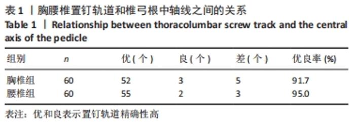
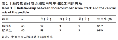
RESULTS AND CONCLUSION: (1) The measurement of 60 vertebral pedicles in the thoracic spine group and the design of pedicle screw track: There were 52 excellent cases, 3 good cases, and 5 poor cases, with an excellent and good rate of 91.67%. The measurement of 60 vertebral pedicles in the lumbar spine group: There were 55 excellent cases, 2 good cases, and 3 poor cases, with an excellent and good rate of 95.00%. No significant difference in the excellent and good rate was detected between thoracic and lumbar spines (P > 0.05). (2) CT scanning was carried out on a total of 30 vertebral pedicles in the navigation experimental group, with an excellent and good rate of screw placement of 96.67%. A total of 30 vertebral pedicles were scanned in the control group, with an excellent and good rate of screw placement of 90.00%. No significant difference in the excellent and good rate was detected between these two groups (P > 0.05). (3) The screw track of computer navigation-assisted percutaneous pedicle screw placement for thoracic and lumbar spines is extremely close to the central axis of the vertebral pedicle or located at the central axis of the vertebral pedicle, and the pedicle screw track designed under navigation is ideal. The excellent and good rate of screw placement among the bone specimens of corpses shows no apparent difference under navigation and direct vision with naked eyes. Therefore, it can be inferred that the pedicle screw track designed with computer navigation-assisted percutaneous placement is feasible.

Key words: computer, navigation, anatomy, minimally invasive, pedicle screw

CLC Number: