[1] 王兵, 熊一静, 罗倩, 等. 儿童及青少年不同骨面型颞下颌关节的研究进展[J]. 口腔医学,2024,44(6):475-480.
[2] MCKIMPSON WM, SPIEGEL S, MUKHANOVA M, et al. Calorie restriction activates a gastric Notch-FOXO1 pathway to expand ghrelin cells. J Cell Biol. 2024;223(10): e202305093.
[3] 彭慧敏, 裴婷婷, 刘畅. 下颌髁突软骨生长发育相关信号通路的研究进展[J]. 口腔生物医学,2018,9(1):53-57.
[4] BENDER ME, LIPIN RB, GOUDY SL. Development of the Pediatric Temporomandibular Joint. Oral Maxillofac Surg Clin North Am. 2018;30(1):1-9.
[5] BECHTOLD TE, SAUNDERS C, DECKER RS, et al. Osteophyte formation and matrix mineralization in a TMJ osteoarthritis mouse model are associated with ectopic hedgehog signaling. Matrix Biol. 2016;52-54:339-354.
[6] BECHTOLD TE, KURIO N, NAH HD, et al. The Roles of Indian Hedgehog Signaling in TMJ Formation. Int J Mol Sci. 2019;20(24):6300.
[7] 李松. 下颌髁状突软骨与生长板软骨生长发育的比较研究[J]. 昆明医科大学学报,2014,35(2):1-4.
[8] METZ CW, BRIDGES CB. Incompatibility of Mutant Races in Drosophila. Proc Natl Acad Sci U S A. 1917;3(12):673-678.
[9] ANDERSSON ER, SANDBERG R, LENDAHL U. Notch signaling: simplicity in design, versatility in function. Development. 2011;138(17):3593-3612.
[10] 王雪淞, 周林, 李林材, 等. Notch信号通路调控间充质干细胞的增殖与分化[J]. 中国组织工程研究,2024,28(19):3076-3083.
[11] 李婷, 程亚楠, 周小平. 阻断Notch信号通路后补益营卫方对衰老皮肤表皮干细胞Notch1、Jagged1、RBP-Jκ和Hes1表达的影响[J]. 中国老年学杂志, 2023,43(4):876-880.
[12] ZACK SR, MEYER A, ZANOTTI B, et al. Notch ligands are biomarkers of anti-TNF response in RA patients. Angiogenesis. 2024;27(2):273-283.
[13] KRISHNA BM, JANA S, SINGHAL J, et al. Notch signaling in breast cancer: From pathway analysis to therapy. Cancer Lett. 2019;461:123-131.
[14] SERRANO MJ, SO S, HINTON RJ. Roles of notch signalling in mandibular condylar cartilage. Arch Oral Biol. 2014;59(7):735-740.
[15] YAN F, FENG J, YANG L, et al. The effect induced by alternated mechanical loading on Notch-1 in mandibular condylar cartilage of growing rabbits. Bone Joint Res. 2021;10(7):437-444.
[16] DOWTHWAITE GP, BISHOP JC, REDMAN SN, et al. The surface of articular cartilage contains a progenitor cell population. J Cell Sci. 2004;117(Pt 6):889-897.
[17] RUTKOWSKI TP, KOHN A, SHARMA D, et al. HES factors regulate specific aspects of chondrogenesis and chondrocyte hypertrophy during cartilage development. J Cell Sci. 2016;129(11):2145-2155.
[18] MATSUMOTO K, LUTHER KB, HALTIWANGER RS. Diseases related to Notch glycosylation. Mol Aspects Med. 2021;79:100938.
[19] LOGEAT F, BESSIA C, BROU C, et al. The Notch1 receptor is cleaved constitutively by a furin-like convertase. Proc Natl Acad Sci U S A. 1998;95(14):8108-8112.
[20] LUCA VC, JUDE KM, PIERCE NW, et al. Structural biology. Structural basis for Notch1 engagement of Delta-like 4. Science. 2015;347(6224): 847-853.
[21] KIM GS, PARK HS, LEE YC. OPTHiS Identifies the Molecular Basis of the Direct Interaction between CSL and SMRT Corepressor. Mol Cells. 2018; 41(9):842-852.
[22] SHI Q, XUE C, ZENG Y, et al. Notch signaling pathway in cancer: from mechanistic insights to targeted therapies. Signal Transduct Target Ther. 2024;9(1):128.
[23] SANALKUMAR R, DHANESH SB, JAMES J. Non-canonical activation of Notch signaling/target genes in vertebrates. Cell Mol Life Sci. 2010;67(17):2957-2968.
[24] JENSEN CH, JOHNSEN RH, ESKILDSEN T, et al. Pericardial delta like non-canonical NOTCH ligand 1 (Dlk1) augments fibrosis in the heart through epithelial to mesenchymal transition. Clin Transl Med. 2024;14(2):e1565.
[25] LIU L, ZHANG L, ZHAO S, et al. Non-canonical Notch Signaling Regulates Actin Remodeling in Cell Migration by Activating PI3K/AKT/Cdc42 Pathway. Front Pharmacol. 2019;10:370.
[26] CHEN S, LAN L, LEI J, et al. Gli1+ Osteogenic Progenitors Contribute to Condylar Development and Fracture Repair. Front Cell Dev Biol. 2022;10:819689.
[27] DENG Z, LIU Y, WANG C, et al. Involvement of PI3K/Akt pathway in rat condylar chondrocytes regulated by PTHrP treatment. Arch Oral Biol. 2014;59(10):1032-1041.
[28] 徐谣, 李文晋. Hedgehog信号通路在下颌骨发育过程中作用机制的研究进展[J]. 口腔疾病防治,2024,32(9):709-714.
[29] JING Y, ZHOU X, HAN X, et al. Chondrocytes Directly Transform into Bone Cells in Mandibular Condyle Growth. J Dent Res. 2015;94(12):1668-1675.
[30] RUSCITTO A, SCARPA V, MOREL M, et al. Notch Regulates Fibrocartilage Stem Cell Fate and Is Upregulated in Inflammatory TMJ Arthritis. J Dental Res. 2020; 99(10):1174-1181.
[31] DONG Y, JESSE AM, KOHN A, et al. RBPjkappa-dependent Notch signaling regulates mesenchymal progenitor cell proliferation and differentiation during skeletal development. Development. 2010;137(9):1461-1471.
[32] MEAD TJ, YUTZEY KE. Notch pathway regulation of chondrocyte differentiation and proliferation during appendicular and axial skeleton development. Proc Natl Acad Sci U S A. 2009;106(34):14420-14425.
[33] TAKABAYASHI H, JI T, PENG L, et al. Regulation of Parietal Cell Homeostasis by Bone Morphogenetic Protein Signaling. Gastro Hep Adv. 2023;2(2):221-231.
[34] SHANG X, WANG J, LUO Z, et al. Notch signaling indirectly promotes chondrocyte hypertrophy via regulation of BMP signaling and cell cycle arrest. Sci Rep. 2016; 6:25594.
[35] 庄欠志, 武秀萍. 甲状旁腺激素相关蛋白在下颌骨髁突软骨生长发育中的作用机制[J]. 口腔医学研究,2021,37(9):791-793.
[36] DELGADO-CALLE J, MCANDREWS K, WU G, et al. The Notch pathway regulates the bone gain induced by PTH anabolic signaling. Faseb J. 2022;36(3):e22196.
[37] FUENTES MA, OPPERMAN LA, BELLINGER LL, et al. Regulation of cell proliferation in rat mandibular condylar cartilage in explant culture by insulin-like growth factor-1 and fibroblast growth factor-2. Arch Oral Biol. 2002;47(9):643-654.
[38] SERRANO MJ, SO S, SVOBODA KK, et al. Cell fate mediators Notch and Twist in mouse mandibular condylar cartilage. Arch Oral Biol. 2011;56(6):607-613.
[39] ISHIDA T, YABUSHITA T, SOMA K. Effects of a liquid diet on temporomandibular joint mechano-receptors. Journal of dental research, 2009;88(2):187-191.
[40] 刘晨燕, 冯剑颖, 谷志远. 咀嚼负荷对幼兔髁突软骨细胞BrdU标记影响的研究[J]. 口腔医学,2015,35(4):250-253+286.
[41] WEI J, WANG Y, TU S, et al. Circadian rhythm disruption upregulating Per1 in mandibular condylar chondrocytes mediating temporomandibular joint osteoarthritis via GSK3β/β-CATENIN pathway. J Transl Med. 2024;22(1):662.
[42] LU K, MA F, YI D, et al. Molecular signaling in temporomandibular joint osteoarthritis. J Orthop Translat. 2022;32:21-27.
[43] MAHJOUB M, SASSI N, DRISS M, et al. Expression patterns of Notch receptors and their ligands in human osteoarthritic and healthy articular cartilage. Tissue Cell. 2012;44(3):182-194.
[44] 兰玲莉, 张伟娜, 蒋扬眉, 等. Notch信号通路在颞下颌骨关节炎中的表达[J]. 口腔疾病防治,2017,25(3):143-152.
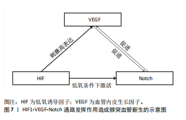
[45] CHEN Y, ZHAO B, ZHU Y, et al. HIF-1-VEGF-Notch mediates angiogenesis in temporomandibular joint osteoarthritis. Am J Transl Res. 2019;11(5):2969-2982.
[46] MORENO-DOMíNGUEZ A, COLINAS O, ARIAS-MAYENCO I, et al. Hif1α-dependent mitochondrial acute O(2) sensing and signaling to myocyte Ca(2+) channels mediate arterial hypoxic vasodilation. Nat Commun. 2024;15(1):6649.
[47] RIBATTI D, D’AMATI A. Bone angiocrine factors. Front Cell Dev Biol. 2023;11:1244372.
[48] SHIRAKURA M, TANIMOTO K, EGUCHI H, et al. Activation of the hypoxia-inducible factor-1 in overloaded temporomandibular joint, and induction of osteoclastogenesis. Biochem Biophys Res Commun. 2010;393(4):800-805.
[49] YANG H, ZHOU Y, YING B, et al. Effects of human umbilical cord mesenchymal stem cell-derived exosomes in the rat osteoarthritis models. Stem Cells Transl Med. 2024;13(8):803-811.
[50] AIDA Y, MAENO M, SUZUKI N, et al. The effect of IL-1beta on the expression of matrix metalloproteinases and tissue inhibitors of matrix metalloproteinases in human chondrocytes. Life Sci. 2005;77(25):3210-3221.
[51] LIU L, CHEN Y, HAN Y, et al. Qing Hua Chang Yin ameliorates chronic colitis in mice by inhibiting PERK-ATF4-CHOP pathway of ER stress and the NF-κB signalling pathway. Pharm Biol. 2024;62(1):607-620.
[52] Retraction: Down-regulation of Notch-1 Inhibits Invasion by Inactivation of Nuclear Factor-κB, Vascular Endothelial Growth Factor, and Matrix Metalloproteinase-9 in Pancreatic Cancer Cells. Cancer Res. 2018;78(18):5476.
[53] QIU J, HUA B, YE X, et al. Intra-articular injection of kartogenin promotes fibrocartilage stem cell chondrogenesis and attenuates temporomandibular joint osteoarthritis progression. Front Pharmacol. 2023;14:1159139. |