Chinese Journal of Tissue Engineering Research ›› 2012, Vol. 16 ›› Issue (49): 9241-9246.doi: 10.3969/j.issn.2095-4344.2012.49.022
Previous Articles Next Articles
In vitro establishment of a neural stem cell aging model and investigation of its biological characteristics
Peng Bin1, 2, Wang Chao-li2, Feng Li2, Wang Ya-ping1
- 1Laboratory of Stem Cell and Tissue Engineering, Chongqing Medical University, Chongqing 400016, China; 2Cytochemical Research Laboratory, North Sichuan Medical College, Nanchong 637007, Sichuan Province, China
-
Received:2012-03-07Revised:2012-04-09Online:2012-12-02Published:2013-01-16 -
Contact:, Professor, Doctoral supervisor, Laboratory of Stem Cell and Tissue Engineering, Chongqing Medical University, Chongqing 400016, China ypwangcq@yahoo.cn -
About author:Peng Bin☆, Doctor, Associate professor, Laboratory of Stem Cell and Tissue Engineering, Chongqing Medical University, Chongqing 400016, China, Cytochemical Research Laboratory, North Sichuan Medical College, Nanchong 637007, Sichuan Province, China binpeng0817@foxmail.com -
Supported by:Project of Education of Sichuan Province, No. 10ZA176*
CLC Number:
Cite this article
Peng Bin, Wang Chao-li, Feng Li, Wang Ya-ping. In vitro establishment of a neural stem cell aging model and investigation of its biological characteristics[J]. Chinese Journal of Tissue Engineering Research, 2012, 16(49): 9241-9246.
share this article
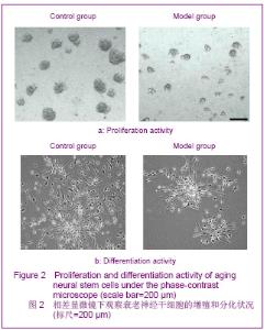
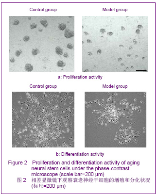
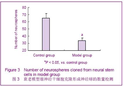
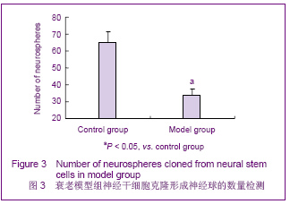
2.1 神经干细胞鉴定 分离纯化的第3代神经球呈明显的Nestin阳性染色。 2.2 衰老模型组神经干细胞的增殖能力 与对照组比较,衰老模型组神经干细胞经100 μmol/L t-BHP诱导 2 h,倒置显微镜下观察其增殖形成神经球的数量和体积均显著下降,见图2;MTT法检测其吸光度值较对照组显著下降了26%,神经球计数显示其克隆形成的神经球的数量显著下降了48%,见图3。 2.3 衰老模型组神经干细胞分化能力检测 衰老模型组神经干细胞经100 μmol/L t-BHP诱导2 h,再用含体积分数10%小牛血清的DMEM/F12诱导其分化培养7 d后,与对照组比较,其分化形成的神经元的数密度[(66.7±3.9)/mm2]较对照组[(172.3±12.2)/mm2]显著降低了61%(P < 0.05),见图2。 "
"
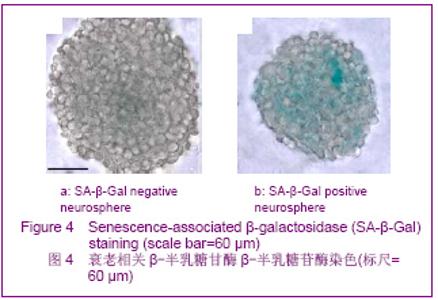
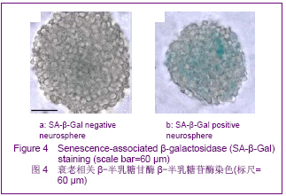
2.4 衰老模型组SA-β-半乳糖苷酶染色阳性神经球百分比 β-半乳糖甘酶染色显示阳性细胞着蓝色,见图4。衰老模型组神经干细胞经100 μmol/L t-BHP诱导2 h后继续培养7 d,其克隆形成的神经球较小,且其中β-半乳糖苷酶阳性神经球百分比(70.5±4.8)%较对照组(3.41± 0.3)%显著增加了19倍(P < 0.05)。"
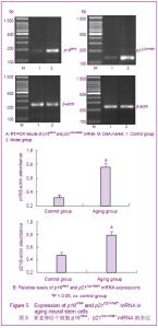
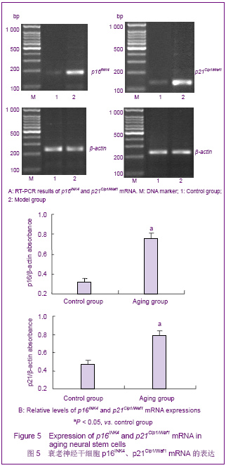
2.5 衰老模型组神经干细胞p16INK4a、p21Cip1/Waf1 mRNA的表达水平 见图5。 RT-PCR结果显示p16INK4a、p21Cip1/Waf1基因在对照组和衰老模型组均有表达。利用p16INK4a、p21Cip1/Waf1 mRNA与β-actin灰度比值作为对照组与衰老模型组p16INK4a、p21Cip1/Waf1相对表达水平。对照组p16INK4a、p21Cip1/Waf1基因的表达明显很低,衰老模型组p16INK4a、p21Cip1/Waf1基因的相对表达水平较对照组分别显著升高了137%和68%"
| [1] Wang YP, Wu H, Wang JW, et al. Beijing Kexue Chubanshe. 2009:1-22.王亚平,吴宏,王建伟,等.干细胞衰老与疾病[M].北京:科学出版社, 2009:1-22.[2] Rossi DJ, Bryder D, Weissman IL. Hematopoietic stem cell aging: mechanism and consequence. Experimental Gerontology. 2007;42(5):385-390.[3] Gazit R,Weissman H,Rossi DJ.Hematopoietic stem cells and the aging hematopoietic system. Semin Hematol. 2008; 45(4): 218-224.[4] Gu YZ, Qiang YZ, Ni XT, et al. Zhongguo Zuzhigongcheng Yanjiu Yu Linchuang Kangfu. 2008, 12(34):6731-6734.古彦铮,强亦忠,倪祥庭,等.干细胞衰老学说[J].中国组织工程研究与临床康复,2008, 12(34):6731-6734.[5] Yu ZJ. Zhongguo Shiyong Yiyao. 2008;3(28):180-183. 余资江.脑衰老与神经干细胞移植治疗研究进展[J].中国实用医药, 2008, 3(28):180-183.[6] Duan JH, Zhu XL, Li XJ. Guoji BingliKexue Yu Linchuang Zazhi. 2011,31(4):291-295.段建辉,祝晓玲,李学军.神经干细胞衰老基础与临床研究进展[J]. 国际病理科学与临床杂志,2011,31(4):291-295.[7] Olariu A, Cleaver KM, Cameron HA. Decreased neurogenesis in aged rats results from loss of granule cell precursors without lengthening of the cell cycle. J. Comp. Neurol. 2007; 501(4): 659-667.[8] Rao MS,Hattiangady B,Shetty AK.The window and mechanisms of major age-related decline in the production of new neurons within the dentate gyrus of the hippocampus. Aging Cell.2006;5 (6):545-558.[9] Zhao CH, Chen XC, Jin JS, et al. Zhongguo Shenglibingli Zazhi. 2005;21(2):238-242.赵朝晖,陈晓春,金建生,等.三丁基过氧化氢诱导WI-38细胞衰老的细胞周期调控机制[J].中国生理病理杂志, 2005,21(2):238- 242.[10] Zhou Y, Yang B, Yao X, et al. Establishment of an aging model of Sca-1+ hematopoietic stem cell and studies on its relative biological mechanisms. In Vitro Cell Dev Biol Anim. 2011; 47(2):149-156.[11] Zhou Y, Yao B, Yao X, et al. Jiepou Xuebao. 2010;41(5):699- 705.周玥,杨斌,姚欣,等.小鼠造血干细胞体外衰老模型的构建及相关生物学研究[J].解剖学报,2010, 41(5):699-705.[12] Gu EY, Ha F, Li LF, et al. Zhongguo Zuzhigongcheng Yanjiu Yu Linchuang Kangfu. 2009,13(10):1948-1950.顾恩妍,哈福,李林凤,等.神经干细胞的鉴定方法[J].中国组织工程研究与临床康复,2009,13(10):1948-1950.[13] Tan XJ, Hu CL, Cai WQ. Guoji Naoxueguan Jibing Zazhi. 2006;14(2):107-109.谭新杰,胡长林,蔡文琴.新生大鼠海马神经干细胞的分离、培养、分化和鉴定[J].国际脑血管病杂志,2006,14(2):107-109.[14] Itoh T, Satou T, H ash im oto S, et a.l Isolation of neu ral stem cells from dam aged rat cerebral cortex after traum at ic b rain in ju ry. Neu roreport. 2005;16: 1687-1691.[15] Peng B, Wang CL, Feng L, et al. Zhongguo Xibao Shengwuxue Xuebao. 2011;33(10): 1116-1122.彭彬,王朝丽,冯丽,等.人参皂苷Rg1调控神经干细胞衰老作用及机制研究[J].中国细胞生物学学报,2011,33(10):1116-1122.[16] Zhao L, Zhang ZY, Tong TJ. Shengli Kexue Jinzhan. 2000; 31(3):205-210.赵亮,张宗玉,童坦君.生物体衰老与复制衰老——体内与体外研究[J].生理科学进展,2000, 31(3):205-210.[17] Rafalski VA, Brunet A. Energy metabolism in adult neural stem cell fate. Prog Neurobiol.2011;93( 2): 182- 203.[18] Pendergrass WR, Lane MA, Bodkin NL, et al. Cellular proliferation potential during aging and caloric restriction in rhesus monkeys (M acaca mulatta). J Cell Physio l.1999;180 (1):123-130.[19] Wang Y, Schulte BA, LaRue AC, et al. Total body irradiation selectively induces murine hematopoietic stem cell senescence. Blood. 2006;107(1):358-366.[20] Meng A, Wang Y, Brown SA, et al. Ionizing radiation and busulfan inhibit murine bone marrow cell hematopoietic function via apoptosis-dependent and independent mechanisms. Experimental Hematology.2003;31(12):1348- 1356.[21] Zheng WJ, Tong TJ, Zhang ZY. Shengmingdehuaxue. 2002; 22(4) :314- 316.郑文婕,童坦君,张宗玉.细胞衰老的重要通路:p16INK/ Rb和p19ARF/ p53/ p21Cip1信号途径[J].生命的化学, 2002,22(4): 314-316.[22] Shilkaitis A, Green A, Punj V, et al. Dehydroepiandrosterone inhibits the progression phase of mammary carcinogenesis by inducing celhlar senescence via a p16 - dependent but p53- independent. Breast Cancer Res.2005;7(6):R1132-1140.[23] Molofsky AV, Slutsky SG, Joseph NM, et al. Increasing p16INK4a expression decreases forebrain progenitors and neurogenesis during aging. Nature.2006; 443(7110):448-452.[24] Janakiraman K, Chad T, Matthew R, et al. Ink4a/Arf expression is a biomarker of aging. J Clin Invest.2004; 114(9): 1299-1307.[25] Guney I,Wu S,Sedivy JM.Reduced c-Myc signaling triggers telomereindependent senescence by regulating Bmi 1 and p16 (INK4a). Proc Natl Acad Sci USA.2006;103(10): 3645-3650.[26] Cammarano MS, Nekrasova T, Noel B, et al. Pak4 induces premature senescence via a pathway requiring p16 INK4/P19 ARF and mitogen activated protein kinase signaling. Mol Cell Biol.2005;25(21): 9532-9542.[27] Gong YH, Yue JP, Wu XD, et al. NSPc1 is a cell growth regulator that acts as a transcriptional repressor of p21Waf1/Cip1 via the RACE element. Nucleic Acids Res. 2006;34(21): 6158-1669. |
| [1] | Wang Quansheng, Chen Songtao, Zhang Yongqing, Qiao Hua. Preparation and performance evaluation of essence with mesenchymal stem cell conditioned medium [J]. Chinese Journal of Tissue Engineering Research, 2026, 30(31): 8092-8099. |
| [2] | Zhang Xiaoxu, Tian Zhenli, Xie Tingting. Roles of pregnane X receptor in sodium arsenite-induced oxidative stress and inflammatory injury in human normal hepatocytes [J]. Chinese Journal of Tissue Engineering Research, 2026, 30(24): 6259-6266. |
| [3] | Zhang Ligang, Liu Tao, Yi Jie, Zhang Nini, Yao Li, Huang Guilin, Hu Xiaohua, Dai Min. Function of human amniotic mesenchymal stem cell exosomes in repairing submandibular gland epithelial cells after radiation injury in SD rats [J]. Chinese Journal of Tissue Engineering Research, 2026, 30(13): 3250-3257. |
| [4] | Wumiti·Taxi, Wang Lining, Li Muzhe, Sun Jie, Chen Shuangliu, Zhu Yihua, Zhou Shijie, Ma Yong, Guo Yang. Wenshen Tongluo Zhitong decoction regulates the bone fat differentiation balance of bone marrow mesenchymal stem cells through exosomal miR-342-3p [J]. Chinese Journal of Tissue Engineering Research, 2026, 30(13): 3258-3269. |
| [5] | Lyu Ruyue, Gu Lulu, Liu Qian, Zhou Siyi, Li Beibei, Xue Letian, Sun Peng. Regulatory mechanisms of exosome secretion and its application prospects in biomedicine [J]. Chinese Journal of Tissue Engineering Research, 2026, 30(1): 184-193. |
| [6] | Xu Canli, He Wenxing, Wang Yuping, Ba Yinying, Chi Li, Wang Wenjuan, Wang Jiajia. Research context and trend of TBK1 in autoimmunity, signaling pathways, gene expression, tumor prevention and treatment [J]. Chinese Journal of Tissue Engineering Research, 2026, 30(在线): 1-11. |
| [7] | Liu Xun, Ouyang Hougan, Pan Rongbin, Wang Zi, Yang Fen, Tian Jiaxuan . Optimal parameters for physical interventions in bone marrow mesenchymal stem cell differentiation [J]. Chinese Journal of Tissue Engineering Research, 2025, 29(31): 6727-6732. |
| [8] | Hu Enxi, He Wenying, Tao Xiang, Du Peijing, Wang Libin. Regulation of THZ1, an inhibitor of cyclin-dependent kinase 7, on stemness of glioma stem cells and its mechanism [J]. Chinese Journal of Tissue Engineering Research, 2025, 29(25): 5374-5381. |
| [9] | Lin Meiyu, Zhao Xilong, Gao Jing, Zhao Jing, Ruan Guangping. Action mechanism and progress of stem cells against ovarian granulosa cell senescence [J]. Chinese Journal of Tissue Engineering Research, 2025, 29(25): 5414-5421. |
| [10] | Tian Zhenli, Zhang Xiaoxu, Fang Xingyan, Xie Tingting. Effects of sodium arsenite on lipid metabolism in human hepatocytes and regulatory factors [J]. Chinese Journal of Tissue Engineering Research, 2025, 29(23): 4956-4964. |
| [11] | Han Fang, Shu Qing, Jia Shaohui, Tian Jun. Electrotactic migration and mechanisms of stem cells [J]. Chinese Journal of Tissue Engineering Research, 2025, 29(23): 4984-4992. |
| [12] | Hu Chen, Jiang Ying, Chen Jia, Qiao Guangwei, Dong Wen, Ma Jian. Preparation and characterization of alendronate/chitosan/polyvinyl alcohol composite hydrogel films [J]. Chinese Journal of Tissue Engineering Research, 2025, 29(22): 4720-4730. |
| [13] | Yang Chao, Luo Zongping. Small molecule drug TD-198946 enhances osteogenic differentiation of rat bone marrow mesenchymal stem cells [J]. Chinese Journal of Tissue Engineering Research, 2025, 29(13): 2648-2654. |
| [14] | Li Xiaofeng, Zhao Duo, Ouyang Qin, Pang Zixiang, Li Yuquan, Chen Qianfen. Protective effect of mangiferin on oxidative stress injury in rat bone marrow mesenchymal stem cells [J]. Chinese Journal of Tissue Engineering Research, 2025, 29(13): 2669-2674. |
| [15] | Hu Zezun, Yang Fanlei, Xu Hao, Luo Zongping. Effect of surface roughness of polydimethylsiloxane on osteogenic differentiation of bone marrow mesenchymal stem cells under stretching conditions [J]. Chinese Journal of Tissue Engineering Research, 2025, 29(10): 1981-1989. |
| Viewed | ||||||
|
Full text |
|
|||||
|
Abstract |
|
|||||