Chinese Journal of Tissue Engineering Research ›› 2026, Vol. 30 ›› Issue (31): 8135-8145.doi: 10.12307/2026.436
Previous Articles Next Articles
Epimedium Sagittatum Maxim.-extracellular vesicles ameliorate vascular smooth muscle cell calcification: a proteomic analysis
Hu Kai1, 2, Chen Yulin1, 2, Yan Jing1, He Yingying1, 2, Meng Yanhui1, 2, Li Runzhen1, 2, Tang Yaoping1, 2, 3
- 1Guangxi University of Chinese Medicine, Nanning 530200, Guangxi Zhuang Autonomous Region, China; 2Ruikang Hospital Affiliated to Guangxi University of Chinese Medicine, Nanning 530200, Guangxi Zhuang Autonomous Region, China; 3Faculty of International Education, Guangxi University of Chinese Medicine, Nanning 530001, Guangxi Zhuang Autonomous Region, China
-
Received:2025-10-15Accepted:2026-01-29Online:2026-11-08Published:2026-05-23 -
Contact:Tang Yaoping, MD, Professor, Chief physician, Guangxi University of Chinese Medicine, Nanning 530200, Guangxi Zhuang Autonomous Region, China; Ruikang Hospital Affiliated to Guangxi University of Chinese Medicine, Nanning 530200, Guangxi Zhuang Autonomous Region, China; Faculty of International Education, Guangxi University of Chinese Medicine, Nanning 530001, Guangxi Zhuang Autonomous Region, China -
About author:Hu Kai, PhD candidate, Attending physician, Guangxi University of Chinese Medicine, Nanning 530200, Guangxi Zhuang Autonomous Region, China; Ruikang Hospital Affiliated to Guangxi University of Chinese Medicine, Nanning 530200, Guangxi Zhuang Autonomous Region, China -
Supported by:Guangxi Key Research and Development Program, No. AB23026143(to TYP); National Natural Science Foundation of China (Regional Science Fund Project), No. 81260856 (to TYP); Guangxi Natural Science Foundation, No. 2024GXNSFDA010031 (to TYP)

CLC Number:
Cite this article
Hu Kai, Chen Yulin, Yan Jing, He Yingying, Meng Yanhui, Li Runzhen, Tang Yaoping. Epimedium Sagittatum Maxim.-extracellular vesicles ameliorate vascular smooth muscle cell calcification: a proteomic analysis[J]. Chinese Journal of Tissue Engineering Research, 2026, 30(31): 8135-8145.
share this article
Add to citation manager EndNote|Reference Manager|ProCite|BibTeX|RefWorks
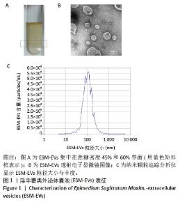
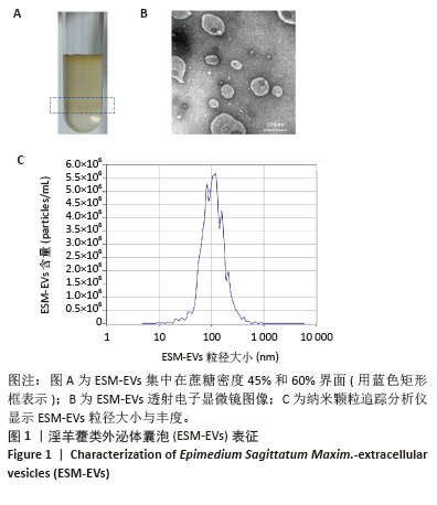
2.1 淫羊藿类外泌体囊泡纳米药物表征 淫羊藿类外泌体囊泡主要集中在蔗糖梯度45%和60%之间(图1A),透射电子显微镜显示淫羊藿类外泌体囊泡呈茶托状,粒径大小在50-200 nm之间(图1B),纳米颗粒追踪分析仪、纳米粒径检测仪与透射电子显微镜结果一致,均显示淫羊藿类外泌体囊泡粒径大小主要集中在50-200 nm之间,颗粒含量约为1.7×10⁸/mL(图1C)。 "
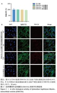
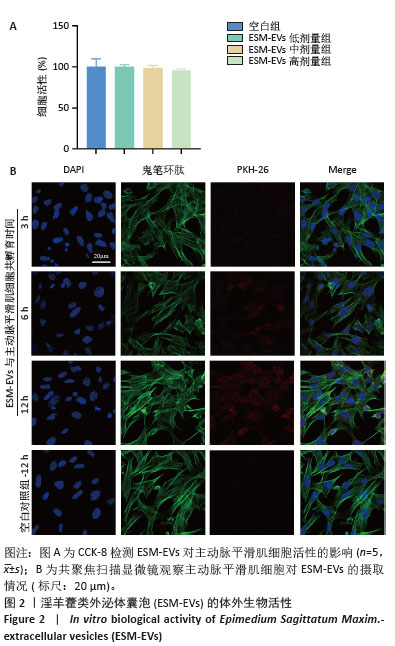
2.2 淫羊藿类外泌体囊泡对主动脉平滑肌细胞活性的影响 CCK-8实验结果显示,淫羊藿类外泌体囊泡在设定的浓度范围内对主动脉平滑肌细胞活性无明显影响(图2A),说明淫羊藿类外泌体囊泡的生物安全性较高,不会引起细胞毒性反应。 2.3 主动脉平滑肌细胞对淫羊藿类外泌体囊泡的摄取情况 孵育3 h,在主动脉平滑肌细胞中可以检测到淫羊藿类外泌体囊泡,表明淫羊藿类外泌体囊泡很快被细胞吸收;孵育12 h,大量主动脉平滑肌细胞已内化淫羊藿类外泌体囊泡(图2B)。 "
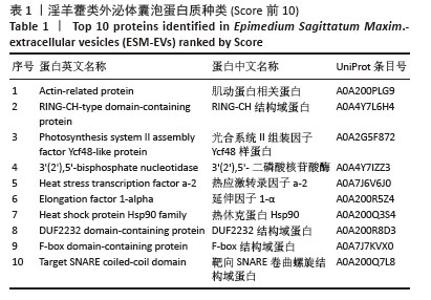
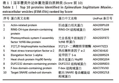
2.4 淫羊藿类外泌体囊泡蛋白质组学结果 通过蛋白组学分析方法,淫羊藿类外泌体囊泡中共鉴定出37种蛋白(表1),其中包括Actin-related protein、Heat shock protein 90、Elongation factor 1-alpha及Histone H4等植物外泌体中常见的重要蛋白。此外,还发现了14-3-3 protein、EF-hand domain、Polyubiquitin等参与细胞骨架稳定、信号转导、蛋白质降解与修饰相关的蛋白质。这些蛋白质共同参与囊泡结构的稳定、细胞间信号的转导、应激反应及代谢调控等多个关键生理过程。 "
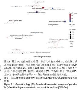
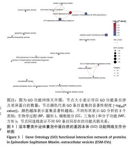
2.5 淫羊藿类外泌体囊泡中蛋白质的GO功能网络互作分析 GO功能网络互作分析发现(图3),这些蛋白在生物学过程中显著富集于细胞对热刺激的响应、肌醇磷酸代谢、谷氨酰胺合成和戊糖磷酸途径等代谢与应激调控过程;在细胞组分方面则集中于RNA聚合酶Ⅱ转录调节复合物、细胞质核周区以及蛋白质复合物;而在分子功能方面主要与转醛醇酶活性、羧化酶/加氧酶激活活性、核苷酸酶活性等相关。同时,网络互作分析提示这些功能并非独立存在,细胞对热刺激的响应与RNA聚合酶Ⅱ介导的转录调控、ATP依赖的蛋白折叠伴侣活性以及蛋白质复合物之间存在密切的相互关系;而谷氨酰胺生物合成过程与谷氨酰胺合成酶活性,肌醇磷酸代谢过程与3’(2’),5’-二磷酸核苷酸酶活性及硫酸盐同化作用之间也表现出明显的功能关联。 "
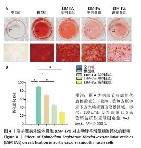
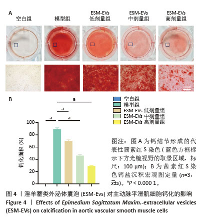
2.6 细胞钙化面积比较 茜素红S染色结果显示,空白组几乎未见红色钙结节沉积,而模型组则有明显且广泛的钙结节沉积(P < 0.001),提示模型成功建立(图4A)。加入淫羊藿类外泌体囊泡处理后,细胞钙化情况明显改善(P < 0.001),钙化面积显著减少(图4B),且随着淫羊藿类外泌体囊泡质量浓度的升高,钙化沉积面积呈明显的剂量依赖性递减趋势,表明淫羊藿类外泌体囊泡对钙化细胞具有明显的抑制作用。 "
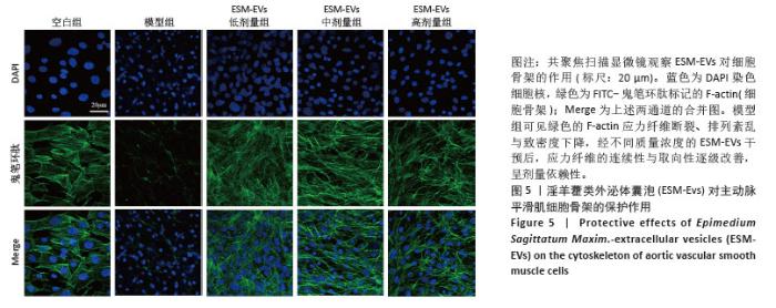
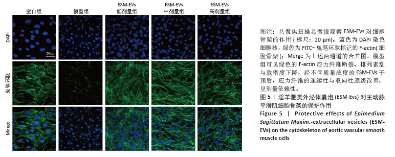
2.7 淫羊藿类外泌体囊泡对细胞骨架的影响 共聚焦扫描显微镜观察鬼笔环肽染色结果显示(图5),空白组细胞骨架结构清晰完整,呈现均匀规则的纤维状分布;模型组则表现出明显的细胞骨架紊乱,大部分细胞仅有DAPI染色的细胞核,而鬼笔环肽染色的细胞骨架结构消失或严重紊乱,提示钙化损伤明显。经低、中、高剂量(5,10,20 mg/L)淫羊藿类外泌体囊泡干预后,细胞骨架结构明显改善,随淫羊藿类外泌体囊泡质量浓度的升高,细胞骨架形态逐渐恢复正常,呈现剂量依赖性改善趋势,提示淫羊藿类外泌体囊泡对钙化诱导的细胞骨架损伤具有显著的保护和修复作用。 "
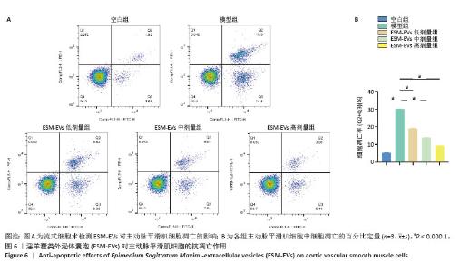
2.8 淫羊藿类外泌体囊泡对主动脉平滑肌细胞的抗凋亡作用 流式细胞术检测结果显示(图6),空白组细胞凋亡比例较低,细胞状态良好;模型组细胞凋亡比例显著增加(P < 0.000 1),细胞受到明显的损伤;经过淫羊藿类外泌体囊泡处理后,细胞凋亡比例明显下降(P < 0.000 1),细胞存活率明显提升,且细胞抗凋亡作用随淫羊藿类外泌体囊泡质量浓度的增加而增强。结果提示,淫羊藿类外泌体囊泡可有效抑制细胞凋亡,对血管平滑肌细胞具有显著的保护作用。"
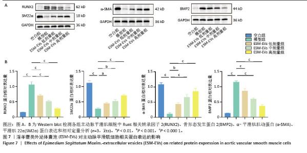
2.9 Runt相关转录因子2、骨形态发生蛋白2、α-平滑肌肌动蛋白、平滑肌22α蛋白表达水平 Western blot结果显示(图7),在钙化诱导下,模型组细胞中Runt相关转录因子2、骨形态发生蛋白2表达显著升高(P < 0.000 1),而α-平滑肌肌动蛋白、平滑肌22α表达明显降低(P < 0.000 1),提示细胞表型发生明显的成骨转化。经过不同质量浓度淫羊藿类外泌体囊泡干预后,Runt相关转录因子2和骨形态发生蛋白2表达明显降低,而α-平滑肌肌动蛋白和平滑肌22α表达则明显升高,提示淫羊藿类外泌体囊泡能够显著抑制细胞的成骨表型转化,维持平滑肌细胞的收缩表型,进而发挥对细胞钙化的抑制作用。"
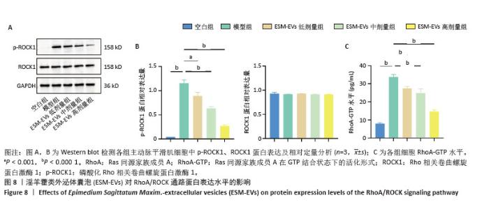
2.10 RhoA/ROCK通路RhoA-GTP、ROCK1、p-ROCK1蛋白表达水平 见图8。ELISA检测结果显示,模型组RhoA-GTP水平显著升高,与空白组相比差异明显(P < 0.000 1)。不同质量浓度淫羊藿类外泌体囊泡干预后,RhoA-GTP水平明显降低(P < 0.000 1)。Western blot结果显示,各组细胞ROCK1总蛋白表达水平无明显差异,而模型组p-ROCK1蛋白表达水平明显升高,提示ROCK通路激活;经淫羊藿类外泌体囊泡干预后,p-ROCK1蛋白表达水平显著下降。以上结果提示,淫羊藿类外泌体囊泡能够有效抑制钙化诱导的RhoA/ROCK信号通路活化。 "
| [1] SOROKIN V, VICKNESON K, KOFIDIS T, et al. Role of Vascular Smooth Muscle Cell Plasticity and Interactions in Vessel Wall Inflammation. Front Immunol. 2020;11:599415. [2] BENZ K, HILGERS KF, DANIEL C, et al. Vascular Calcification in Chronic Kidney Disease: The Role of Inflammation. Int J Nephrol. 2018;2018:4310379. [3] LEE SJ, LEE IK, JEON JH. Vascular Calcification-New Insights Into Its Mechanism. Int J Mol Sci. 2020;21(8):2685. [4] DURHAM AL, SPEER MY, SCATENA M, et al. Role of smooth muscle cells in vascular calcification: implications in atherosclerosis and arterial stiffness. Cardiovasc Res. 2018;114(4):590-600. [5] SINGH A, TANDON S, TANDON C. An update on vascular calcification and potential therapeutics. Mol Biol Rep. 2021;48(1):887-896. [6] AKHMANOVA A, KAPITEIN LC. Mechanisms of microtubule organization in differentiated animal cells. Nat Rev Mol Cell Biol. 2022;23(8):541-558. [7] DOKUMACIOGLU E, DUZCAN I, ISKENDER H, et al. RhoA/ROCK-1 Signaling Pathway and Oxidative Stress in Coronary Artery Disease Patients. Braz J Cardiovasc Surg. 2022;37(2):212-218. [8] LIU H, YIN H, WANG Z, et al. Rho A/ROCK1 signaling-mediated metabolic reprogramming of valvular interstitial cells toward Warburg effect accelerates aortic valve calcification via AMPK/RUNX2 axis. Cell Death Dis. 2023;14(2):108. [9] ZHOU W, YUAN X, LI J, et al. Retinol binding protein 4 promotes the phenotypic transformation of vascular smooth muscle cells under high glucose condition via modulating RhoA/ROCK1 pathway. Transl Res. 2023; 259:13-27. [10] TSUDA T, IMANISHI M, OOGOSHI M, et al. Rho-associated protein kinase and cyclophilin a are involved in inorganic phosphate-induced calcification signaling in vascular smooth muscle cells. J Pharmacol Sci. 2020;142(3): 109-115. [11] SUN Y, HUANG D, ZHANG Y. The bone-vascular axis: the link between osteoporosis and vascular calcification. Mol Cell Biochem. 2025;480(6): 3413-3427. [12] VACHEY C, CANDELLIER A, TOUTAIN S, et al. The Bone-Vascular Axis in Chronic Kidney Disease: From Pathophysiology to Treatment. Curr Osteoporos Rep. 2024;22(1):69-79. [13] WANG ZX, LUO ZW, LI FX, et al. Aged bone matrix-derived extracellular vesicles as a messenger for calcification paradox. Nat Commun. 2022; 13(1):1453. [14] GAO J, XIANG S, WEI X, et al. Icariin Promotes the Osteogenesis of Bone Marrow Mesenchymal Stem Cells through Regulating Sclerostin and Activating the Wnt/β-Catenin Signaling Pathway. Biomed Res Int. 2021; 2021:6666836. [15] 薛春阳,王秀会.淫羊藿苷调节酸性微环境减轻绝经后老年骨质疏松性疼痛[J].中国组织工程研究,2024,28(28):4461-4468. [16] ZENG Y, XIONG Y, YANG T, et al. Icariin and its metabolites as potential protective phytochemicals against cardiovascular disease: From effects to molecular mechanisms. Biomed Pharmacother. 2022;147:112642. [17] SHARMA S, IQUBAL A, KHAN V, et al. Icariin ameliorates oxidative stress-induced inflammation, apoptosis, and heart failure in isoproterenol-challenged Wistar rats. Iran J Basic Med Sci. 2023;26(5):517-525. [18] HUANG X, LEI S, XIONG X, et al. Unveiling the Therapeutic Potential of Herba Epimedii: Enhancing Bone Healing Through Cytoskeletal Regulation of RhoA/Rock1 Pathway. Chem Biodivers. 2024;21(2): e202301383. [19] 韩一旦,张海凤,许云腾,等.淫羊藿苷通过caveolin-1/Hippo信号通路促进BMSCs成骨分化改善骨代谢紊乱的机制研究[J].中国中药杂志, 2025,50(3):600-608. [20] 闵捷,李雅琴,罗云梅,等.淫羊藿苷的心血管保护作用及机制研究进展[J].中国新药与临床杂志,2021,40(11):741-745. [21] 白晓君,刘艳,郜珊珊,等.淫羊藿苷抑制氧化应激诱导主动脉血管平滑肌细胞钙化的机制研究[J].中国中药杂志,2021,46(17):4497-4503. [22] XU Z, XU Y, ZHANG K, et al. Plant-derived extracellular vesicles (PDEVs) in nanomedicine for human disease and therapeutic modalities. J Nanobiotechnology. 2023;21(1):114. [23] LIAN MQ, CHNG WH, LIANG J, et al. Plant-derived extracellular vesicles: Recent advancements and current challenges on their use for biomedical applications. J Extracell Vesicles. 2022;11(12):e12283. [24] NEMATI M, SINGH B, MIR RA, et al. Plant-derived extracellular vesicles: a novel nanomedicine approach with advantages and challenges. Cell Commun Signal. 2022;20(1):69. [25] ALZAHRANI FA, KHAN MI, KAMELI N, et al. Plant-Derived Extracellular Vesicles and Their Exciting Potential as the Future of Next-Generation Drug Delivery. Biomolecules. 2023;13(5):839. [26] KANG SJ, LEE JH, RHEE WJ. Engineered plant-derived extracellular vesicles for targeted regulation and treatment of colitis-associated inflammation. Theranostics. 2024;14(14):5643-5661. [27] MARTÍNEZ FAJARDO C, MOROTE L, MORENO-GIMÉNEZ E, et al. Exosome-like nanoparticles from Arbutus unedo L. mitigate LPS-induced inflammation via JAK-STAT inactivation. Food Funct. 2024; 15(22):11280-11290. [28] DE ROBERTIS M, SARRA A, D’ORIA V, et al. Blueberry-Derived Exosome-Like Nanoparticles Counter the Response to TNF-α-Induced Change on Gene Expression in EA.hy926 Cells. Biomolecules. 2020; 10(5):742. [29] NIU W, XIAO Q, WANG X, et al. A Biomimetic Drug Delivery System by Integrating Grapefruit Extracellular Vesicles and Doxorubicin-Loaded Heparin-Based Nanoparticles for Glioma Therapy. Nano Lett. 2021;21(3): 1484-1492. [30] KIM J, LI S, ZHANG S, et al. Plant-derived exosome-like nanoparticles and their therapeutic activities. Asian J Pharm Sci. 2022;17(1):53-69. [31] GUPTA D, ZICKLER AM, EL ANDALOUSSI S. Dosing extracellular vesicles. Adv Drug Deliv Rev. 2021;178:113961. [32] WELSH JA, GOBERDHAN DCI, O’DRISCOLL L, et al. Minimal information for studies of extracellular vesicles (MISEV2023): From basic to advanced approaches. J Extracell Vesicles. 2024;13(2):e12404. [33] HOU Z, WANG X, YANG Z, et al. Pomegranate-derived exosome-like nanovesicles ameliorate high-fat diet–induced nonalcoholic fatty liver disease via alleviating mitochondrial dysfunction. J Funct Foods. 2023;108:105734. [34] DEMICHEV V, MESSNER CB, VERNARDIS SI, et al. DIA-NN: neural networks and interference correction enable deep proteome coverage in high throughput. Nat Methods. 2020;17(1):41-44. [35] WANG S, LI W, HU L, et al. NAguideR: performing and prioritizing missing value imputations for consistent bottom-up proteomic analyses. Nucleic Acids Res. 2020;48(14):e83. [36] MILACIC M, BEAVERS D, CONLEY P, et al. The Reactome Pathway Knowledgebase 2024. Nucleic Acids Res. 2024;52(D1):D672-D678. [37] PANG Q, WANG P, PAN Y, et al. Irisin protects against vascular calcification by activating autophagy and inhibiting NLRP3-mediated vascular smooth muscle cell pyroptosis in chronic kidney disease. Cell Death Dis. 2022;13(3):283. [38] KÜRTÖSI B, KAZSOKI A, ZELKÓ R. A Systematic Review on Plant-Derived Extracellular Vesicles as Drug Delivery Systems. Int J Mol Sci. 2024; 25(14):7559. [39] KIM M, JANG H, KIM W, et al. Therapeutic Applications of Plant-Derived Extracellular Vesicles as Antioxidants for Oxidative Stress-Related Diseases. Antioxidants (Basel). 2023;12(6):1286. [40] TIWARI A, SONI N, DONGRE S, et al. The role of plant-derived extracellular vesicles in ameliorating chronic diseases. Mol Biol Rep. 2025;52(1):360. [41] FENG W, TENG Y, ZHONG Q, et al. Biomimetic Grapefruit-Derived Extracellular Vesicles for Safe and Targeted Delivery of Sodium Thiosulfate against Vascular Calcification. ACS Nano. 2023;17(24): 24773-24789. [42] 陈钰璘,何莹莹,胡凯,等.瓜蒌类外泌体囊泡防治动脉粥样硬化的作用及机制[J].中国组织工程研究,2026,30(7):1768-1781. [43] SHARMA S, MAHANTY M, RAHAMAN SG, et al. Avocado-derived extracellular vesicles loaded with ginkgetin and berberine prevent inflammation and macrophage foam cell formation. J Cell Mol Med. 2024;28(7):e18177. [44] ZHANG S, XIA J, ZHU Y, et al. Establishing Salvia miltiorrhiza-Derived Exosome-like Nanoparticles and Elucidating Their Role in Angiogenesis. Molecules. 2024;29(7):1599. [45] LÓPEZ DE LAS HAZAS MC, TOMÉ-CARNEIRO J, DEL POZO-ACEBO L, et al. Therapeutic potential of plant-derived extracellular vesicles as nanocarriers for exogenous miRNAs. Pharmacol Res. 2023;198:106999. [46] SHUKEN SR. An Introduction to Mass Spectrometry-Based Proteomics. J Proteome Res. 2023;22(7):2151-2171. [47] LIU Z, CHEN X, YANG S, et al. Integrated mass spectrometry strategy for functional protein complex discovery and structural characterization. Curr Opin Chem Biol. 2023;74:102305. [48] HUNTER CL, BONS J, SCHILLING B. Perspectives and opinions from scientific leaders on the evolution of data-independent acquisition for quantitative proteomics and novel biological applications. Aust J Chem. 2023;76(8): 379-398. [49] YANG Y, LIN L, QIAO L. Deep learning approaches for data-independent acquisition proteomics. Expert Rev Proteomics. 2021;18(12):1031-1043. [50] LIU Y, XIAO S, WANG D, et al. A review on separation and application of plant-derived exosome-like nanoparticles. J Sep Sci. 2024;47(8): e2300669. [51] SUHARTA S, BARLIAN A, HIDAJAH AC, et al. Plant-derived exosome-like nanoparticles: A concise review on its extraction methods, content, bioactivities, and potential as functional food ingredient. J Food Sci. 2021; 86(7):2838-2850. [52] OBSILOVA V, OBSIL T. Structural insights into the functional roles of 14-3-3 proteins. Front Mol Biosci. 2022;9:1016071. [53] PENNINGTON KL, CHAN TY, TORRES MP, et al. The dynamic and stress-adaptive signaling hub of 14-3-3: emerging mechanisms of regulation and context-dependent protein-protein interactions. Oncogene. 2018; 37(42):5587-5604. [54] CHIOSIS G, DIGWAL CS, TREPEL JB, et al. Structural and functional complexity of HSP90 in cellular homeostasis and disease. Nat Rev Mol Cell Biol. 2023;24(11):797-815. [55] LI W, SUN W, YANG CH, et al. Tanshinone II a protects against lipopolysaccharides-induced endothelial cell injury via Rho/Rho kinase pathway. Chin J Integr Med. 2014;20(3):216-223. [56] CHEN J, WANG H, GAO C, et al. Tetramethylpyrazine alleviates LPS-induced inflammatory injury in HUVECs by inhibiting Rho/ROCK pathway. Biochem Biophys Res Commun. 2019;514(1):329-335. [57] XIE L, WANG T, LIN S, et al. Uncaria Rhynchophylla attenuates angiotensin Ⅱ-induced myocardial fibrosis via suppression of the RhoA/ROCK1 pathway. Biomed Pharmacother. 2022;146:112607. [58] ÁLVAREZ-SANTOS MD, ÁLVAREZ-GONZÁLEZ M, ESTRADA-SOTO S, et al. Regulation of Myosin Light-Chain Phosphatase Activity to Generate Airway Smooth Muscle Hypercontractility. Front Physiol. 2020;11:701. [59] YAP C, MIEREMET A, DE VRIES CJM, et al. Six Shades of Vascular Smooth Muscle Cells Illuminated by KLF4 (Krüppel-Like Factor 4). Arterioscler Thromb Vasc Biol. 2021;41(11):2693-2707. [60] CAI X, WANG KC, MENG Z. Mechanoregulation of YAP and TAZ in Cellular Homeostasis and Disease Progression. Front Cell Dev Biol. 2021;9:673599. [61] OH YJ, KIM H, KIM AJ, et al. Reduction of Secreted Frizzled-Related Protein 5 Drives Vascular Calcification through Wnt3a-Mediated Rho/ROCK/JNK Signaling in Chronic Kidney Disease. Int J Mol Sci. 2020;21(10):3539. [62] GU X, MASTERS KS. Role of the Rho pathway in regulating valvular interstitial cell phenotype and nodule formation. Am J Physiol Heart Circ Physiol. 2011;300(2):H448-458. |
| [1] | Zhou Sirui, Xu Yukun, Zhao Kewei. Ideas and methods of anti-melanogenesis of Angelica dahurica extracellular vesicles [J]. Chinese Journal of Tissue Engineering Research, 2026, 30(7): 1747-1754. |
| [2] | Yang Xiao, Bai Yuehui, Zhao Tiantian, Wang Donghao, Zhao Chen, Yuan Shuo. Cartilage degeneration in temporomandibular joint osteoarthritis: mechanisms and regenerative challenges [J]. Chinese Journal of Tissue Engineering Research, 2026, 30(4): 926-935. |
| [3] | Zhang Chuanlin, Wu Yuhuai, Zhou Haixiang, Liu Jianping, Xu Qian, Zhang Lili, Li Cuie. An innovative strategy for engineered exosomal long non-coding RNAs in treatment of osteoporosis [J]. Chinese Journal of Tissue Engineering Research, 2026, 30(31): 8264-8271. |
| [4] | Zhang Enqi, Wu Hongwei, Wu Chengliang, Yang Tianqi, Tang Weijie, Wang Wenhui, Yu Zeming, Zhang Yanwen, Wang Yuebing. Extracellular vesicles as gene therapy vectors: engineering strategies, mechanistic advantages, and clinical applications [J]. Chinese Journal of Tissue Engineering Research, 2026, 30(31): 8210-8218. |
| [5] | Wang Zhengye, Liu Wanlin, Zhao Zhenqun. Mechanism by which vascular endothelial growth factor A targets regulation of angiogenesis in the treatment of steroid-induced osteonecrosis of the femoral head [J]. Chinese Journal of Tissue Engineering Research, 2026, 30(3): 671-679. |
| [6] | Yang Hu, Zheng Yu, Jia Chengming, Wang Tong, Zhang Guangfei, Ji Yaoyao. Immune microenvironment regulates bone regeneration [J]. Chinese Journal of Tissue Engineering Research, 2026, 30(3): 701-710. |
| [7] | Yang Yunhong, Guo Lihua, Tang Han, Lin Lvping, Kuang Hongjun, Zhao Hong. Proteomic analysis of the mechanism of moxibustion intervention in a rat model of atopic dermatitis [J]. Chinese Journal of Tissue Engineering Research, 2026, 30(29): 7581-7591. |
| [8] | Wang Ziqi, Bu Xianzhong, Guo Xiaohui, Li Hanxi, Qian Yuhao, Wang Yixin, Bu Baoxian. Integrated proteomics and transcriptomics analysis of the mechanism of Buyang Huanwu Tang in protecting the acute spinal cord injury rat model [J]. Chinese Journal of Tissue Engineering Research, 2026, 30(25): 6472-6488. |
| [9] | Liu Jinwei, Zhang Dan, Guo Hongli, Chen Huan, Li Jingjing, Cao Weiguo. Combined proteomics and metabolomics analysis of pathological mechanisms in mouse models of coronary heart disease [J]. Chinese Journal of Tissue Engineering Research, 2026, 30(25): 6544-6553. |
| [10] | Zhou Tianle, Wang Wei, Zhang Zhiwen, Liu Ximing. Application and progress of transcriptomics and proteomics techniques in the study of intervertebral disc degeneration [J]. Chinese Journal of Tissue Engineering Research, 2026, 30(23): 5925-5933. |
| [11] | Ding Yan, Nie Hongguang, Sun Yu. Mechanisms and clinical strategies of mesenchymal stem cell-derived extracellular vesicles intervening in cell regulatory networks to treat pulmonary fibrosis [J]. Chinese Journal of Tissue Engineering Research, 2026, 30(19): 5024-5032. |
| [12] | Wang Xinyue, Li Hongli, Guo Chunhui, Chen Jibing, Yu Hua. Changes in the expression of six microRNAs in ovarian tissue from animal models of premature ovarian failure and in peripheral blood of patients with premature ovarian failure [J]. Chinese Journal of Tissue Engineering Research, 2026, 30(18): 4675-4684. |
| [13] | Liang Zhou, Pan Chengzhen, Chen Feng, Zhang Chi, Yang Bo, Wei Zongbo, Meng Jianhua, Zhou Zhu. Identification of diagnostic biomarkers related to osteoporosis exosomes and preliminary drug screening [J]. Chinese Journal of Tissue Engineering Research, 2026, 30(13): 3458-3473. |
| [14] | Liu Jing, Zhao Xiaoli, Xia Tian. Role of extracellular vesicle-mediated intercellular communication in female follicle reproduction, preimplantation embryo development and implantation [J]. Chinese Journal of Tissue Engineering Research, 2026, 30(1): 218-228. |
| [15] | Zhou Yang, Liu Kexin, Wang Deli, Sun Zhang. Regenerative effects of engineered extracellular vesicles on repairing bone defects [J]. Chinese Journal of Tissue Engineering Research, 2025, 29(36): 7839-7847. |
| Viewed | ||||||
|
Full text |
|
|||||
|
Abstract |
|
|||||